Review
. 2019;26(19):3620-3638.
doi: 10.2174/0929867324666170530080636.

The Role of Diet in the Pathogenesis of Cholesterol Gallstones

Affiliations
Review

The Role of Diet in the Pathogenesis of Cholesterol Gallstones

Agostino Di Ciaula et al. Curr Med Chem. 2019.

Abstract

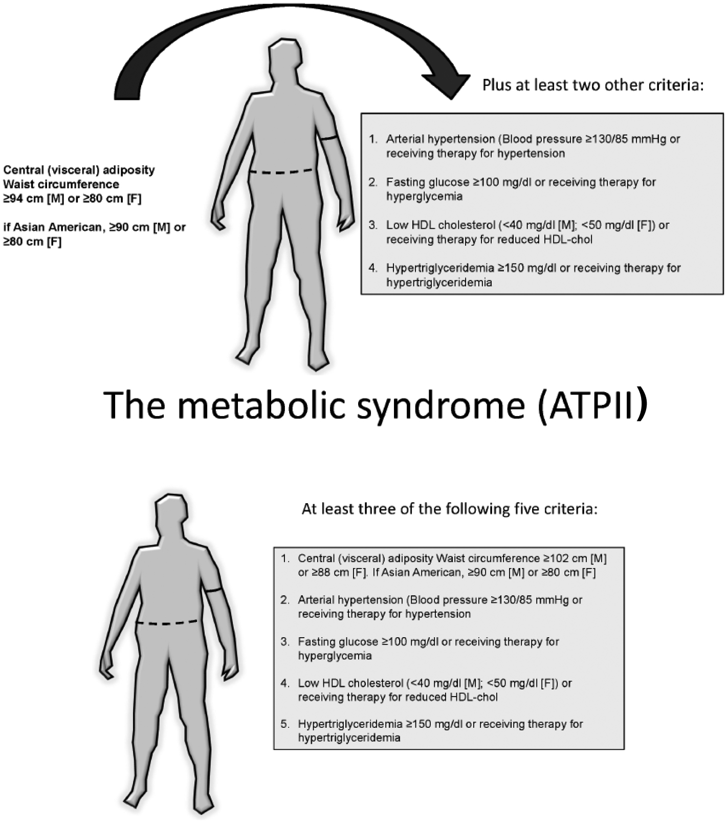
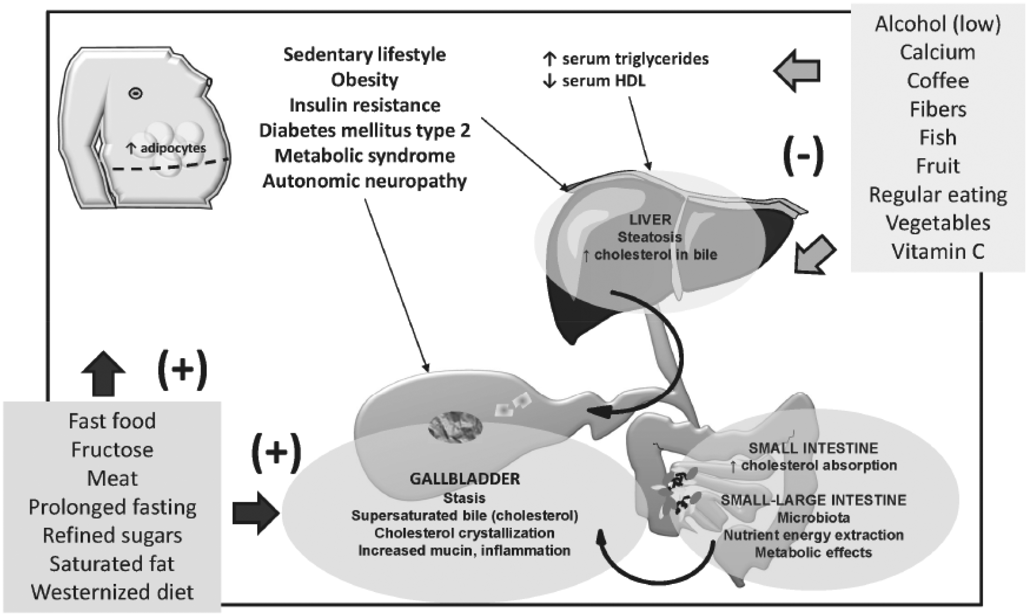
Cholesterol gallstone disease is a major health problem in Westernized countries and depends on a complex interplay between genetic factors, lifestyle and diet, acting on specific pathogenic mechanisms. Overweigh, obesity, dyslipidemia, insulin resistance and altered cholesterol homeostasis have been linked to increased gallstone occurrence, and several studies point to a number of specific nutrients as risk- or protective factors with respect to gallstone formation in humans. There is a rising interest in the identification of common and modifiable dietetic factors that put the patients at risk of gallstones or that are able to prevent gallstone formation and growth. In particular, dietary models characterized by increased energy intake with highly refined sugars and sweet foods, high fructose intake, low fiber contents, high fat, consumption of fast food and low vitamin C intake increase the risk of gallstone formation. On the other hand, high intake of monounsaturated fats and fiber, olive oil and fish (ω-3 fatty acids) consumption, vegetable protein intake, fruit, coffee, moderate alcohol consumption and vitamin C supplementation exert a protective role. The effect of some confounding factors (e.g., physical activity) cannot be ruled out, but general recommendations about the multiple beneficial effects of diet on cholesterol gallstones must be kept in mind, in particular in groups at high risk of gallstone formation.

Keywords: Caloric intake; diet; fibers; macronutrients; obesity; weight loss..


Conflict of interest statement

CONFLICT OF INTEREST

The authors declare no conflict of interest, financial or otherwise.

Figures

Fig. (1).
Fig. (1).
a) Definition of the metabolic syndrome according to the International Diabetes Federation (IDF) [42, 191]. b) Definition of the metabolic syndrome according to the National Cholesterol Education Program (NCEP) Adult Treatment Panel III (ATP III) [36].
Fig. (2).
Fig. (2).
The potential deleterious metabolic effects of excess fructose consumption in diet are shown by the study of metabolic pathways of fructose in the liver. a) A first step is the conversion of fructose to fructose-1-phosphate and then to glyceraldehyde, dihydroxyacetone phosphate, and glyceraldehyde-3-phosphate. This first important step paves the way to the synthesis of glycogen and the synthesis of triglycerides, this latter pathway, a potential risk for liver steatosis. b) The conversion of fructose to glycogen is shown in the liver, anticipated by gluconeogenic precursors. Once liver glycogen is accumulated, the following pathway re-direct the fructose intermediates towards the synthesis of triglyceride. c) Conversion of fructose to triglyceride in the liver. Especially in the presence of excess fructose intake, the steps leading to glycerol synthesis and pyruvate synthesis are followed by construction of the backbone of triglyceride.
Fig. (2).
Fig. (2).
The potential deleterious metabolic effects of excess fructose consumption in diet are shown by the study of metabolic pathways of fructose in the liver. a) A first step is the conversion of fructose to fructose-1-phosphate and then to glyceraldehyde, dihydroxyacetone phosphate, and glyceraldehyde-3-phosphate. This first important step paves the way to the synthesis of glycogen and the synthesis of triglycerides, this latter pathway, a potential risk for liver steatosis. b) The conversion of fructose to glycogen is shown in the liver, anticipated by gluconeogenic precursors. Once liver glycogen is accumulated, the following pathway re-direct the fructose intermediates towards the synthesis of triglyceride. c) Conversion of fructose to triglyceride in the liver. Especially in the presence of excess fructose intake, the steps leading to glycerol synthesis and pyruvate synthesis are followed by construction of the backbone of triglyceride.
Fig. (3).
Fig. (3).
The structure of the primary bile acids, secondary bile acids and “tertiary” bile acid ursodeoxycholic acid. Bile acids are synthesized from cholesterol in the liver as soluble amphiphiles. The biliary bile acid pool in humans is mainly made of the primary bile acids, i.e. the 3,7,12-trihydroxy cholic acid and the 3,7-dihydroxy chenodeoxyholic acid. After being secreted in bile and entering the recirculation in the intestine, the primary bile acids are biotransformed by colonic bacteria into secondary bile acids, i.e. the 3,12-dihydroxy deoxycholic acid (from cholic acid) and the3- monohydroxy lithocholic acid (from chenodeoxycholic acid). “Tertiary” bile acids are the result of modification of secondary bile acids by intestinal flora or hepatocytes. These are the sulfate ester of lithocholic acid and the 3,7-dihydroxy ursodeoxycholic acid (UDCA), and the 7β-epimer of chenodeoxycholic acid. Bile acids are highly soluble, detergent-like amphiphilic molecules; the hydrophilic (polar) areas of bile acids are the hydroxyl groups and conjugation side chain of either glycine or taurine and their hydrophobic (nonpolar) area is the ringed steroid nucleus [64, 192].
Fig. (3).
Fig. (3).
The structure of the primary bile acids, secondary bile acids and “tertiary” bile acid ursodeoxycholic acid. Bile acids are synthesized from cholesterol in the liver as soluble amphiphiles. The biliary bile acid pool in humans is mainly made of the primary bile acids, i.e. the 3,7,12-trihydroxy cholic acid and the 3,7-dihydroxy chenodeoxyholic acid. After being secreted in bile and entering the recirculation in the intestine, the primary bile acids are biotransformed by colonic bacteria into secondary bile acids, i.e. the 3,12-dihydroxy deoxycholic acid (from cholic acid) and the3- monohydroxy lithocholic acid (from chenodeoxycholic acid). “Tertiary” bile acids are the result of modification of secondary bile acids by intestinal flora or hepatocytes. These are the sulfate ester of lithocholic acid and the 3,7-dihydroxy ursodeoxycholic acid (UDCA), and the 7β-epimer of chenodeoxycholic acid. Bile acids are highly soluble, detergent-like amphiphilic molecules; the hydrophilic (polar) areas of bile acids are the hydroxyl groups and conjugation side chain of either glycine or taurine and their hydrophobic (nonpolar) area is the ringed steroid nucleus [64, 192].
Fig. (4).
Fig. (4).
The structure of the potent gastrointestinal hormone cholecystokinin (CCK) acting on the smooth muscle contractility at the level of the gallbladder, upon fat stimulation in food [2, 193].The figure depicts the 8-amino acid C-terminal fragment of cholecystokinin, and also known as CCK-8. From National Center for Biotechnology Information. PubChem Compound Database; CID=9833444, https://pubchem.ncbi.nlm.nih.gov/compound/9833444 (accessed May 12, 2016).
Fig. (5).
Fig. (5).
The structure of ω−3 fatty acids eicosapentaenoic acid (top) and docosahexaenoic acid (bottom).
Fig. (6).
Fig. (6).
Dietary factors and lifestyles may act as risk factors (+) or protective factors (−) on typical pathogenic factors of cholesterol cholelithiasis, involving the liver, the intestine and the gallbladder.

References

    1. Attili AF; Capocaccia R; Carulli N; Festi D; Roda E; Barbara L; Capocaccia L; Menotti A; Okolicsanyi L; Ricci G; Lalloni L; Mariotti S; Sama C; Scafato E Factors associated with gallstone disease in the MICOL experience. Multicenter Italian Study on Epidemiology of Cholelithiasis. Hepatology, 1997, 26, 809–818. - PubMed
    1. Portincasa P; Moschetta A; Palasciano G Cholesterol gallstone disease. Lancet, 2006, 368, 230–239. - PubMed
    1. Everhart JE; Ruhl CE Burden of digestive diseases in the United States part I: overall and upper gastrointestinal diseases. Gastroenterology, 2009, 136, 376–386. - PubMed
    1. Portincasa P; Wang DQH Gallstones. In: Podolsky KD, Camilleri M, Fitz JG, Kalloo AN, Shanahan F, Wang TC, editors. Yamada’s Textbook of Gastroenterology. 6th ed. UK: Wiley-Blackwell; 2015. p. 1808–1834.
    1. Portincasa P; Wang DQH Gallstones. In: Podolsky KD, Camilleri M, Fitz JG, Kalloo AN, Shanahan F, Wang TC, editors. Yamada’s Atlas of Gastroenterology. 5th ed. UK: Wiley-Blackwell; 2016. p. 335–353.