Case Reports
. 2017 May 29;5(1):40.
doi: 10.1186/s40478-017-0447-3.

Rho-associated protein kinase 2 (ROCK2): a new target of autoimmunity in paraneoplastic encephalitis

Affiliations
Case Reports

Rho-associated protein kinase 2 (ROCK2): a new target of autoimmunity in paraneoplastic encephalitis

Stoyan Popkirov et al. Acta Neuropathol Commun. .

Abstract

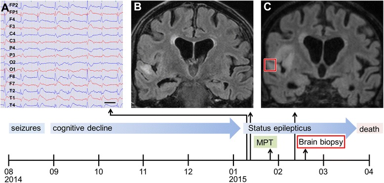
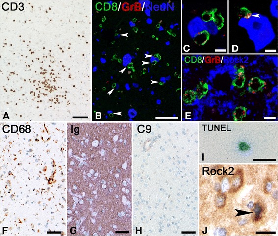
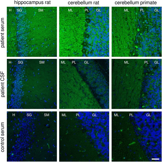
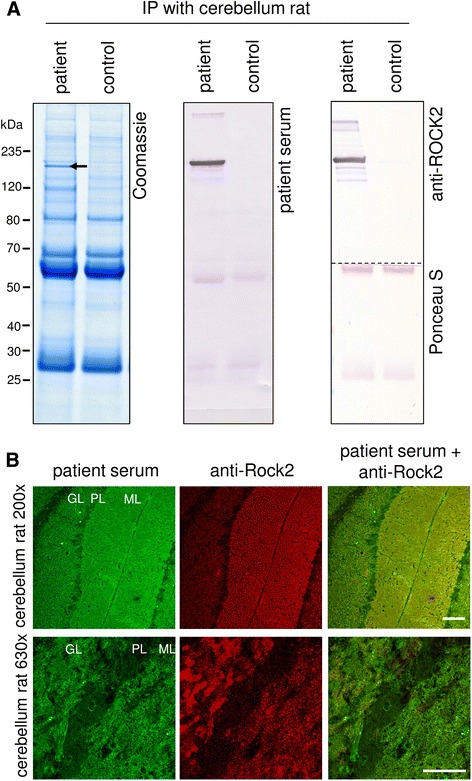
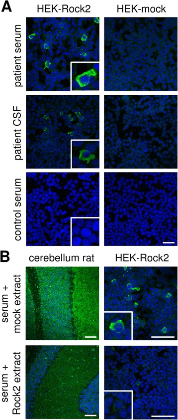
Onconeural antibodies are associated with cancer and paraneoplastic encephalitis. While their pathogenic role is still largely unknown, their high diagnostic value is undisputed. In this study we describe the discovery of a novel target of autoimmunity in an index case of paraneoplastic encephalitis associated with urogenital cancer.A 75-year-old man with a history of invasive bladder carcinoma 6 years ago with multiple recurrences and a newly discovered renal cell carcinoma presented with seizures and progressive cognitive decline followed by super-refractory status epilepticus. Clinical and ancillary findings including brain biopsy suggested paraneoplastic encephalitis. Immunohistochemistry of the brain biopsy was used to characterize the inflammatory response. Indirect immunofluorescence assay (IFA) was used for autoantibody screening. The autoantigen was identified by histo-immunoprecipitation and mass spectrometry and was validated by expressing the recombinant antigen in HEK293 cells and neutralization tests. Sera from 125 control patients were screened using IFA to test for the novel autoantibodies.IFA analysis of serum revealed a novel autoantibody against brain tissue. An intracellular enzyme, Rho-associated protein kinase 2 (ROCK2), was identified as target-antigen. ROCK2 was expressed in affected brain tissue and archival bladder tumor samples of this patient. Brain histopathology revealed appositions of cytotoxic CD8+ T cells on ROCK2-positive neurons. ROCK2 antibodies were not found in the sera of 20 patients with bladder cancer and 17 with renal cancer, both without neurological symptoms, 49 healthy controls, and 39 patients with other antineuronal autoantibodies. In conclusion, novel onconeural antibodies targeting ROCK2 are associated with paraneoplastic encephalitis and should be screened for when paraneoplastic neurological syndromes, especially in patients with urogenital cancers, occur.

Keywords: Autoantibody; Paraneoplastic encephalitis; Rho-associated protein kinase 2; Status epilepticus; Urogential cancer.


Figures

Fig. 1
Fig. 1
Clinical course, EEG and MRI of the index patient. Time axis of disease development with main symptoms, treatments and findings. a EEG excerpt in average potential reference montage shows right-sided periodic lateralized epileptiform discharges (PLED); bar: 1 s. b Coronal T2-FLAIR MRI on admission shows hyperintensity in the right superior temporal gyrus, compatible with inflammation. c Coronally reconstructed 3D-FLAIR MRI shows progression after 30 days. Abbreviations: MPT, methylprednisolone pulse therapy; T2-FLAIR, T2-weighted fluid attenuation inversion recovery; 3D-FLAIR, three dimensionally acquired fluid attenuation inversion recovery. Red square indicates site of brain biopsy
Fig. 2
Fig. 2
Immunohistochemistry of brain biopsy suggesting T cell-mediated inflammation. a Staining for CD3 shows the presence of large numbers of T cells in the parenchyma. Bar: 10 μm. b Confocal triple staining for CD8 (green), GrB (red) and NeuN (blue) shows multiple appositions (arrowheads) of cytotoxic T cells on neurons. Bar: 20 μm. c and d show the same triple staining for CD8 (green), GrB (red) and NeuN (blue) as (b). The staining in (c) shows that multiple CD8+/GrB+ T cells can be found in apposition to neurons. The staining in (d) again shows GrB translocation (arrowhead), now facing the neuronal membrane. Bars: 5 μm. e Triple staining for CD8 (green), GrB (red) and ROCK2 (blue) reveals that neurons with appositions of T cells are Rock2 positive. Bar: 20 μm. f Staining for CD68 shows moderate microglia activation. Bar: 5 μm. g Staining for anti-human-Ig shows staining in the parenchyma as a sign of immunoglobulin leakage. Bar: 20 μm (h) Staining for C9neo fails to reveal parenchymal staining. Bar: 50 μm. i TUNEL stain reveals the presence of single degenerating cells in the parenchyma. Bar: 20 μm. j Staining for ROCK2 shows the presence of multiple ROCK2+ neurons. The neuron indicated by the arrowhead shows a condensed nucleus suggesting apoptosis. Bar: 10 μm
Fig. 3
Fig. 3
Immunofluorescence staining of central nervous system tissue. Cryosections were incubated with patient’s serum, control serum (each 1:32) or patient’s CSF (undiluted) in the first step, and with Alexa488-labelled goat anti-human immunoglobulin G (green) in the second step. Nuclei were counterstained by incubation with TO-PRO-3 iodide (blue). A fine-granular to homogeneous staining of molecular layer of both rat and monkey cerebellum as well as rat hippocampus and plaster-like staining of cerebellum granular layer excluding Purkinje cells was obtained. Scale bar = 50 μm; all figures same magnification. H = hilus, SM = stratum moleculare, SG = stratum granulosum, ML = molecular layer, PL = Purkinje cell layer, GL = granule cell layer
Fig. 4
Fig. 4
Identification of Rho-associated protein kinase 2 as the target antigen. a Lysates of rat cerebellum were incubated with patient or control sera (1:33). Immunocomplexes were isolated with protein-G-coated magnetic beads, eluted by SDS and subjected to SDS-PAGE analysis followed by (left) staining with colloidal Coomassie, (middle) Western blot using patient serum or (right) Western blot using polyclonal rabbit anti-ROCK2 and Ponceau S staining. Arrow indicates the position of the immunoprecipitated antigen at about 160 kDa. b Double immunofluorescence staining of cerebellar tissues with patient serum (1:50, green) and rabbit anti-ROCK2 antibody (1:250, red). The anti-ROCK2 antibody produced fluorescence patterns matching those generated by the patient’s serum. Scale bar = 100 μm ML = molecular layer, PL = Purkinje cell layer, GL = granule cell layer
Fig. 5
Fig. 5
Verification of ROCK2 as the novel autoantigen by indirect immunofluorescence. a: Indirect immunofluorescence using acetone-fixed ROCK2- or mock-transfected HEK293 cells incubated with patient’s serum, control serum (each 1:320) or patient’s CSF (1:10) (green). Scale bar = 50 μm; all figures same magnification. b: Neutralisation of immunofluorescence reaction on cerebellum rat and ROCK2-transfected HEK293 cells. Patient serum (green) was pre-incubated with extracts of HEK293 cells transfected with ROCK2 or with empty vector as control. The extract containing ROCK2 abolished the immune reaction. Nuclei were counterstained by incubation with TO-PRO-3 iodide (blue). Inserts show enlargement of positive and negative ROCK2-transfected cells. Scale bar = 100 μm

References

    1. Abe H, Kamai T, Hayashi K, et al. The Rho-kinase inhibitor HA-1077 suppresses proliferation/migration and induces apoptosis of urothelial cancer cells. BMC Cancer. 2014;14:412. doi: 10.1186/1471-2407-14-412. - DOI - PMC - PubMed
    1. Abe H, Kamai T, Tsujii T, et al. Possible role of the RhoC/ROCK pathway in progression of clear cell renal cell carcinoma. Biomed Res. 2008;29:155–161. doi: 10.2220/biomedres.29.155. - DOI - PubMed
    1. Bauer J, Bien CG. Neuropathology of autoimmune encephalitides. Handb Clin Neurol. 2016;133:107–120. doi: 10.1016/B978-0-444-63432-0.00007-4. - DOI - PubMed
    1. Bauer J, Lassmann H. Neuropathological techniques to investigate central nervous system sections in multiple sclerosis. Methods Mol Biol. 2016;1304:211–229. doi: 10.1007/7651_2014_151. - DOI - PubMed
    1. Bell BB, Tognoni PG, Bihrle R. Limbic encephalitis as a paraneoplastic manifestation of renal cell carcinoma. J Urol. 1998;160:828. doi: 10.1016/S0022-5347(01)62798-8. - DOI - PubMed

Publication types

MeSH terms