Review
. 2017 Jul 7;429(14):2127-2147.
doi: 10.1016/j.jmb.2017.05.017. Epub 2017 May 26.

RNA G-Quadruplexes in Biology: Principles and Molecular Mechanisms

Affiliations
Review

RNA G-Quadruplexes in Biology: Principles and Molecular Mechanisms

Marta M Fay et al. J Mol Biol. .

Abstract

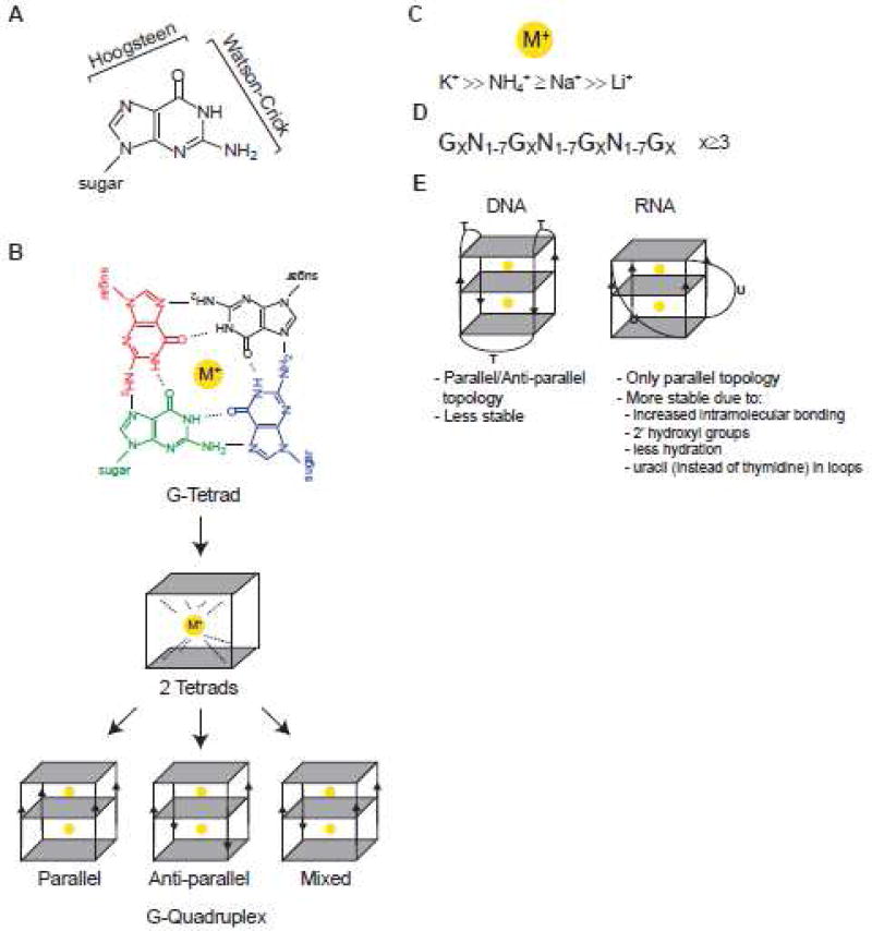
G-quadruplexes (G4s) are extremely stable DNA or RNA secondary structures formed by sequences rich in guanine. These structures are implicated in many essential cellular processes, and the number of biological functions attributed to them continues to grow. While DNA G4s are well understood on structural and, to some extent, functional levels, RNA G4s and their functions have received less attention. The presence of bona fide RNA G4s in cells has long been a matter of debate. The development of G4-specific antibodies and ligands hinted on their presence in vivo, but recent advances in RNA sequencing coupled with chemical footprinting suggested the opposite. In this review, we will critically discuss the biology of RNA G4s focusing on the molecular mechanisms underlying their proposed functions.

Keywords: G-quadruplex; RNA; RNA regulation; RNA structure.


Figures

Figure 1
Figure 1. G-quartets and G-quadruplexes
A. Guanine atoms participating in Watson-Crick and Hoogsteen base pairing interactions. B. Upper: The structure of G-quartet (G-tetrad) showing four-fold symmetrical arrangement of coplanar guanines. Hydrogen bonds between each pair of guanines involves four donor/acceptor atoms (the N1, N7, N2 and O6 atoms). Overall arrangement has a total of eight hydrogen bonds (four N2-H…N7 and four N1-H…O6 bonds). Four carbonyl oxygen (O6) atoms form a negatively charged core in the center of G-quartet that favors binding of monovalent cations (M+). The sugar moieties are removed for clarity. Middle: 2 tetrads coordinated by a monovalent cation are shown. Lower: G-quadruplex structures with different topologies are shown. Note that G4 can be inter- and intra-molecular. C. Preferential binding of monovalent cations to G4s. D. Simplified prediction algorithm used to identify potential G4 motif (GX-N1–7-GX-N1–7-GXN1–7-GX, where x is 3–6 nucleotides and N corresponds to any nucleotide (A, G, T, C or U)). E. Summary of the main differences between DNA and RNA G4s.
Figure 2
Figure 2. Possible mechanisms of action underlying RNA G4 functions
A. G4s act as specific binding sites for regulatory or structural proteins. B. G4s act as barriers or kinetic traps for movement of proteins or protein machines along RNA. C. G4s regulate the formation of alternative secondary structures on RNA, recognized by different proteins. Arrows indicate possible bidirectional shift of equilibrium between G4 and hairpin conformation. D. Examples of proposed G4-specific binding proteins.
Figure 3
Figure 3. Proposed roles of RNA G4s in transcriptional regulation
A. R-loops, structures that contain an RNA-DNA hybrid and displaced single-stranded DNA, can form during transcription when RNA emerging from the transcription machinery hybridizes with the DNA template (RNA transcript shown in red). B. As few as two tandem G-tracks (guanine-rich sequences) on a non-template DNA strand are capable of forming hybrid G4 with guanine-rich transcript. C–D. G4s can act as terminator sequences to cause Pol II transcription to pause. Hybrid DNA/RNA G4s are very stable and require assistance of specialized enzymes to unwind G4s and assist with transcription termination. In mammals, helicase senataxin (SETX) cooperates with exoribonuclease Xrn2 to resolve hybrid G4s, promote degradation of 3'-end RNA cleavage product (red) and release of Pol II from DNA.
Figure 4
Figure 4. Proposed roles of RNA G4s in 3'-end mRNA processing
A. Three primary sequence elements that define the polyadenylation site. These pre-mRNA cis-elements include the hexamer AAUAAA polyadenylation signal, CA cleavage site and G/U-rich downstream element. B. Under optimal conditions, multi-subunit cleavage/polyadenylation machinery (CFI/CFII/CstF/CPSF) assembles on these cis-elements to promote efficient 3'-end processing and global transcription (left panel). 3'-end processing of specific stress-responsive mRNAs, such as encoding DNA damage factor p53, is however inhibited. The downstream G/U-rich element of TP53 pre-mRNA assembles a G4 that is recognized by hnRNP H/F. Binding of hnRNP H/F interferes with efficient recruitment of cleavage/polyadenylation machinery and inhibits 3'-end processing of TP53 pre-mRNA (right panel). C. Under DNA damage, specific factors sequester the essential polyadenylation factor CSTF into an inactive complex thus inhibiting 3'-end processing (left panel). In contrast, hnRNP H/F bound to G4 of TP53 pre-mRNA associates with CstF thus protecting it from sequestration and promoting TP53 3'-end processing and expression of p53 (right panel).
Figure 5
Figure 5. Proposed roles of RNA G4s in splicing regulation
A. Exonic and intronic regions of a putative pre-mRNA are shown as blue (Exon (X) and Exon (X+1)) or green (Intron X), respectively. Intronic G4s can either enhance or silence intron splicing thus acting as intronic splicing enhancer (such as in intron of TP53 pre-mRNA) or intronic splicing silencer (such as in intron of hTERT pre-mRNA). B. Exon-located G4s can act as exonic splicing enhancers. FMRP recognizes G4s in its own pre-mRNA (FMR1) to regulate its splicing pattern and production of protein isoforms.
Figure 6
Figure 6. Proposed roles of RNA G4s in mRNA localization
A. In specialized cells such as neurons, processed G4-containing mRNAs are exported from the nucleus to the cytoplasm where specific RBPs (e.g. hnRNP U, FUS/TLS and FMRP) recognize and bind to their G4 structures. B. G4-bound RBPs assemble into large mRNPs (such as neuronal granules) that specialize in mRNA transport using molecular motors such as microtubule-associated kinesins. C. Upon arrival to the destination site (e.g. dendritic synapses), G4-containing mRNPs remodel allowing local translation.
Figure 7
Figure 7. Proposed roles of RNA G4s in protein synthesis
A. Proposed roles of G4s in mRNA translation. G4s can be found in both non-coding (5'- and 3'-UTRs) and coding (ORF) regions of mRNA where they can inhibit or stimulate translation. In 5'-UTRs, G4s are commonly found near mRNA 5'-cap structures, where they inhibit translation initiation by mechanisms that may involve interference with cap binding or inhibition of 43S pre-initiation complex scanning (e.g. in NRas mRNA). G4-mediated stimulation of translation is proposed in an IRES-like manner (e.g. in 5'-UTRs of VEGF or FGF2 mRNAs), although strong experimental evidences are still lacking. In 3'-UTRs, G4s can both inhibit and stimulate translation by recruitment of translational silencers or stimulators, the identity of which is still unknown. As in non-coding parts, G4s located in the ORF can both block (most likely acting as roadblocks for elongating ribosomes) or promote translation by recruitment of specific protein complexes (such as RBP Aven and RNA helicase DHX36). B. Hypothetical role of G4s in repeat-associated non-AUG (RAN) translation, an unconventional translation mechanism. In ALS patients, hexameric r(GGGGCC)n repeats located in intron of C9ORF72 gene are amplified (i). These repeats are capable of forming higher order G4 structures that make them extremely stable (ii). Some r(GGGGCC)n transcripts undergo RAN translation via direct recruitment of translationally competent ribosomal complexes to produce di-peptide proteins (iii).

References

    1. Bang I. Examination or the guanyle acid. Biochemische Zeitschrift. 1910;26:293–311.
    1. Gellert M, Lipsett MN, Davies DR. Helix formation by guanylic acid. Proc Natl Acad Sci U S A. 1962;48:2013–8. - PMC - PubMed
    1. Ralph RK, Connors WJ, Khorana HG. Secondary structure and aggregation in deoxyguanosine oligonucleotides. J. Amer. Chem. Soc. 1962;84:2265–2266.
    1. Forman SL, Fettinger JC, Pieraccini S, Gottarelli G, Davis JT. Toward Artificial Ion Channels: A Lipophilic G-Quadruplex. J. Am. Chem. Soc. 2000;122:4060–4067.
    1. Pinnavaia TJ, Marshall CL, Mettler CM, Fisk CL, Miles HT, Becker ED. Alkali metal ion specificity in the solution ordering of a nucleotide, 5'-guanosine monophosphate. J. Am. Chem. Soc. 1978;100:3625–3627.

Publication types