Review
. 2017 Sep;1864(9):1494-1506.
doi: 10.1016/j.bbamcr.2017.05.018. Epub 2017 May 26.

ER-plasma membrane junctions: Why and how do we study them?

Affiliations
Review

ER-plasma membrane junctions: Why and how do we study them?

Chi-Lun Chang et al. Biochim Biophys Acta Mol Cell Res. 2017 Sep.

Abstract

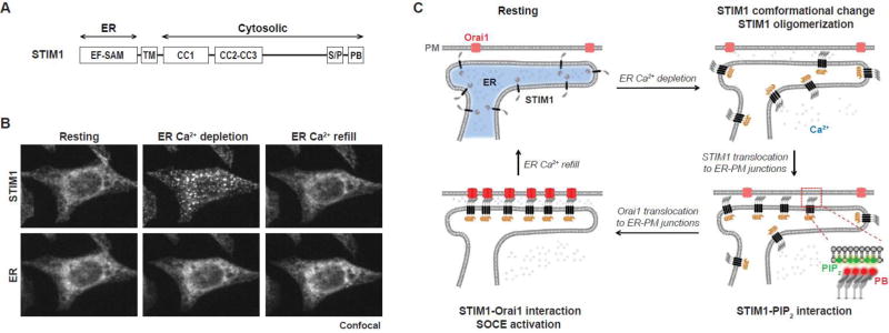
Endoplasmic reticulum (ER)-plasma membrane (PM) junctions are membrane microdomains important for communication between the ER and the PM. ER-PM junctions were first reported in muscle cells in 1957, but mostly ignored in non-excitable cells due to their scarcity and lack of functional significance. In 2005, the discovery of stromal interaction molecule 1 (STIM1) mediating a universal Ca2+ feedback mechanism at ER-PM junctions in mammalian cells led to a resurgence of research interests toward ER-PM junctions. In the past decade, several major advancements have been made in this emerging topic in cell biology, including the generation of tools for labeling ER-PM junctions and the unraveling of mechanisms underlying regulation and functions of ER-PM junctions. This review summarizes early studies, recently developed tools, and current advances in the characterization and understanding of ER-PM junctions. This article is part of a Special Issue entitled: Membrane Contact Sites edited by Christian Ungermann and Benoit Kornmann.


Figures

Fig. 1
Fig. 1. Activation of SOCE at ER-PM junctions by STIM1
(A) A diagram of the domain structure of human STIM1. EF-SAM, EF hand and sterile alpha motif; TM, transmembrane; CC, coiled-coil domain; S/P, serine and proline rich region; PB, polybasic motif. (B) Bottom-section confocal images of a HeLa cell co-expressing YFP-STIM1 and a CFP-tagged ER luminal marker. The cell was imaged at the resting state (left), after 5 µM 2',5'-di (tert-butyl)-1,4-benzohydroquinone (BHQ) treatment to deplete ER Ca2+ store (middle), and 3 min after BHQ washout to allow ER Ca2+ store refill (right). (C) Schematic representation of SOCE activation. In the resting state, STIM1 binds to Ca2+ and localizes diffusely throughout the ER. Following ER Ca2+ depletion, dissociation of Ca2+ from STIM1 triggers STIM1 conformational change and oligomerization. STIM1 oligomers translocate to ER-PM junctions by binding to PM PIP2 via its PB domain. STIM1 at ER-PM junctions subsequently recruits Orai1 and activates SOCE to sustain elevated cytosolic Ca2+ levels and refill ER Ca2+ store.
Fig. 2
Fig. 2. Visualization of ER-PM junctions using ER-labeling or MAPPER
(A) TIRF images (top) of a HeLa cell co-expressing an YFP-tagged ER luminal marker and mCherry-STIM1. The cell was imaged at time zero (left), 5 min after 5 µM BHQ treatment to induce STIM1 translocation (middle), and 5 min after BHQ washout (right). Arrows indicate stable ER puncta corresponding to ER-PM junctions. Schematic diagrams (bottom) depicting STIM1 translocation to ER-PM junctions. The scale bar represents 2 µm. (B) Domain structure of MAPPER. GFP, green fluorescence protein; TM, transmembrane; FRB, FKBP12-rapamycin binding; PB, polybasic motif from the small G protein Rit. (C) Bottom-section confocal images of a HeLa cell co-expressing MAPPER and a mCherry-tagged ER luminal marker. The scale bar represents 2 µm. © Chang et al. and Cell Reports, 2013. Originally published in Cell Reports, 5 (2013) 813–825. (D) TIRF image of a HeLa cell co-expressing MAPPER and a mCherry-tagged ER luminal marker. The scale bar represents 2 µm. © Chang et al. and Cell Reports, 2013. Originally published in Cell Reports, 5 (2013) 813–825. (E) STIM1 translocation induced by 1µM thapsigargin (TG) monitored by TIRF microscopy in a HeLa cell co-expressing MAPPER and mCherry-STIM1. The scale bar represents 2 µm. © Chang et al. and Cell Reports, 2013. Originally published in Cell Reports, 5 (2013) 813–825. (F) Density of ER-PM junctions labeled by ER marker or MAPPER in HeLa cells monitored by TIRF microscopy. Mean ± SD is shown. © Chang et al. and Cell Reports, 2013. Originally published in Cell Reports, 5 (2013) 813–825.
Fig. 3
Fig. 3. Proteins that localize at yeast cortical ER
Domain structure of yeast proteins localized at the cortical ER. Black bar, transmembrane; PB, polybasic motif; HS, hydrophobic segments; SMP, synaptotagmin-like mitochondrial lipid binding protein domain; MSP, major sperm protein domain; PH, pleckstrin homology; FFAT, two phenylalanines in an acidic tract motif; ORD, OSBP-related domain; BAR, Bin/Amphiphysin/Rvs domain; PHg, GRAM domains in the Pleckstrin homology domain; StART, Steroidogenic Acute Regulatory Transfer domain; cER, the cortical ER.
Fig. 4
Fig. 4. Proteins that localize at ER-PM junctions in non-excitable mammalian cells
Domain structure of proteins that localize at ER-PM junctions in mammalian cells. HS, hydrophobic segments; SMP, synaptotagmin-like mitochondrial lipid binding protein domain; EF-SAM, EF hand and sterile alpha motif; Black bar, transmembrane; CC, coiled-coil domain; S/P, serine and proline rich region; PB, polybasic motif; MSP, major sperm protein domain; PITP, PI transfer protein domain; FFAT, two phenylalanines in an acidic tract motif; LNS2, Lipin/Ned1/Smp2 domain. ER-PM, ER-PM junction; ER-Mito, ER-Mitochondria contacts.
Fig. 5
Fig. 5. Model depicting dynamic localization of proteins at ER-PM junctions during receptor activation
E-Syt2 and E-Syt3 readily localize at ER-PM junctions by binding to PM PIP2 in the resting state. During mild receptor activation, STIM2 responds to a small decrease in ER Ca2+ level to activate Orai1 at ERPM junctions for SOCE. E-Syt1 responds to an increase cytosolic Ca2+ level to mediate a decrease in gap distance at ER-PM junctions. Nir3 responds to a small increase in PA at the PM resulting from PIP2 hydrolysis to mediate PI/PA exchange at ER-PM junctions for PM PIP2 homeostasis. Following further or intense receptor activation, STIM1 and Nir2 translocate to ER-PM junctions to support the homeostatic regulation of the PIP2-Ca2+ signaling system with STIM2, Nir3, and E-Syt1.

Similar articles

Cited by

References

    1. Baumann O, Walz B. Endoplasmic reticulum of animal cells and its organization into structural and functional domains. International review of cytology. 2001;205:149–214. - PubMed
    1. West M, Zurek N, Hoenger A, Voeltz GK. A 3D analysis of yeast ER structure reveals how ER domains are organized by membrane curvature. The Journal of cell biology. 2011;193:333–346. - PMC - PubMed
    1. Nixon-Abell J, Obara CJ, Weigel AV, Li D, Legant WR, Xu CS, Pasolli HA, Harvey K, Hess HF, Betzig E, Blackstone C, Lippincott-Schwartz J. Increased spatiotemporal resolution reveals highly dynamic dense tubular matrices in the peripheral ER. Science. 2016;354 - PMC - PubMed
    1. Berridge MJ, Lipp P, Bootman MD. The versatility and universality of calcium signalling. Nat Rev Mol Cell Biol. 2000;1:11–21. - PubMed
    1. Carrasco S, Meyer T. STIM proteins and the endoplasmic reticulum-plasma membrane junctions. Annu Rev Biochem. 2011;80:973–1000. - PMC - PubMed

Publication types

LinkOut - more resources