. 2017 Oct;67(4):716-726.
doi: 10.1016/j.jhep.2017.05.021. Epub 2017 May 26.

CD39 limits P2X7 receptor inflammatory signaling and attenuates sepsis-induced liver injury

Affiliations

CD39 limits P2X7 receptor inflammatory signaling and attenuates sepsis-induced liver injury

Luiz Eduardo Baggio Savio et al. J Hepatol. 2017 Oct.

Abstract

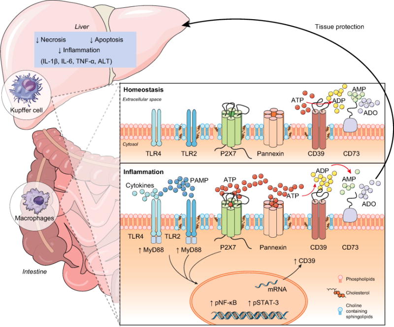
Background & aims: The severity of sepsis can be linked to excessive inflammatory responses resulting in hepatic injury. P2X7 receptor activation by extracellular ATP (eATP) exacerbates inflammation by augmenting cytokine production; while CD39 (ENTPD1) scavenges eATP to generate adenosine, thereby limiting P2X7 activation and resulting in A2A receptor stimulation. We aim to determine how the functional interaction of P2X7 receptor and CD39 control the macrophage response, and consequently impact on sepsis and liver injury.

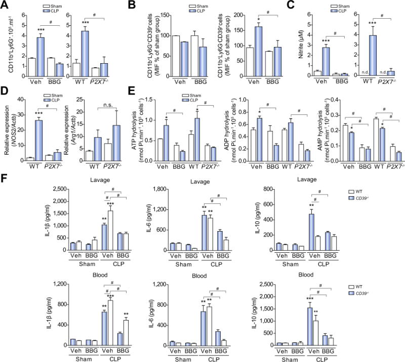
Methods: Sepsis was induced by cecal ligation and puncture in C57BL/6 wild-type (WT) and CD39-/- mice. Several in vitro assays were performed using peritoneal or bone marrow derived macrophages to determine CD39 ectonucleotidase activity and its role in sepsis-induced liver injury.

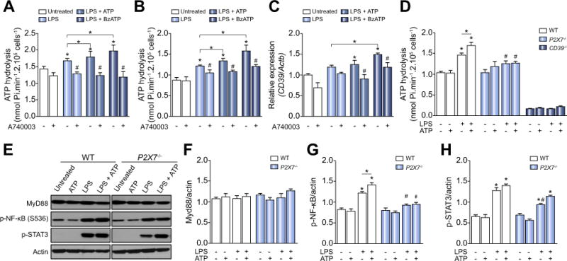
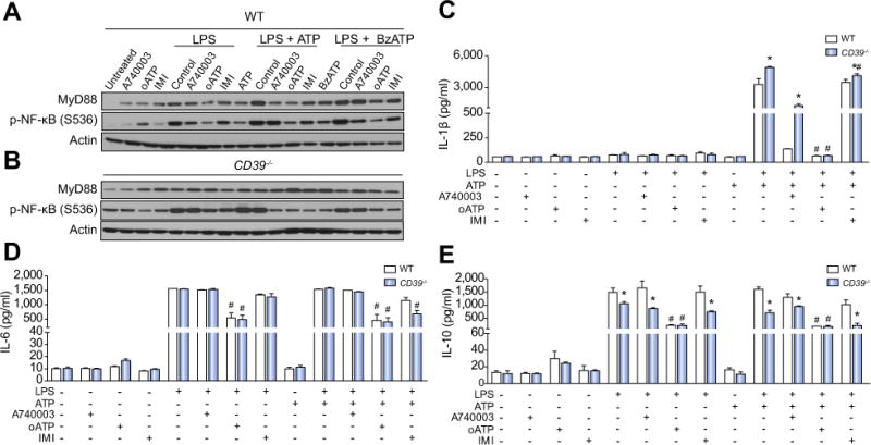
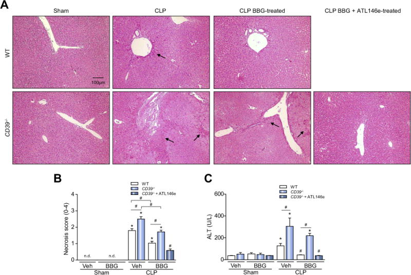
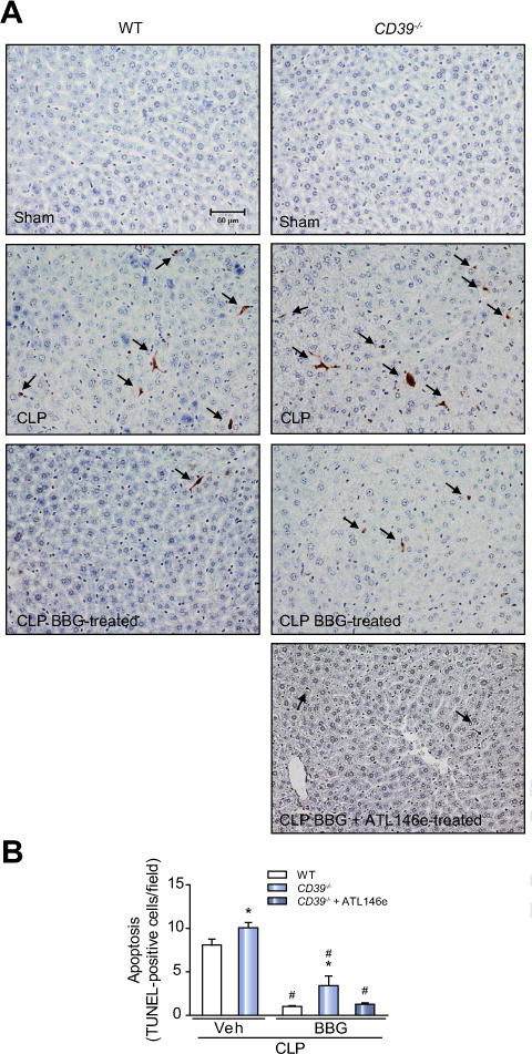
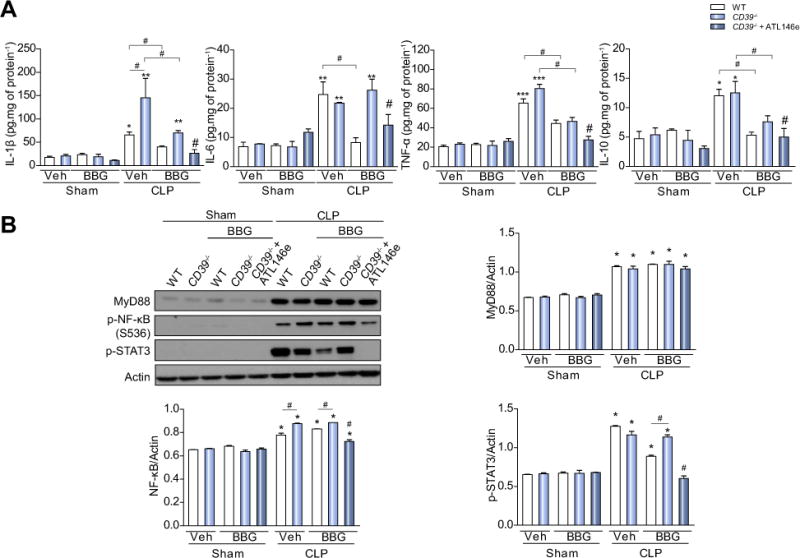
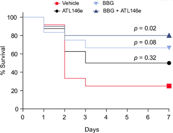
Results: CD39 expression in macrophages limits ATP-P2X7 receptor pro-inflammatory signaling. P2X7 receptor paradoxically boosts CD39 activity. Inhibition and/or deletion of P2X7 receptor in LPS-primed macrophages attenuates cytokine production and inflammatory signaling as well as preventing ATP-induced increases in CD39 activity. Septic CD39-/- mice exhibit higher levels of inflammatory cytokines and show more pronounced liver injury than WT mice. Pharmacological P2X7 blockade largely prevents tissue damage, cell apoptosis, cytokine production, and the activation of inflammatory signaling pathways in the liver from septic WT, while only attenuating these outcomes in CD39-/- mice. Furthermore, the combination of P2X7 blockade with adenosine A2A receptor stimulation completely inhibits cytokine production, the activation of inflammatory signaling pathways, and protects septic CD39-/- mice against liver injury.

Conclusions: CD39 attenuates sepsis-associated liver injury by scavenging eATP and ultimately generating adenosine. We propose boosting of CD39 would suppress P2X7 responses and trigger adenosinergic signaling to limit systemic inflammation and restore liver homeostasis during the acute phase of sepsis. Lay summary: CD39 expression in macrophages limits P2X7-mediated pro-inflammatory responses, scavenging extracellular ATP and ultimately generating adenosine. CD39 genetic deletion exacerbates sepsis-induced experimental liver injury. Combinations of a P2X7 antagonist and adenosine A2A receptor agonist are hepatoprotective during the acute phase of abdominal sepsis.

Keywords: Adenosine; Ectonucleotidases; Extracellular ATP; Kupffer cells; Systemic inflammation.


Conflict of interest statement

Conflict of interest

The authors who have taken part in this study declared that they do not have anything to disclose regarding funding or conflict of interest with respect to this manuscript.

Please refer to the accompanying ICMJE disclosure forms for further details.

Figures

Fig. 1
Fig. 1. P2X7 receptor boosts CD39 activity and contributes to NF-κB and STAT3 activation in LPS-primed macrophages
(A) ATP hydrolysis by LPS-primed peritoneal macrophages and (B) BMDM stimulated with P2X7 receptor agonists pretreated or not with A740003. (C) CD39 gene expression in LPS-primed BMDM. (D) ATP hydrolysis by P2X7 and CD39 deficient LPS-primed macrophages stimulated with ATP. (E) Representative Western blot membranes with densitometric analysis of (F) MyD88, (G) p-NF-κB, and (H) p-STAT3 protein expression in LPS-primed macrophages derived from WT or P2X7−/− mice stimulated with 500 μM ATP. Data are expressed as mean ± SEM of three independent experiments performed in triplicates analyzed by one-way ANOVA, Turkey tests. Asterisks represent statistically significant differences (p <0.05; *) compared to unstimulated control group. The number signs (#) represent statistically significant difference (p <0.05) when comparing groups of animals and WT vs. P2X7−/−.
Fig. 2
Fig. 2. P2X7 receptor blockade decreases LPS-mediated NF-κB activation and cytokine production in WT and CD39−/− BMDM
(A) MyD88 and p-NF-κB protein expression (B) IL-1β, (C) IL-6, and (D) IL-10 production in WT and CD39−/− LPS-primed BMDM stimulated with ATP (500 μM) or BzATP (100 μM). For P2X7 receptor inhibition, samples were pretreated with the P2X7 receptor antagonists oATP (300 μM, for 2 h) or A740003 (0.1 μM, for 30 min) or with imipramine (IMI, 30 μM for 30 min) before priming. Data are expressed as mean ± SEM of three independent experiments analyzed by one-way ANOVA, Turkey tests. Asterisks represent statistically significant differences (p <0.05; *) when comparing WT to CD39−/− or groups. Number signs (#) represent statistically significant differences (p <0.05) when the group pretreated with antagonists were compared to the same stimulated group non-pretreated.
Fig. 3
Fig. 3. P2X7 receptor blockade attenuates systemic inflammatory responses in septic WT and CD39−/− mice
Peritoneal lavage fluid was collected 3 h after surgery and the (A) number of neutrophils (CD11b+Ly6G+) and monocyte/macrophages (CD11b+Ly6G) was quantified by flow cytometry, as well as, (B) CD39 expression in these cells. (C) Nitric oxide production and (D) Nos2 expression in peritoneal cavity cells from WT or P2X7−/− or BBG-pretreated septic mice. (E) ATP, ADP, and AMP hydrolysis by peritoneal cavity cells from WT or P2X7−/− or BBG-pretreated septic mice. (F) Cytokine production in peritoneal lavage fluid and blood from WT or CD39−/− mice. Data are expressed as mean ± SEM of four independent experiments (min. n = 3 mice/experiment) analyzed by one-way ANOVA, Turkey tests. Statistically significant differences between Sham and CLP, and between CLP groups (CLP vehicle vs. BBG, CLP WT vs. P2X7−/− or CD39−/−) are indicated by asterisks (*, p <0.05; **, p <0.01; ***, p <0.001) and by the number sign (#, p <0.05), respectively.
Fig. 4
Fig. 4. P2X7 receptor pharmacological inhibition and adenosine A2A receptor activation completely protects the liver from WT and CD39−/− mice during sepsis
The liver was harvested 24 h after surgery for the evaluation of histopathological changes. Representative photomicrographs of hepatic parenchyma in (A) WT and CD39−/− mice are shown. Arrows show the necrotic areas. (B) Necrosis score; values represent mean ± SEM of four animals per group, considering 10 random fields per mice. (C) Alanine aminotransferase (ALT) levels in septic mice. Data are expressed as mean ± SEM of two independent experiments (n = 6) analyzed by one-way ANOVA, Turkey tests. Statistically significant differences between Sham and CLP, and between CLP groups (CLP vehicle vs. CLP BBG, CLP vehicle vs. CLP BBG + ATL146e) are indicated by asterisks (*, p <0.05) and by number sign (#, p <0.05), respectively.
Fig. 5
Fig. 5. P2X7 receptor inhibition decreases the number of apoptotic cells in the liver from septic WT or CD39−/− mice
The liver was harvested 24 h after surgery. (A) Photomicrographs show the hepatic TUNEL-positive cells (arrows) in septic WT and CD39−/− mice. (B) Number of TUNEL-positive cells per field. Values represent mean ± SEM of four animals per group, considering five random fields per mice. Data are expressed as mean ± SEM of two independent experiments (n = 6) analyzed by one-way ANOVA, Turkey tests. Statistically significant differences between Sham and CLP, and between CLP groups (CLP vehicle vs. CLP BBG, CLP vehicle vs. CLP BBG + ATL146e) are indicated by asterisks (*, p <0.05) and by the number sign (#, p <0.05), respectively.
Fig. 6
Fig. 6. P2X7 receptor blockade and concurrent adenosine A2A receptor activation inhibit inflammatory cytokine production and NF-κB/STAT3 activation in the liver from septic WT or CD39−/− mice
The liver was harvested 24 h after surgery and (A) cytokine (IL-1β, IL-6, TNF-α, and IL-10) production were determined by ELISA. (B) MyD88, p-NF-κB and p-STAT3 protein levels in the liver from septic mice treated or not treated with BBG or BBG + ATL146e. Data are presented as mean ± SEM from three independent experiments (n = 6) analyzed by one-way ANOVA, Turkey tests. Statistically significant differences between Sham and CLP, and between CLP groups (CLP vehicle vs. CLP BBG, CLP vehicle vs. CLP BBG + ATL146e) are indicated by asterisks (*, p <0.05; **, p <0.01; ***, p <0.001) and by the number sign (#, p <0.05), respectively.
Fig. 7
Fig. 7. P2X7 receptor blockade and adenosine A2A receptor activation prolong time to euthanasia and death of septic mice
Survival curves of septic WT mice after pharmacological treatments (ATL146e, BBG or BBG + ATL146e) showing time to euthanasia or death. The statistical significances were analyzed using the Kaplan-Meier method and Log-rank test (n = 8–12).
Fig. 8
Fig. 8. Schematic representation of P2X7 receptor and CD39 interactions on monocyte-macrophages that modulate liver injury during inflammation
The recognition of PAMPs – such as bacterial LPS – by PRRs can induce ATP release activating the P2X7 receptor. P2X7 receptor activation induces large-scale ATP release that can occur mainly via pannexin hemichannels boosting purinergic signaling. CD39 functions to generate AMP from extracellular ATP to limit P2X7 activation and inhibit pro-inflammatory responses. Then, CD73 hydrolyzes AMP to adenosine (ADO) providing for anti-inflammatory effects and tissue protection. This purinergic regulatory mechanism is functional in limiting liver injury and restoring homeostasis during sepsis.

References

    1. Angus DC, Linde-Zwirble WT, Lidicker J, Clermont G, Carcillo J, Pinsky MR. Epidemiology of severe sepsis in the United States: analysis of incidence, outcome, and associated costs of care. Crit Care Med. 2001;29:1303–1310. - PubMed
    1. Singer M, Deutschman CS, Seymour CW, Shankar-Hari M, Annane D, Bauer M, et al. The third international consensus definitions for sepsis and septic shock (Sepsis-3) JAMA. 2016;315:801–810. - PMC - PubMed
    1. Nesseler N, Launey Y, Aninat C, Morel F, Malledant Y, Seguin P. Clinical review: The liver in sepsis. Crit Care. 2012;16:235. - PMC - PubMed
    1. Possamai LA, Thursz MR, Wendon JA, Antoniades CG. Modulation of monocyte/macrophage function: a therapeutic strategy in the treatment of acute liver failure. J Hepatol. 2014;61:439–445. - PubMed
    1. Antoniades CG, Khamri W, Abeles RD, Taams LS, Triantafyllou E, Possamai LA, et al. Secretory leukocyte protease inhibitor: a pivotal mediator of anti-inflammatory responses in acetaminophen-induced acute liver failure. Hepatology. 2014;59:1564–1576. - PubMed

Publication types

MeSH terms