Review
. 2017 May 29;18(6):1153.
doi: 10.3390/ijms18061153.

The Methods of Choice for Extracellular Vesicles (EVs) Characterization

Affiliations
Review

The Methods of Choice for Extracellular Vesicles (EVs) Characterization

Rafal Szatanek et al. Int J Mol Sci. .

Abstract

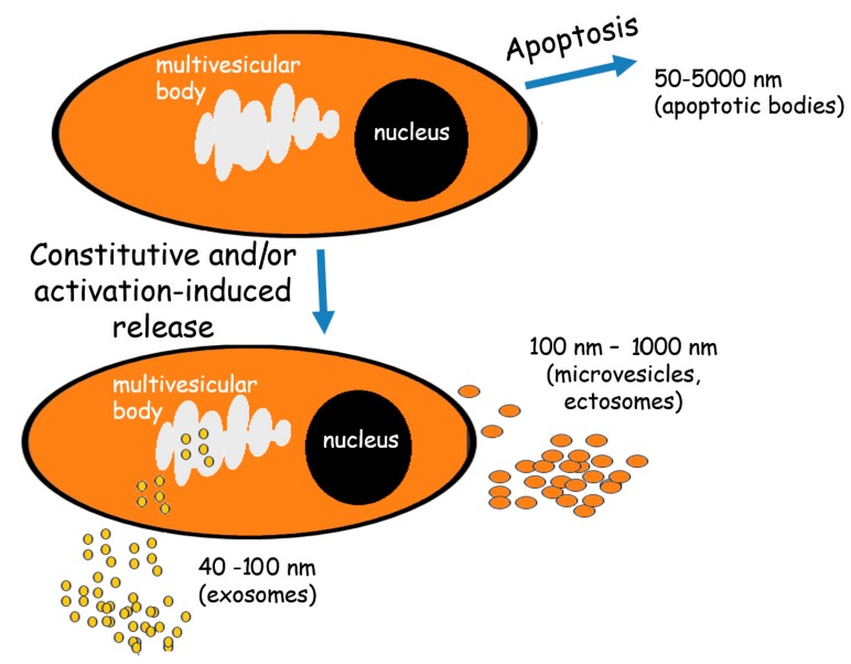
In recent years, extracellular vesicles (EVs) have become a subject of intense study. These membrane-enclosed spherical structures are secreted by almost every cell type and are engaged in the transport of cellular content (cargo) from parental to target cells. The impact of EVs transfer has been observed in many vital cellular processes including cell-to-cell communication and immune response modulation; thus, a fast and precise characterization of EVs may be relevant for both scientific and diagnostic purposes. In this review, the most popular analytical techniques used in EVs studies are presented with the emphasis on exosomes and microvesicles characterization.

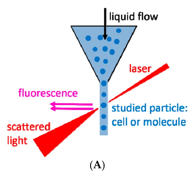
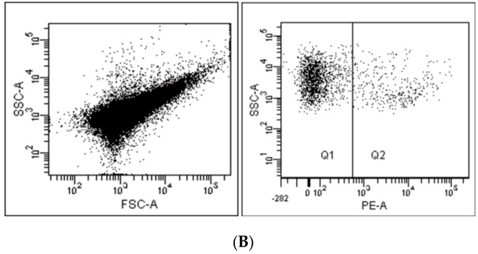
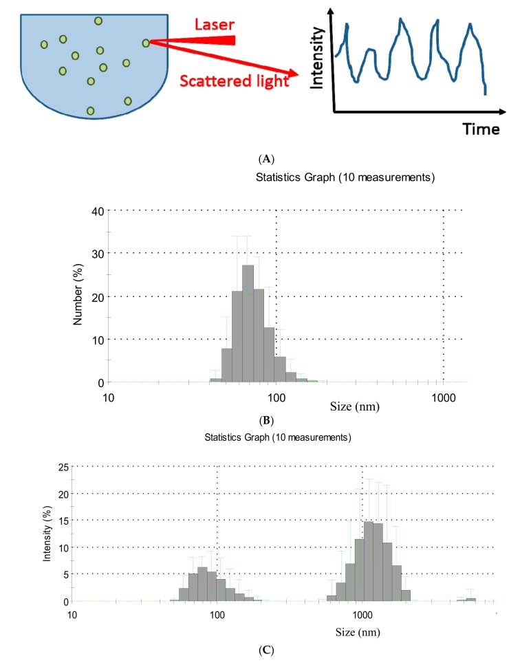
Keywords: atomic force microscopy (AFM); cryo-electron microscopy (Cryo-EM); dynamic light scattering (DLS); exosomes; extracellular vesicles (EVs); flow cytometry; microvesicles (MVs); nanoparticle tracking analysis (NTA); stimulated emission depletion microscopy (STED); transmission electron microscopy (TEM).


Conflict of interest statement

The authors declare no conflict of interest.

Figures

Figure 1
Figure 1
The extracellular vesicles (EVs) release. Alive cells release both exosomes and microvesicles either constitutively and/or under activation. Exosomes are formed from multivesicular bodies while microvesicles arise through direct budding from the plasma membrane. The cells undergoing apoptosis release apoptotic bodies formed by random blebbing.
Figure 2
Figure 2
(A) The principal of flow cytometry; (B) An exemplary analysis of microvesicles (MVs) derived from HPC-4 cell line. Morphology of MVs according to forward scatter/side scatter (FSC/SSC) (left) and surface expression of Her-2/neu antigen detected by fluorochrome (phycoerithrin-PE) conjugated antibody (right). Plots from FACSCanto flow cytometer (BD Biosciences, San Jose, CA, USA).
Figure 2
Figure 2
(A) The principal of flow cytometry; (B) An exemplary analysis of microvesicles (MVs) derived from HPC-4 cell line. Morphology of MVs according to forward scatter/side scatter (FSC/SSC) (left) and surface expression of Her-2/neu antigen detected by fluorochrome (phycoerithrin-PE) conjugated antibody (right). Plots from FACSCanto flow cytometer (BD Biosciences, San Jose, CA, USA).
Figure 3
Figure 3
(A) The principle of the dynamic light scattering; (B,C) Exemplary spectra of dynamic light scattering (DLS) measurements of EVs present in human plasma of gastric cancer patient: (B) The most numerous EVs population in the sample; (C) EVs size distribution.
Figure 4
Figure 4
(A) A graphic representation of the nanoparticle tracking analysis (NTA) principle; (B) An image of EVs secreted by tumors cells of the gastric cancer cell line GC1401 acquired by the NTA system; (C) The corresponding EVs size distribution.
Figure 4
Figure 4
(A) A graphic representation of the nanoparticle tracking analysis (NTA) principle; (B) An image of EVs secreted by tumors cells of the gastric cancer cell line GC1401 acquired by the NTA system; (C) The corresponding EVs size distribution.
Figure 5
Figure 5
A graphic illustration of transmission electron microscopy (TEM) (A) and cryo-TEM (B) principles; TEM image of extracellular vesicles collected from plasma of gastric cancer patients (C).
Figure 6
Figure 6
(A) Schematic illustration of atomic force microscopy; (B) The size distribution of EVs derived from HPC-4 cell line, obtained by the analysis of scanned topography image (inset).
Figure 6
Figure 6
(A) Schematic illustration of atomic force microscopy; (B) The size distribution of EVs derived from HPC-4 cell line, obtained by the analysis of scanned topography image (inset).

References

    1. Zomer A., Vendrig T., Hopmans E.S., van Eijndhoven M., Middeldorp J.M., Pegtel D.M. Exosomes. Commun. Integr. Biol. 2010;3:447–450. doi: 10.4161/cib.3.5.12339. - DOI - PMC - PubMed
    1. Valadi H., Ekström K., Bossios A., Sjöstrand M., Lee J.J., Lötvall J.O. Exosome-mediated transfer of mRNAs and microRNAs is a novel mechanism of genetic exchange between cells. Nat. Cell Biol. 2007;9:654–659. doi: 10.1038/ncb1596. - DOI - PubMed
    1. Simpson R.J., Jensen S.S., Lim J.W.E. Proteomic profiling of exosomes: Current perspectives. Proteomics. 2008;8:4083–4099. doi: 10.1002/pmic.200800109. - DOI - PubMed
    1. Subra C., Laulagnier K., Perret B., Record M. Exosome lipidomics unravels lipid sorting at the level of multivesicular bodies. Biochimie. 2007;89:205–212. doi: 10.1016/j.biochi.2006.10.014. - DOI - PubMed
    1. Théry C., Zitvogel L., Amigorena S. Exosomes: Composition, biogenesis and function. Nat. Rev. Immunol. 2002;2:569–579. - PubMed