. 2017 May 15:8:295.
doi: 10.3389/fphys.2017.00295. eCollection 2017.

Caveolin-1: Functional Insights into Its Role in Muscarine- and Serotonin-Induced Smooth Muscle Constriction in Murine Airways

Affiliations

Caveolin-1: Functional Insights into Its Role in Muscarine- and Serotonin-Induced Smooth Muscle Constriction in Murine Airways

Maryam Keshavarz et al. Front Physiol. .

Abstract

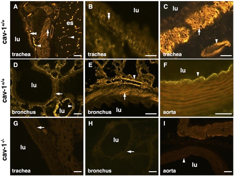
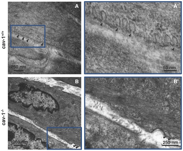
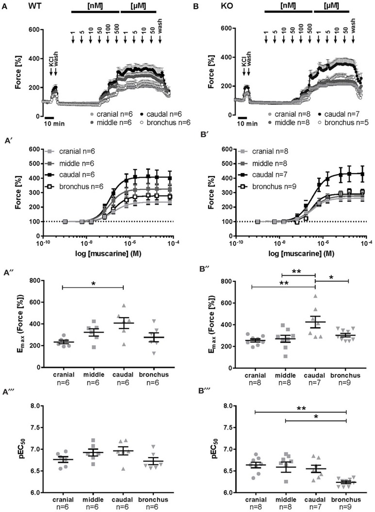
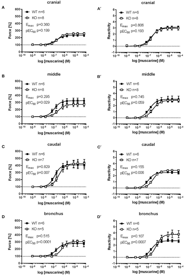
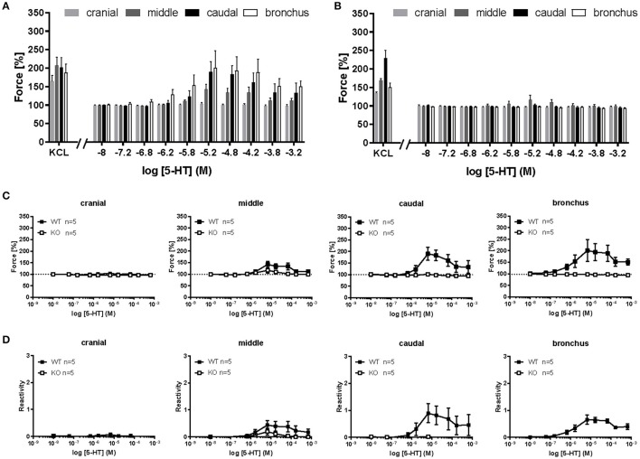
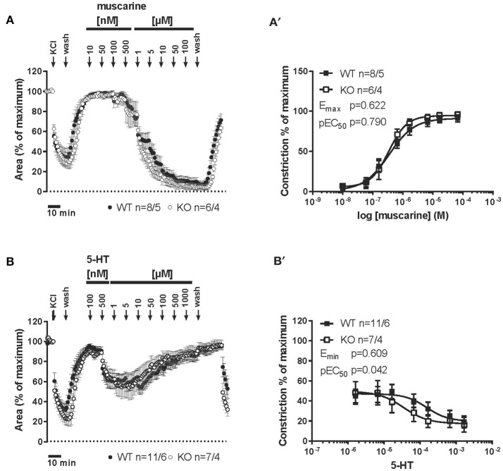
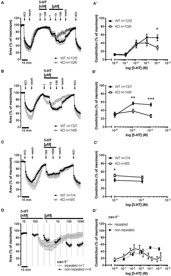
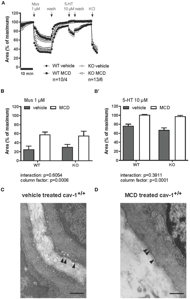
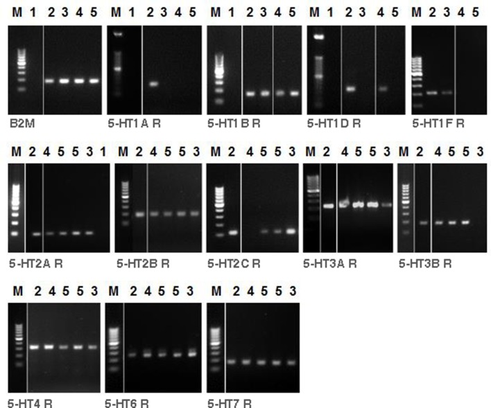
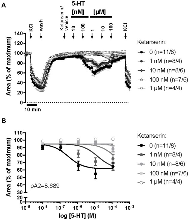
An increased bronchoconstrictor response is a hallmark in the progression of obstructive airway diseases. Acetylcholine and 5-hydroxytryptamine (5-HT, serotonin) are the major bronchoconstrictors. There is evidence that both cholinergic and serotonergic signaling in airway smooth muscle (ASM) involve caveolae. We hypothesized that caveolin-1 (cav-1), a structural protein of caveolae, plays an important regulatory role in ASM contraction. We analyzed airway contraction in different tracheal segments and extra- and intrapulmonary bronchi in cav-1 deficient (cav-1-/-) and wild-type mice using organ bath recordings and videomorphometry of methyl-beta-cyclodextrin (MCD) treated and non-treated precision-cut lung slices (PCLS). The presence of caveolae was investigated by electron microscopy. Receptor subtypes driving 5-HT-responses were studied by RT-PCR and videomorphometry after pharmacological inhibition with ketanserin. Cav-1 was present in tracheal epithelium and ASM. Muscarine induced a dose dependent contraction in all airway segments. A significantly higher Emax was observed in the caudal trachea. Although, caveolae abundancy was largely reduced in cav-1-/- mice, muscarine-induced airway contraction was maintained, albeit at diminished potency in the middle trachea, in the caudal trachea and in the bronchus without changes in the maximum efficacy. MCD-treatment of PLCS from cav-1-/- mice reduced cholinergic constriction by about 50%, indicating that cholesterol-rich plasma domains account for a substantial portion of the muscarine-induced bronchoconstriction. Notably, cav-1-deficiency fully abrogated 5-HT-induced contraction of extrapulmonary airways. In contrast, 5-HT-induced bronchoconstriction was fully maintained in cav-1-deficient intrapulmonary bronchi, but desensitization upon repetitive stimulation was enhanced. RT-PCR analysis revealed 5-HT1B, 5-HT2A, 5-HT6, and 5-HT7 receptors as the most prevalent subtypes in the airways. The 5-HT-induced-constriction in PCLS could be antagonized by ketanserin, a 5-HT2A receptor inhibitor. In conclusion, the role of cav-1, caveolae, and cholesterol-rich plasma domains in regulation of airway tone are highly agonist-specific and dependent on airway level. Cav-1 is indispensable for serotonergic contraction of extrapulmonary airways and modulates cholinergic constriction of the trachea and main bronchus. Thus, cav-1/caveolae shall be considered in settings such as bronchial hyperreactivity in common airway diseases and might provide an opportunity for modulation of the constrictor response.

Keywords: 5-HT; airway smooth muscle; bronchus; caveolin-1; contraction; muscarine.


Figures

Figure 1
Figure 1
Localization of cav-1 in murine thoracic organs using immunohistochemistry. Images are representative for cav-1+/+ (n = 3 animals) and cav-1−/− mice (n = 4 animals). (A–C) Cav-1-immunoreactivity is observed in endothelial cells of the esophagus and trachea and in tracheal basal epithelium and both tracheal and bronchial SM. (D,E) Alveolar region of the lung, bronchial SM and cells present in the alveolar wall (endothelial cells, epithelial cells) are immunoreactive for cav-1. (F) Aorta. (G–I) Controls for the specificity of the anti-cav-1 antibody. The cav-1-labeling is not present in cav-1−/− trachea, lung and aorta. Arrowhead, endothelial cell; double arrowhead, basal epithelium; arrow, SM; lu, lumen; es, esophagus; bar, 50 μm.
Figure 2
Figure 2
Ultrastructure of smooth muscle from cav-1+/+ and cav-1−/− mice. Transmission electron microscopy. Images are representative for tracheae of cav-1−/− (n = 6 animals) and cav-1+/+ mice (n = 4 animals). (A,A') Cav-1+/+ mice. Tracheal smooth muscle cells exhibit numerous caveolae that are located in groups at the plasma membrane. (B,B') Cav-1−/− mice. Tracheal smooth muscle cells have low number of caveolae (arrowheads) at the plasma membrane. Caveolae = arrowhead.
Figure 3
Figure 3
Muscarine-mediated changes in constrictor force and reactivity in different parts of the trachea and extrapulmonary bronchi from cav-1+/+ and cav-1−/− mice. Organ bath recordings, each point represents the mean of number of animals (n) ± SEM. (A,B) Muscarine induces concentration-dependent contraction of ASM in cav-1+/+ (A) and cav-1−/− (B) mouse strains. (A',B') Constrictor force in all parts of the airways of cav-1+/+ (A') and cav-1−/− (B') mice. Following equilibration, baseline tension was adjusted to 0.5 g for all airway segments in all preparations. Baseline was set as 100% and the maximum response at each concentration was calculated. Sigmoidal concentration-response curves were plotted according to the Hill equation. Maximal responses (Emax, A”,B”) and pEC50 (A”',B”') values for muscarine were estimated for each individual experiment. In cav-1+/+ the caudal part responded with a stronger contraction, whereas there was no difference in pEC50 values between segments. In cav-1−/− the caudal part responded with a stronger contraction whereas the pEC50 decreased in the bronchus. *p ≤ 0.05; **p < 0.01, One-way ANOVA followed by Tukey's multiple comparisons test with the exception of (B”') which was analyzed by Kruskall-Wallis test followed by Dunn's multiple comparisons test.
Figure 4
Figure 4
Comparison of force and reactivity between both mouse strains. Airway reactivity is expressed as contraction effect induced by muscarine compared to the correspondent contraction induced by KCl. Each point represents the mean of number of animals (n) ± SEM. Please note that when SEM is smaller than the symbol size it is not displayed in the figure. Data points were plotted as sigmoidal concentration-response curves and values for Emax and pEC50 were estimated for each individual experiment and analyzed with Student's unpaired t-test except for data shown in (B,B'), which was analyzed by Mann-Whitney U-test. The cranial part of the cav-1−/− trachea shows no significant difference compared to cav-1+/+ in both force (A) and reactivity (A') levels. The middle (B,B') and caudal (C,C') part of the cav-1+/+ trachea were more sensitive to muscarine as indicated by a right-shift in the concentration-response curves and significant decrease in the pEC50 values for muscarine in both force and reactivity in the cav-1−/− mice strain. The main bronchus (D,D') of cav-1+/+ mice was also more sensitive to muscarine compared to cav-1−/− mice.
Figure 5
Figure 5
5-HT-mediated changes in constrictor force and reactivity of different parts of the trachea and extrapulmonary bronchi from cav-1+/+ and cav-1−/− mice. Organ bath recordings, each point represents the mean of number of animals (n) ± SEM. (A,B) Changes in the force were recorded after cumulative application of different concentrations of 5-HT. Following equilibration, baseline tension was adjusted to 0.5 g for all airway segments in all preparations. Baseline was set as 100% and the maximum response at each concentration was evaluated. 5-HT induces concentration-dependent contraction in main bronchus, tracheal middle and caudal parts in cav-1+/+ mice (A), whereas the cranial part of cav-1+/+ mice and all tracheal parts and bronchi of cav-1−/− mice are not responsive to 5-HT (A,B). (C,D) Comparison of constrictor response between both mouse strains. Note that means are not defined (and left blank) when values are 0 or negative.
Figure 6
Figure 6
Muscarine- and 5-HT-mediated changes in the luminal area of peripheral bronchi from cav-1+/+ and cav-1 −/− mice. Videomorphometric analyses of PCLS. Data are presented as mean of number of bronchi (n)/number of animals ± SEM. (A) Muscarine induced concentration-dependent decreases in the luminal airway area in cav-1+/+ and cav-1−/− mice. Bronchi of cav-1+/+ and cav-1−/− mice responded to 100 nM-100 μM muscarine in a dose dependent pattern with sustained contraction. (A') Sigmoidal concentration-response curves of concentration vs. luminal area reduction of peripheral bronchi of cav-1+/+ and cav-1−/− mice were plotted according to the Hill equation. Values for Emax and pEC50 were estimated for each individual experiment and analyzed with Student's unpaired t-test. (B) 5-HT induced concentration-dependent decreases in luminal airway area in cav-1+/+ and cav-1−/− mice. Bronchi of cav-1+/+ and cav-1−/− mice responded to 500 nM-1 mM 5-HT in a dose dependent pattern with sustained contraction followed by relaxation. (B') Sigmoidal concentration-response curves of the relaxation starting at 5 μM of peripheral bronchi of cav-1+/+ and cav-1−/− mice were plotted according to the Hill equation. Values for Emin and pEC50 were estimated for each individual experiment and analyzed with Student's unpaired t-test.
Figure 7
Figure 7
5-HT-mediated changes in the luminal area of peripheral bronchi from cav-1+/+ and cav-1 −/− mice. Videomorphometric analyses of PCLS. (A,A') Bronchi of cav-1+/+ and cav-1−/− mice responded to increasing 5-HT doses. 5-HT-induced constriction in cav-1−/− mice PCLS was significantly reduced at μM concentrations of 5-HT compared to cav-1+/+ mice strain. We applied KCl (60 mM) as a viability control for 5 min at 2 points of experiment. (B,B') Bronchi of cav-1+/+ and cav-1−/− mice responded to increasing 5-HT doses. 5-HT-induced bronchoconstriction in PCLS was significantly reduced at μM concentrations in cav-1−/− mice compared to cav-1+/+ mice. (C,C') Bronchi of cav-1+/+ and cav-1−/− mice responded to 5-HT with a rapid sustained constriction. (D,D') Comparison of the response to 5-HT after repetitive stimulation (in half logarithmic mode) with the direct (non-repetitive) 5-HT response. The intrapulmonary bronchi of cav-1−/− mice exhibit a decrease in 5-HT-induced bronchoconstriction after repetitive 5-HT application. Data are presented as mean of number of bronchi (n)/number of animals ± SEM. *p ≤ 0.05; **, p < 0.01; ***p < 0.001, Student's unpaired t-test with the exception of values at 1 mM 5-HT in (C') which was analyzed by Mann-Whitney U-test.
Figure 8
Figure 8
Muscarine- and 5-HT-mediated changes in the luminal area of peripheral bronchi from cav-1+/+ and cav-1−/− mice after vehicle (___) or MCD (- - - -) treatment. Changes in the luminal area of mouse peripheral airways were recorded by videomorphometry after application of 1 μM muscarine (Mus), 10 μM 5-HT and 60 mM KCl for vehicle (___) or MCD (- - - -) treatment. Data are presented as means ± SEM; n = number of bronchi/animals with baseline value set as 100%. (A) The bronchi from cav-1+/+ and cav-1−/− mice constrict in response to Mus and 5-HT. In both mouse strains, caveolae disruption by MCD reduces the response to Mus whereas the response to 5-HT is fully abrogated. No differences in the response to KCl occur after MCD-treatment in either mouse strain. (B) Bar graphs of the maximum response after application of 1 μM Mus and 10 μM 5-HT. Luminal area of peripheral bronchi of cav-1+/+ and cav-1−/− mice after vehicle (gray) or MCD (white) treatment. Data was analyzed by 2-way ANOVA. Whereas the p-value of the column factor indicates that MCD is effective in both mouse strains, there is no statistically significant interaction. (C) Transmission electron microscopy of intrapulmonary bronchi derived from PCLS included in videomorphometric experiments. Vehicle-treated murine ASM containing areas with caveolae (arrowheads) in the plasma membrane of cav-1+/+ mice. (D) Cell surface region of an equivalent bronchial SM after caveolae disruption by MCD. Arrows point to plasma membrane without caveolae and bar = 500 nm.
Figure 9
Figure 9
RT-PCR analysis of 5-HT receptor subtypes in tracheal muscle, main bronchus and lung homogenate of C57BL/6J mice. The presence of mRNA coding for 5-HT receptor subtypes are shown. M = marker. ß-2-Microglobulin (B2M) as housekeeping gene control was run for different samples. H2O (1) as control reactions included the absence of template, brain (2) as positive control, lung (3), tracheal muscle (4) and main bronchus (5).
Figure 10
Figure 10
5-HT-mediated changes in the luminal area of peripheral bronchi from cav-1−/− mice after treatment with vehicle or with the 5-HT2A receptor antagonist, ketanserin. (A) Changes in the luminal area of mouse peripheral airways was recorded by videomorphometry in PCLS after cumulative application of different concentrations of 5-HT and KCl. 5-HT induces a decrease in luminal airway area in cav-1−/− mice. (B) Sigmoidal concentration-response curves for 5-HT were plotted according to the Hill equation in the presence of the different doses of ketanserin and the pA2-value for ketanserin (8.689) was estimated by Schild-analysis. Hill-slope and Schild-slope were fixed to 1.0. Data are presented as mean of number of animals (n) ± SEM.

Similar articles

Cited by

References

    1. Bai Y., Zhang M., Sanderson M. J. (2007). Contractility and Ca2+ signaling of smooth muscle cells in different generations of mouse airways. Am. J. Respir. Cell Mol. Biol. 36, 122–130. 10.1165/rcmb.2006-0036OC - DOI - PMC - PubMed
    1. Bains S. N., Tourkina E., Atkinson C., Joseph K., Tholanikunnel B., Chu H. W., et al. . (2012). Loss of caveolin-1 from bronchial epithelial cells and monocytes in human subjects with asthma. Allergy 67, 1601–1604. 10.1111/all.12021 - DOI - PMC - PubMed
    1. Bastiani M., Parton R. G. (2010). Caveolae at a glance. J. Cell Sci. 123(Pt 22), 3831–3836. 10.1242/jcs.070102 - DOI - PubMed
    1. Bhatnagar A., Sheffler D. J., Kroeze W. K., Compton-Toth B., Roth B. L. (2004). Caveolin-1 interacts with 5-HT2A serotonin receptors and profoundly modulates the signaling of selected Galphaq-coupled protein receptors. J. Biol. Chem. 279, 34614–34623. 10.1074/jbc.M404673200 - DOI - PubMed
    1. Buels K. S., Jacoby D. B., Fryer A. D. (2012). Non-bronchodilating mechanisms of tiotropium prevent airway hyperreactivity in a guinea-pig model of allergic asthma. Br. J. Pharmacol. 165, 1501–1514. 10.1111/j.1476-5381.2011.01632.x - DOI - PMC - PubMed

LinkOut - more resources