Review
. 2017 Jul;33(7):1125-1141.
doi: 10.1007/s00381-017-3442-x. Epub 2017 May 29.

Antenatal management of fetal neurosurgical diseases

Affiliations
Review

Antenatal management of fetal neurosurgical diseases

Sergio Cavalheiro et al. Childs Nerv Syst. 2017 Jul.

Abstract

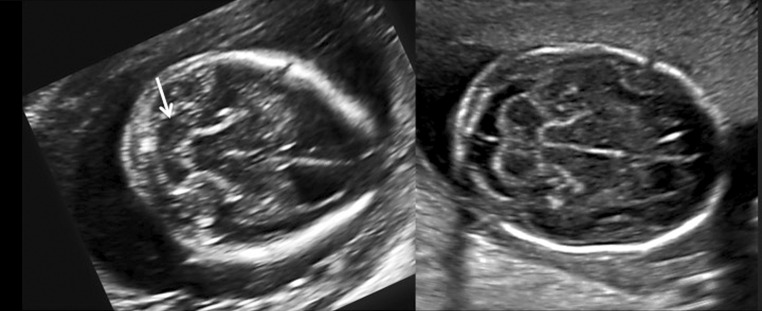
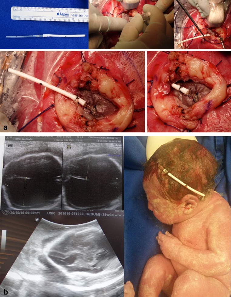
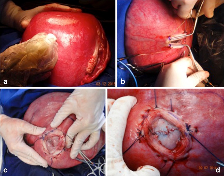
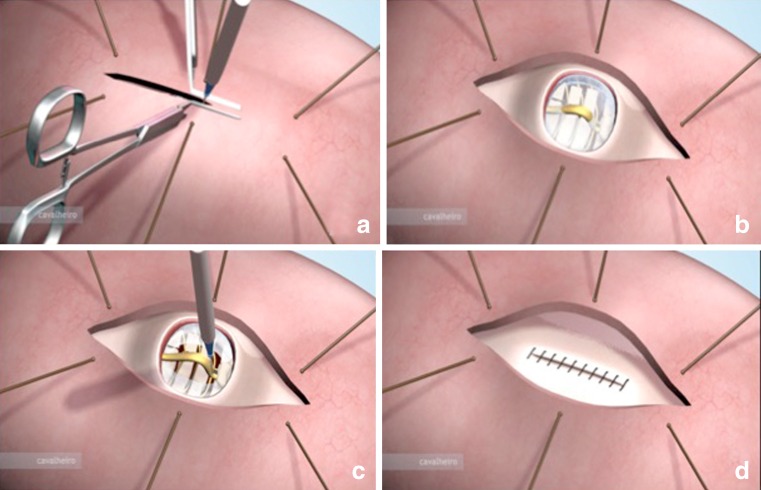
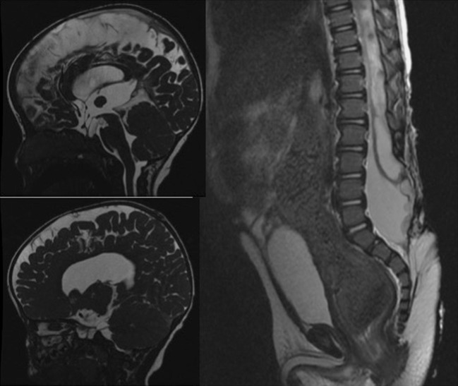
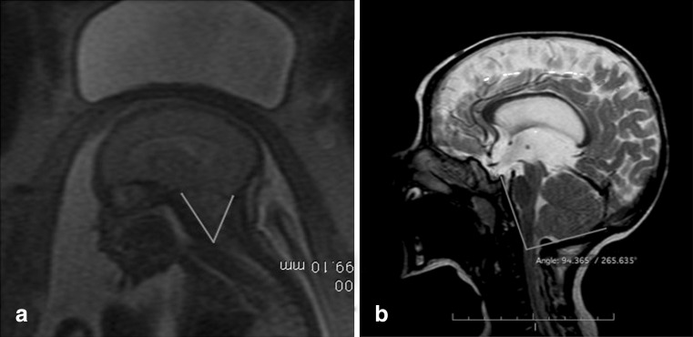
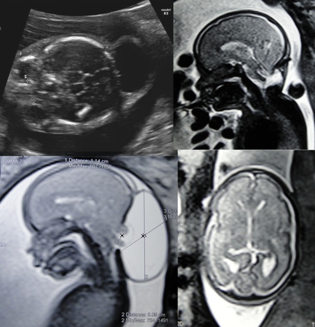
The advance in the imaging tools during the pregnancy (ultrasound and magnetic resonance) allowed the early diagnose of many fetal diseases, including the neurological conditions. This progress brought the neurosurgeons the possibility to propose treatments even before birth. Myelomeningocele is the most recognized disease that can be treated during pregnancy with a high rate of success. Additionally, this field can be extended to other conditions such as hydrocephalus and encephaloceles. However, each one of these diseases has nuances in the diagnostic evaluation that should fit the requirements to perform the fetal procedure and overbalance the benefits to the patients. In this article, the authors aim to review the neurosurgical aspects of the antenatal management of neurosurgical conditions based on the experience of a pediatric neurosurgery center.

Keywords: Cephalocentesis; Encephalocele; Hydrocephalus; Myelomeningocele; Ventricle-amniotic shunt.


Conflict of interest statement

None.

Figures

Fig. 1
Fig. 1
Algorithm for treatment of fetal hydrocephalus
Fig. 2
Fig. 2
Scheme of ventriculoamniotic shunt placement. a Ultrasound-guided transabdominal puncture reaching the occipital horn of the lateral ventricle. b Catheter insertion and intraventricular portion released after partial removal of the trocar. c Complete removal of the trocar to release the catheter in the amniotic cavity and decrease hydrocephaly. d Double pigtail catheter. e Patient at birth exposing the ventriculoamniotic shunt used during the uterine life to treat a fetal hydrocephalus; in detail can be seen that the catheter was still working. f example of a fetal hydrocephalus due to aqueductal stenosis treated by ventriculoamniotic shunt. The pre-and post-MRIs show the reduction of the ventricular cavities after procedure
Fig. 3
Fig. 3
Two-dimensional ultrasound showing: a Meningocele. b Myelomeningocele with a neural tissue inside and the respective intraoperative view. c Rachischisis
Fig. 4
Fig. 4
Two-dimensional ultrasonography. Counting of vertebral bodies by determining the 12th thoracic vertebra that corresponds to the last rib
Fig. 5
Fig. 5
The occipitum-dens line described to evaluate the degree of the brainstem and tonsils herniation into to the cervical spine canal
Fig. 6
Fig. 6
Two-dimensional ultrasonography. The “lemon sign”—the skull in a shape of a lemon due frontal scalloping bone caused by intracranial hypotension
Fig. 7
Fig. 7
Left image shows the “banana sign” that represents the inversion of the cerebellum curvature (arrow). Right image shows a comparative normal posterior fossa and cerebellum and normal frontal shape
Fig. 8
Fig. 8
Illustrative case of simultaneous MMC correction and ventriculoamniotic shunt. a Insertion of the ventricular catheter with peel-away technique (Valve type Accu-flo Codman® low pressure). b Pre- and postoperative ultrasound evidencing the catheter insertion and complete reduction of the ventricle cavity. Neonate picture at the 36th week of pregnancy showing the shunt still working at a good place
Fig. 9
Fig. 9
Intraoperative view of open fetal surgery for myelomeningocele. a Intraoperative ultrasound monitoring. b Cardiac forceps for the uterus hemostasis during its opening. c Exposure of the myelomeningocele. d Intraoperative view after the myelomeningocele closure
Fig. 10
Fig. 10
Illustration of the uterus opening. a Using electric cautery and hemostatic forceps. b Exposure of the myelomeningocele. c Cutting the apical ligament producing spinal cord detachment. d Final aspect of skin closure
Fig. 11
Fig. 11
T2-WI MRI of the lumbosacral and skull in a male patient that is 8 months—age that underwent a fetal surgery at 25 weeks of pregnancy showing an absence of intracranial hypertension, patency of the aqueduct, cerebellum in the normal position, and subdural cerebrospinal fluid effusion as a benign hydrocephalus. In the sagittal view is represented a large spinal dysraphism from L2 to S4, showing syringomyelia, tethered cord, and epidermoid tumor
Fig. 12
Fig. 12
a Illustration of the complex myelomeningocele and Chiari type II. b Involution of the Chiari type II due to the expansion of the posterior fossa
Fig. 13
Fig. 13
a Preoperative MRI showing the fetal clivus-supraocciput angle before the myelomeningocele correction. b Opening of the clivus-supraocciput angle after the myelomeningocele correction
Fig. 14
Fig. 14
Head circumference curve of patients that was submitted to fetal myelomeningocele correction and followed without ventriculoperitoneal shunt (red curve) compared to patients that was submitted to postnatal correction of the myelomeningocele and followed with ventriculoperitoneal shunt (blue curve)
Fig. 15
Fig. 15
Illustrative case of a fetus that presented occipital encephalocele and trending to microcephaly that underwent to fetal correction at the 26th week of gestation. Left pictures illustrate the preoperative ultrasound and MRI evidencing the occipital encephalocele sac. Right pictures illustrate the postoperative MRI showing the correction of the Chiari type III

References

    1. Charpentier A (1987) Hydrocephalus. In: Grandin EH, Charpentier A (eds) Cyclopedia of obstetrics and gynecology. W. Wood & Company, New York, p 262
    1. Barke MW, Scarbough JI, O’Gorman L, Thompson WB., Jr Intrauterine ventriculography of the hydrocephalic fetus. Obstet Gynecol. 1966;28:568–570. - PubMed
    1. Kellner KR, Cruz AC, Gelman SR, Vries JK, Spellacy WN. Percutaneous fetal ventriculography: a simplified technique to aid in management of the hydrocephalic fetus. J Reprod Med. 1980;24:225–228. - PubMed
    1. Frigoletto FD, Jr, Birnholz JC, Greene MF. Antenatal treatment of hydrocephalus by ventriculoamniotic shunting. JAMA. 1982;248:2496–2497. doi: 10.1001/jama.1982.03330190060034. - DOI - PubMed
    1. Clewell WH, Johnson ML, Meier PR, Newkirk JB, Zide SL, Hendee RW, Bowes WA, Jr, Hecht F, O’Keeffe D, Henry GP, Shikes RH. A surgical approach to the treatment of fetal hydrocephalus. N Engl J Med. 1982;306:1320–1325. doi: 10.1056/NEJM198206033062202. - DOI - PubMed

LinkOut - more resources