. 2017 Sep;13(3):331-338.
doi: 10.1007/s11302-017-9565-4. Epub 2017 May 29.

The P2Y1 receptor-mediated leukocyte adhesion to endothelial cells is inhibited by melatonin

Affiliations

The P2Y1 receptor-mediated leukocyte adhesion to endothelial cells is inhibited by melatonin

Tassya Cataldi Cardoso et al. Purinergic Signal. 2017 Sep.

Abstract

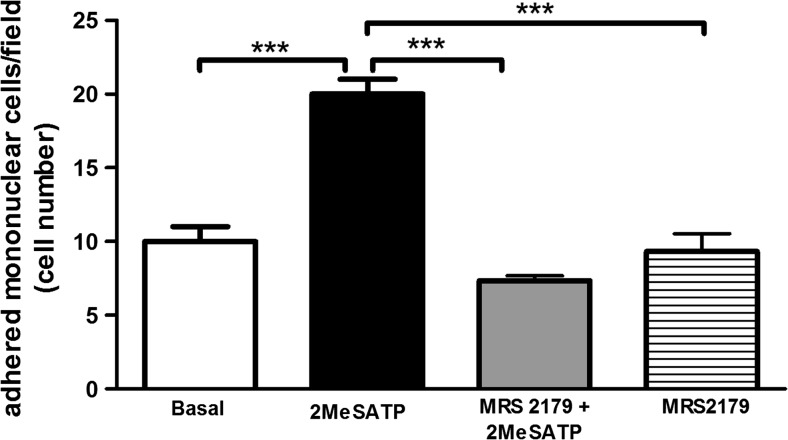
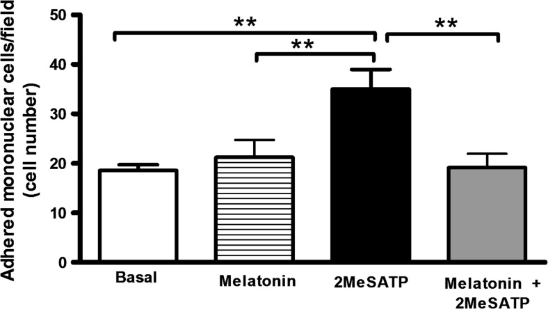
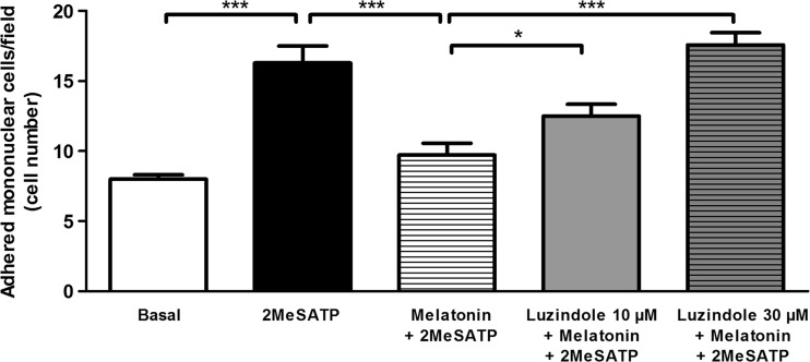
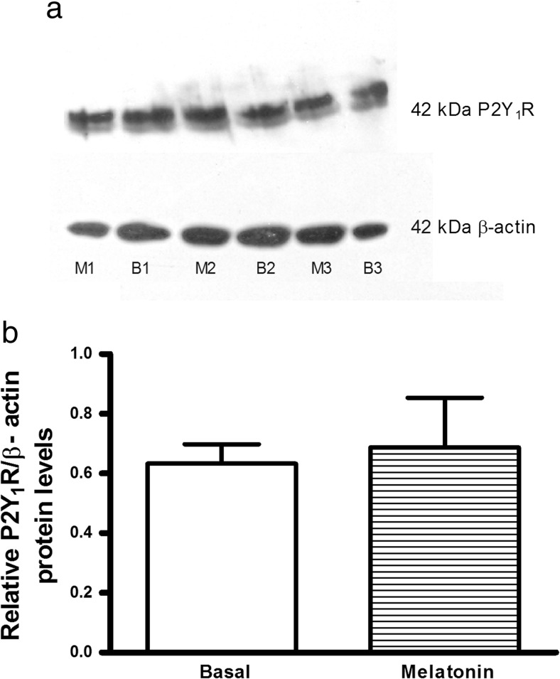
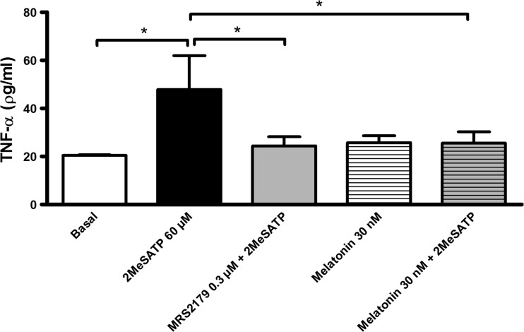
Extracellular ATP (released by endothelial and immune cells) and its metabolite ADP are important pro-inflammatory mediators via the activation of purinergic P2 receptors (P2Y and P2X), which represent potential new targets for anti-inflammatory therapy. Endothelial P2Y1 receptor (P2Y1R) induces endothelial cell activation triggering leukocyte adhesion. A number of data have implicated melatonin as a modulator of immunity, inflammation, and endothelial cell function, but to date no studies have investigated whether melatonin modulates endothelial P2YR signaling. Here, we evaluated the putative effect of melatonin on P2Y1R-mediated leukocyte adhesion to endothelial cells and TNF-α production, using mesenteric endothelial cells and fresh peripheral blood mononuclear cells isolated from rats. Endothelial cells were treated with the P2Y1R agonist 2MeSATP, alone or in combination with melatonin, and then exposed to mononuclear cells. 2MeSATP increased leukocyte adhesion to endothelial cells and TNF-α production in vitro, and melatonin inhibited both effects without altering P2Y1R protein expression. In addition, assays with the Ca2+ chelator BAPTA-AM indicate that the effect of melatonin on 2MeSATP-stimulated leukocyte adhesion depends on intracellular Ca2+ modulation. P2Y1R is considered a potential target to control chronic inflammation. Therefore, our data unveiled a new endothelial cell modulator of purinergic P2Y1 receptor signaling.

Keywords: Endothelial cells; Inflammation; Melatonin; P2Y1 receptor; Purinergic signaling.


Conflict of interest statement

Funding

This study was funded by National Council for Scientific and Technological Development (CNPq, Brazil, grant number 455436/2014-2).

Conflict of interest

Tassya Cataldi Cardoso declares that she has no conflict of interest.

Thaís Emanuelle Pompeu declares that she has no conflict of interest.

Claudia Lucia Martins Silva declares that she has no conflict of interest.

Ethical approval

All procedures performed in studies involving animals were in accordance with the ethical standards of the institution or practice at which the studies were conducted.

Figures

Fig. 1
Fig. 1
Endothelial P2Y1R activation stimulates leukocyte adhesion to mesenteric endothelial cells. Rat endothelial cells were left untreated (“basal” group) or were treated with the P2Y1R agonist 2MeSATP (60 μM; black bar) for 4 h, followed by the addition of mononuclear cells. Alternatively, endothelial cells were pre-incubated with the P2Y1R antagonist MRS2179 (0.3 μM) for 30 min before treatment with 2MeSATP (gray bar). Leukocyte adhesion to endothelial cells was estimated by direct counting by light microscopy. Data are expressed as mean ± SEM. N = 3 independent experiments performed in triplicates. ***P < 0.001 vs. 2MeSATP, by one-way ANOVA followed by Newman-Keuls test
Fig. 2
Fig. 2
Melatonin inhibits P2Y1R-mediated leukocyte adhesion to mesenteric endothelial cells. Rat endothelial cells were left untreated (basal group) or were treated with the P2Y1R agonist 2MeSATP (60 μM, for 4 h), followed by the addition of mononuclear cells (black bar). Alternatively, endothelial cells were pre-incubated with melatonin (30 nM) for 30 min prior to treatment with 2MeSATP (in the presence of melatonin; gray bar). Mononuclear cell adhesion to endothelial cells was estimated by direct counting by light microscopy. Data are expressed as mean ± SEM. N = 7–9 replicates performed, with 2–3 independent experiments. **P < 0.01 vs. 2MeSATP, by one-way ANOVA followed by Newman-Keuls test
Fig. 3
Fig. 3
Melatonin MT receptors mediate the inhibitory effect of melatonin on P2Y1R-dependent leukocyte adhesion to mesenteric endothelial cells. Rat endothelial cells were left untreated (basal group) or were treated with the P2Y1R agonist 2MeSATP (60 μM, for 4 h), followed by the addition of mononuclear cells (black bar). Alternatively, endothelial cells were pre-incubated with the MT receptor antagonist luzindole (10 μM (gray bar) or 30 μM (hatched gray bar)) for 30 min prior to incubation with melatonin (30 nM, in the presence of luzindole) for a further 30 min, and before treatment with 2MeSATP (4 h). Data are expressed as mean ± SEM. N = 7 replicates, from 3 independent experiments. ***P < 0.001 vs. 2MeSATP or melatonin plus 2MeSATP; *P < 0.05 vs. melatonin plus 2MeSATP, by one-way ANOVA followed by Newman-Keuls test
Fig. 4
Fig. 4
Melatonin does not alter endothelial P2Y1 receptor (P2Y1R) expression. Western blotting analysis of P2Y1R expression in endothelial cells. a P2Y1R and β-actin protein expression in rat mesenteric endothelial cells (20 μg of protein/lane, in 10% SDS-PAGE gels). b Densitometry analysis of P2Y1R bands (relative to β-actin) in blots (n = 3 independent experiments using three different cultures) from endothelial cells left untreated (basal (B1, B2, B3)) or treated with 30 nM melatonin (M1, M2, M3) for 4 h, showing that melatonin treatment did not alter P2Y1R expression. Data are expressed as mean ± SEM (P = 0.78, by Student’s t-test)
Fig. 5
Fig. 5
The P2Y1 receptor-mediated TNF-α production by endothelial cells is inhibited by melatonin. Mesenteric endothelial cells were treated with 60 μM 2MeSATP (4 h; black bar) in the absence or presence of the P2Y1R antagonist MRS2179 (0.3 μM; gray bar) or melatonin (30 nM; hatched gray bar), both added 30 min before the agonist. Alternatively, cells were left untreated (basal; white bar). Data are expressed as mean ± SEM. *P < 0.005 vs. 2MeSATP (by one-way ANOVA followed by Newman-Keuls test; n = 3–5 different cultures)

References

    1. Muller WA. Transendothelial migration: unifying principles from the endothelial perspective. Immunol Rev. 2016;273:61–75. doi: 10.1111/imr.12443. - DOI - PMC - PubMed
    1. Idzko M, Ferrari D, Eltzschig HK. Nucleotide signalling during inflammation. Nature. 2014;509:310–317. doi: 10.1038/nature13085. - DOI - PMC - PubMed
    1. Acuña-Castroviejo D, Escames G, Venegas C, Díaz-Casado ME, Lima-Cabello E, López LC, et al. Extrapineal melatonin: sources, regulation, and potential functions. Cel Mol Life Sci. 2014;71:2997–3025. doi: 10.1007/s00018-014-1579-2. - DOI - PMC - PubMed
    1. Silva CL, Tamura EK, Macedo SM, Cecon E, Bueno-Alves L, Farsky SH, et al. Melatonin inhibits nitric oxide production by microvascular endothelial cells in vivo and in vitro. Br J Pharmacol. 2007;151(2):195–205. doi: 10.1038/sj.bjp.0707225. - DOI - PMC - PubMed
    1. Tamura EK, Cecon E, Monteiro AW, Silva CL, Markus RP. Melatonin inhibits LPS-induced NO production in rat endothelial cells. J Pineal Res. 2009;46(3):268–274. doi: 10.1111/j.1600-079X.2008.00657.x. - DOI - PubMed

Publication types

MeSH terms

LinkOut - more resources