. 2017 May 30:8:15563.
doi: 10.1038/ncomms15563.

Central and peripheral clocks are coupled by a neuropeptide pathway in Drosophila

Affiliations

Central and peripheral clocks are coupled by a neuropeptide pathway in Drosophila

Mareike Selcho et al. Nat Commun. .

Abstract

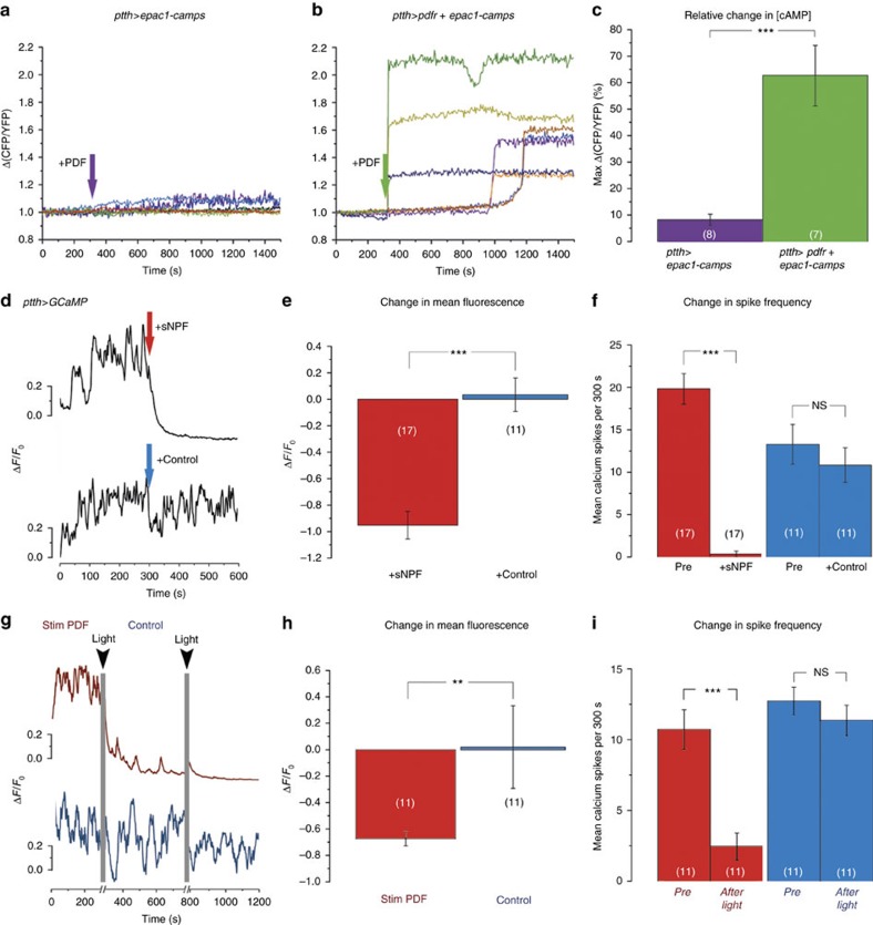
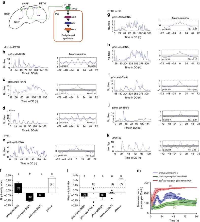
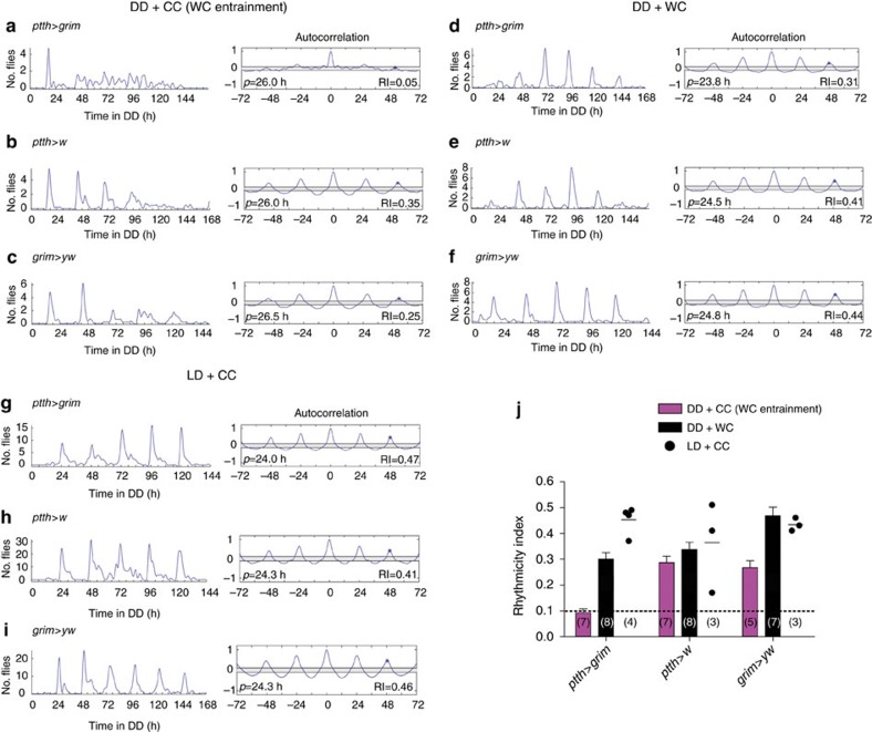
Animal circadian clocks consist of central and peripheral pacemakers, which are coordinated to produce daily rhythms in physiology and behaviour. Despite its importance for optimal performance and health, the mechanism of clock coordination is poorly understood. Here we dissect the pathway through which the circadian clock of Drosophila imposes daily rhythmicity to the pattern of adult emergence. Rhythmicity depends on the coupling between the brain clock and a peripheral clock in the prothoracic gland (PG), which produces the steroid hormone, ecdysone. Time information from the central clock is transmitted via the neuropeptide, sNPF, to non-clock neurons that produce the neuropeptide, PTTH. These secretory neurons then forward time information to the PG clock. We also show that the central clock exerts a dominant role on the peripheral clock. This use of two coupled clocks could serve as a paradigm to understand how daily steroid hormone rhythms are generated in animals.


Conflict of interest statement

The authors declare no competing financial interests.

Figures

Figure 1
Figure 1. Anatomical relationship between PDF- and PTTH-positive neurons.
(a) PDF and ptth-gal4 expression in the fly brain before emergence. PDF-positive (magenta) and PTTH neurons (green) arborize in the superior protocerebrum. (b) Arborization pattern of PTTH neurons shown alone. (c) Higher magnification of a 1 μm Z-stack of the superior lateral protocerebrum boxed in a, showing the close proximity between the arborizations of PTTH neurons and the endings of the PDF-expressing sLNvs. (d) Sagittal view of the medial region of a pharate fly head and anterior thorax. (e) Confocal projection of the white box shown in d. PTTH neurons innervate the PG where they form a dense varicose mesh of putative release sites (arrowhead). ant, antenna; lab, labellum; cns, central nervous system; ros, rostrum; thx, thorax. Scale bars in a,b and e: 50 μm, in c: 25 μm.
Figure 2
Figure 2. PTTH neurons respond to sNPF but not to PDF.
(a,b) Bath application of PDF (10−5 M) did not elicit an increase in cAMP levels in PTTH neurons (a) unless they ectopically expressed PDFR (b); each trace represents the response from a single ROI of PTTH cell bodies (N=8 ROIs from 7 pharate adult brains for a, 7 ROIs from 5 pharate adult brains for b). (c) Summary of results shown in a,b, indicating the average maximum response upon PDF application (±s.e.m.). (d) Bath application of sNPF (10−5 M; red arrow) caused rapid reduction in spontaneous calcium oscillations in PTTH neurons (upper trace); no such response occurred when challenged with solvent alone (control; blue arrow, lower trace). (e,f) Average (±s.e.m.) fluorescence change (e) and change in spike frequency (f) of GCaMP6 fluorescence induced by sNPF (red) versus control (blue) (+sNPF N=17 ROIs from 11 pharate adult brains; control: N=11 ROIs from 8 pharate adult brains). See also Supplementary Fig. 2. (g) Light stimulation of brains in which PDF neurons express ChR2-XXL caused a reduction in spontaneous calcium oscillations in PTTH neurons (red trace; time of light stimulation indicated by grey vertical lines); no such effect was obtained when the pdf-LexA transgene was omitted (blue trace; control). (h,i) Average (±s.e.m.) change in intensity (h) and change in spike frequency (i) of GCaMP6 fluorescence induced in PTTH neurons by light stimulation in flies expressing ChR2-XXL in PDF neurons (red) versus control (blue). (Stim PDF: N=11 ROIs from 6 pharate adult brains; control: N=11 ROIs from 7 pharate adult brains). (NS=P>0.05; **P<0.01; ***P<0.001; t-test: e;f,i blue; Wilcoxon rank sum test: c,h;f, i red).
Figure 3
Figure 3. Brain and PG clocks are coupled by sNPF and PTTH.
(a) Diagram showing the neuronal and molecular components that couple the brain clock (left) to the PG clock (right). sLNv neurons (purple line) contain sNPF and project dorsally to the vicinity of PTTH neurons (green). PTTH acts on the PG to activate the TORSO intracellular transduction pathway. (b) Knockdown of PDFR in PTTH neurons did not alter the circadian rhythmicity of emergence; by contrast, knockdown of sNPF receptor in these neurons rendered arrhythmic the pattern of adult emergence (c). Records show time course of emergence of a single population in DD (left) and corresponding autocorrelation analysis (right); principal periodicity and associated RI is indicated. (e) Reduction of PTTH neuropeptide in PTTH neurons eliminated the circadian rhythmicity of emergence. (f) Average RI values (±s.e.m.) for results shown in b,c,e and in control d; dashed lines mark RI cutoff value of 0.1. Different letters indicate statistically different groups (P<0.05; one-way ANOVA, Tukey's post hoc multiple comparison analyses). Numbers in parenthesis indicate number of separate experiments. (gj) Knockdown of PTTH transduction pathway in the PG caused the expression of an arrhythmic pattern of adult emergence. (l) Average RI values (±s.e.m.) for results shown in gj and in control k, shown as described for f; individual values are indicated when N<5 and average indicated by short horizontal line. Flies bearing only UAS-RNAi transgenes express normal rhythmicities of emergence (Supplementary Fig. 4). See also Supplementary Fig. 3. (m) Knockdown of torso expression in the PG eliminated circadian fluctuations of cre-driven bioluminescence in the PG. Values plotted correspond to average±s.e.m.; numbers in parenthesis indicate number of records averaged. See also Supplementary Fig. 7. In all experiments RNAi knockdown was enhanced by co-expression of dcr2.
Figure 4
Figure 4. PTTH neurons are required for entrainment by temperature cycles.
(ac) Left: pattern of emergence in DD from cultures entrained in DD with a 12 h:12 h 25 °C/16 °C (WC) temperature cycle in which PTTH neurons were selectively killed (a) and in controls (b,c); Right: corresponding autocorrelation analysis with value of RI indicated. (df) Left: pattern of emergence under DD+WC regime when PTTH neurons were selectively killed and in controls (e,f). (gi) Left: pattern of emergence under LD+CC regime when PTTH neurons were selectively killed (g) and in controls (h,i). (j) Average RI (±s.e.m.) for genotypes and conditions shown in ai; dashed line marks RI cutoff value of 0.1. Numbers in parenthesis indicate number of separate experiments. Individual values are indicated when N<5 and average indicated by short horizontal line.
Figure 5
Figure 5. phm-gal80 effectively suppresses tim-gal4 expression in the PG.
Pattern of GFP (left; a,d,g) and corresponding DAPI (right; c,f,i) and merged images (center; b,e,h) of pre-pupal brains (Br) expressing GFP under the control of tim-gal4 (a), tim-gal4+phm-gal80 (d) and phm-gal4 (g). Gain in a,b was set to visualize expression within the brain; arrowheads indicate LNs. Insets in a,b show expression in PG using the same gain used for g. In the ring gland, tim-gal4 drives expression in the PG and the corpora allata (labelled CA in a,d). Expression in the PG is suppressed when the tim-gal4 driver is combined with phm-gal80 (d). Scale bar (for ai): 100μm.
Figure 6
Figure 6. torso is required only in the PG for circadian rhythmicity of eclosion.
(a) Driving torso RNAi in all clocks using tim-gal4 driver rendered the pattern of emergence arrhythmic. Left: pattern of eclosion in DD; right: autocorrelation analysis of record, with dominant periodicity and value of RI indicated. (b) Suppressing tim-gal4 action in the PG combining tim-gal4 with phm-gal80 restored the circadian rhythm of eclosion indicating that torso function is only required in the PG clock for a circadian rhythmicity of emergence. (c) Individual values and average RI (±s.e.m.) for genotypes shown in a,b; dashed line marks RI cutoff value of 0.1. Different letters indicate statistically different groups (P<0.05; one-way ANOVA, Tukey's post hoc multiple comparison analyses). Numbers in parenthesis indicate number of separate experiments. In all experiments RNAi knockdown was enhanced by co-expression of dcr2.
Figure 7
Figure 7. Stopping clock in brain and/or PG leads to arrhythmic adult emergence.
Pattern of emergence when all clocks (a), the brain clock (b) or the PG (c) clock, have been stopped. Schematic shown on the left represents the brain and PG clocks (large and small circle, respectively); red indicates which clock has been stopped; middle: pattern of eclosion in DD; right: autocorrelation analysis of record, with dominant periodicity and value of RI indicated. (e) Average RI (±s.e.m.) for genotypes shown in ad and for controls; individual values are indicated when N<5 and average indicated by short horizontal line. Dashed line marks RI cutoff value of 0.1. Different letters indicate statistically different groups (P<0.05; one-way ANOVA, Tukey's post hoc multiple comparison analyses). Numbers in parenthesis indicate number of separate experiments. phm-gal80 effectively suppresses tim-gal4 expression in the PG (cf. Fig. 5d). (Strictly speaking, in (b) gene expression is driven in all clocks except the PG. However, since circadian rhythmicity of emergence depends only on the clocks in the brain and in the PG, such experiment is equivalent to driving gene expression only in the brain.)
Figure 8
Figure 8. Hierarchical relationship between the brain clock and the PG clock.
(ac) Pattern of emergence when all clocks (a) or only the brain (b) or the PG (c) clock have been slowed down. Schematic shown on the left represents the brain and PG clocks (large and small circle, respectively); orange colouring indicates which clock has been slowed down; middle: pattern of eclosion in DD; right: MESA analysis of record, with dominant periodicity indicated. (d) Average periodicities (±s.e.m.) for genotypes shown in ac and for controls; circles indicate periodicity for each separate experiment (N=5–8); average is indicated by horizontal line; different letters indicate statistically different groups (P<0.05; one-way ANOVA, Tukey's post hoc multiple comparison analyses). (eg) Pattern of emergence when all clocks (e), or only the brain (f) or the PG (g) clock have been sped up (indicated in green). (h) Average periodicities (±s.e.m.) for genotypes shown in eg and for controls, represented as described in d. phm-gal80 effectively suppresses tim-gal4 expression in the PG (cf. Fig. 5d). (Strictly speaking, in (b,f) gene expression is driven in all clocks except the PG. However, since circadian rhythmicity of emergence depends only on the clocks in the brain and in the PG, such experiment is equivalent to driving gene expression only in the brain).

Similar articles

Cited by

References

    1. Takeda N. & Maemura K. Circadian clock and vascular disease. Hypertens. Res. 33, 645–651 (2010). - PubMed
    1. Scheer F. A., Hilton M. F., Mantzoros C. S. & Shea S. A. Adverse metabolic and cardiovascular consequences of circadian misalignment. Proc. Natl Acad. Sci. USA 106, 4453–4458 (2009). - PMC - PubMed
    1. Tsang A. H., Astiz M., Friedrichs M. & Oster H. Endocrine regulation of circadian physiology. J. Endocrinol. 230, R1–R11 (2016). - PubMed
    1. Yoo S. H. et al.. PERIOD2::LUCIFERASE real-time reporting of circadian dynamics reveals persistent circadian oscillations in mouse peripheral tissues. Proc. Natl Acad. Sci. USA 101, 5339–5346 (2004). - PMC - PubMed
    1. Albrecht U. Timing to perfection: the biology of central and peripheral circadian clocks. Neuron 74, 246–260 (2012). - PubMed

Publication types

LinkOut - more resources