

Differential responses of the dorsomedial prefrontal cortex and right posterior superior temporal sulcus to spontaneous mentalizing
- PMID: 28556306
- PMCID: PMC6866721
- DOI: 10.1002/hbm.23626
Differential responses of the dorsomedial prefrontal cortex and right posterior superior temporal sulcus to spontaneous mentalizing
Abstract
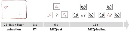
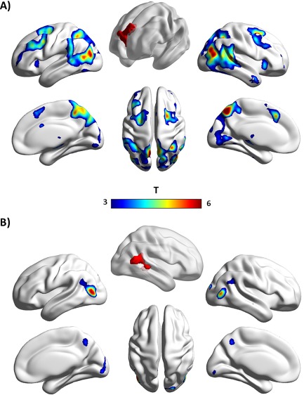
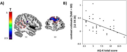
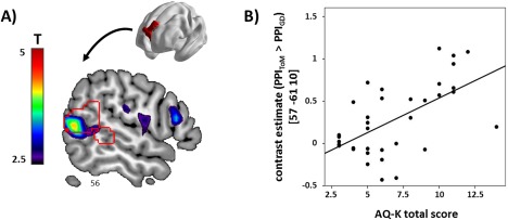
Previous research suggests a role of the dorsomedial prefrontal cortex (dmPFC) in metacognitive representation of social information, while the right posterior superior temporal sulcus (pSTS) has been linked to social perception. This study targeted these functional roles in the context of spontaneous mentalizing. An animated shapes task was presented to 46 subjects during functional magnetic resonance imaging. Stimuli consisted of video clips depicting animated shapes whose movement patterns prompt spontaneous mentalizing or simple intention attribution. Based on their differential response during spontaneous mentalizing, both regions were characterized with respect to their task-dependent connectivity profiles and their associations with autistic traits. Functional network analyses revealed highly localized coupling of the right pSTS with visual areas in the lateral occipital cortex, while the dmPFC showed extensive coupling with instances of large-scale control networks and temporal areas including the right pSTS. Autistic traits were related to mentalizing-specific activation of the dmPFC and to the strength of connectivity between the dmPFC and posterior temporal regions. These results are in good agreement with the hypothesized roles of the dmPFC and right pSTS for metacognitive representation and perception-based processing of social information, respectively, and further inform their implication in social behavior linked to autism. Hum Brain Mapp 38:3791-3803, 2017. © 2017 Wiley Periodicals, Inc.
Keywords: autistic traits; dorsomedial prefrontal cortex; fMRI; posterior superior temporal sulcus; spontaneous mentalizing.
© 2017 Wiley Periodicals, Inc.
Conflict of interest statement
AM‐L has received consultant fees from AstraZeneca, Elsevier, F. Hoffmann‐La Roche, Gerson Lehrman Group, Lundbeck, Outcome Europe Sárl, Outcome Sciences, Roche Pharma, Servier International, and Thieme Verlag; and has received lecture fees including travel expenses from Abbott, AstraZeneca, Aula Médica Congresos, BASF, Boehringer Ingelheim, Groupo Ferrer International, Janssen‐Cilag, Lilly Deutschland, LVR Klinikum Düsseldorf, Otsuka Pharmaceuticals, and Servier Deutschland.
TB served in an advisory or consultancy role for Actelion, Hexal Pharma, Lilly, Medice, Novartis, Oxford outcomes, PCM scientific, Shire, and Viforpharma. He received conference support or speaker's fee by Medice, Novartis, and Shire. He is/has been involved in clinical trials conducted by Shire & Viforpharma. He received royalities from Hogrefe, Kohlhammer, CIP Medien, and Oxford University Press. This work is unrelated to the above grants and relationships.
LP received conference support or speaker's fee by Shire, Lilly, Novartis, and Medice. She receives royalities from Hogrefe and Schattauer.
The other authors report no biomedical financial interests or other potential conflicts of interest.
Figures

Similar articles
-
Brain Activation and Aberrant Effective Connectivity in the Mentalizing Network of Preadolescent Children at Familial High Risk of Schizophrenia or Bipolar Disorder.Biol Psychiatry Cogn Neurosci Neuroimaging. 2025 Jan;10(1):68-79. doi: 10.1016/j.bpsc.2024.08.004. Epub 2024 Aug 23. Biol Psychiatry Cogn Neurosci Neuroimaging. 2025. PMID: 39182726
-
The selfless mind: How prefrontal involvement in mentalizing with similar and dissimilar others shapes empathy and prosocial behavior.Cognition. 2016 Dec;157:24-38. doi: 10.1016/j.cognition.2016.08.003. Epub 2016 Aug 29. Cognition. 2016. PMID: 27568587
-
Dissecting functional contributions of the social brain to strategic behavior.Neuron. 2021 Oct 20;109(20):3323-3337.e5. doi: 10.1016/j.neuron.2021.07.025. Epub 2021 Aug 17. Neuron. 2021. PMID: 34407389
-
Interacting minds--a biological basis.Science. 1999 Nov 26;286(5445):1692-5. doi: 10.1126/science.286.5445.1692. Science. 1999. PMID: 10576727 Review.
-
Mapping the mentalizing brain: An ALE meta-analysis to differentiate the representation of social scenes and ages on theory of mind.Neurosci Biobehav Rev. 2024 Dec;167:105918. doi: 10.1016/j.neubiorev.2024.105918. Epub 2024 Oct 9. Neurosci Biobehav Rev. 2024. PMID: 39389437 Review.
Cited by
-
Neural and behavioral effects of oxytocin administration during theory of mind in schizophrenia and controls: a randomized control trial.Neuropsychopharmacology. 2019 Oct;44(11):1925-1931. doi: 10.1038/s41386-019-0417-5. Epub 2019 May 18. Neuropsychopharmacology. 2019. PMID: 31103018 Free PMC article. Clinical Trial.
-
Overlapping and specific neural correlates for empathizing, affective mentalizing, and cognitive mentalizing: A coordinate-based meta-analytic study.Hum Brain Mapp. 2021 Oct 1;42(14):4777-4804. doi: 10.1002/hbm.25570. Epub 2021 Jul 29. Hum Brain Mapp. 2021. PMID: 34322943 Free PMC article. Review.
-
Superior temporal sulcus folding, functional network connectivity, and autistic-like traits in a non-clinical population.Mol Autism. 2024 Oct 8;15(1):44. doi: 10.1186/s13229-024-00623-3. Mol Autism. 2024. PMID: 39380071 Free PMC article.
-
Iranian Brain Imaging Database: A Neuropsychiatric Database of Healthy Brain.Basic Clin Neurosci. 2021 Jan-Feb;12(1):115-132. doi: 10.32598/bcn.12.1.1774.2. Epub 2021 Jan 1. Basic Clin Neurosci. 2021. PMID: 33995934 Free PMC article.
-
Behavioral and neuro-cognitive bases for emergence of norms and socially shared realities via dynamic interaction.Commun Biol. 2022 Dec 15;5(1):1379. doi: 10.1038/s42003-022-04329-1. Commun Biol. 2022. PMID: 36522539 Free PMC article.
References
-
- Abell F, Happé F, Frith U (2000): Do triangles play tricks? Attribution of mental states to animated shapes in normal and abnormal development. Cogn Dev 15:1–16.
-
- Apperly IA, Butterfill SA (2009): Do humans have two systems to track beliefs and belief‐like states? Psychol Rev 116:953–970. - PubMed
Publication types
MeSH terms
LinkOut - more resources
Full Text Sources
Other Literature Sources
