. 2017 Aug;246(8):625-634.
doi: 10.1002/dvdy.24524. Epub 2017 Jun 28.

A role for primary cilia in aortic valve development and disease

Affiliations

A role for primary cilia in aortic valve development and disease

Katelynn A Toomer et al. Dev Dyn. 2017 Aug.

Abstract

Background: Bicuspid aortic valve (BAV) disease is the most common congenital heart defect, affecting 0.5-1.2% of the population and causing significant morbidity and mortality. Only a few genes have been identified in pedigrees, and no single gene model explains BAV inheritance, thus supporting a complex genetic network of interacting genes. However, patients with rare syndromic diseases that stem from alterations in the structure and function of primary cilia ("ciliopathies") exhibit BAV as a frequent cardiovascular finding, suggesting primary cilia may factor broadly in disease etiology.

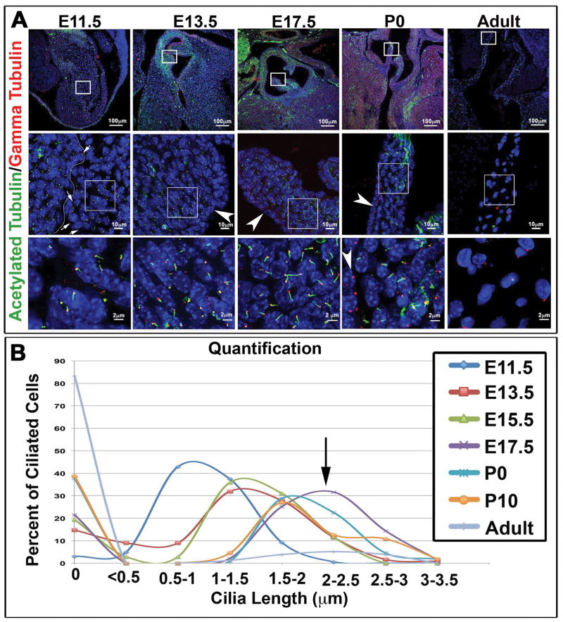
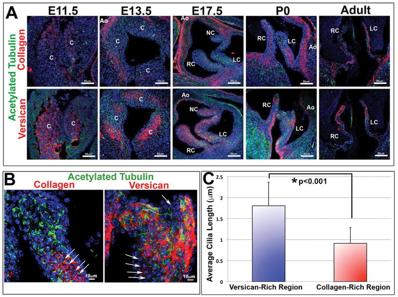
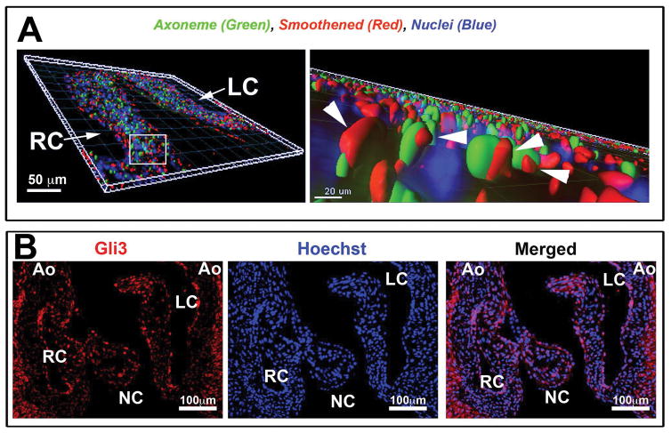
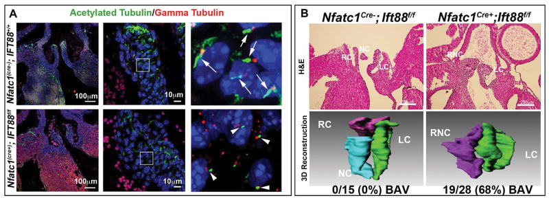
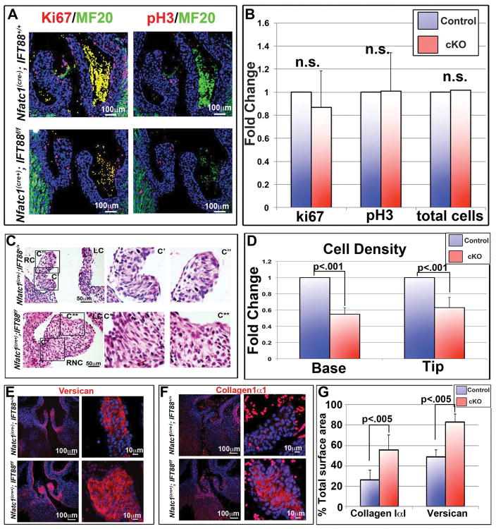
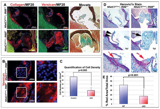
Results: Our data are the first to demonstrate that primary cilia are expressed on aortic valve mesenchymal cells during embryonic development and are lost as these cells differentiate into collagen-secreting fibroblastic-like cells. The function of primary cilia was tested by genetically ablating the critical ciliogenic gene Ift88. Loss of Ift88 resulted in abrogation of primary cilia and increased fibrogenic extracellular matrix (ECM) production. Consequentially, stratification of ECM boundaries normally present in the aortic valve were lost and a highly penetrant BAV phenotype was evident at birth.

Conclusions: Our data support cilia as a novel cellular mechanism for restraining ECM production during aortic valve development and broadly implicate these structures in the etiology of BAV disease in humans. Developmental Dynamics 246:625-634, 2017. © 2017 Wiley Periodicals, Inc.

Keywords: bicuspid aortic valve; cardiac development; extracellular matrix; primary cilia.


Figures

Figure 1
Figure 1. Expression of primary cilia during aortic valve development
(A) IHC for the ciliary axoneme (green), basal body (red), and nuclei (blue) show expression of cilia throughout valve development. Cilia appear shorter during early embryonic stages and longer right before and after birth. Arrows at E11.5 depict very short axonemes on endothelial cells (line depicting the edge of the endocardium is denoted) whereas arrowheads at the over timepoints depict lack of primary cilia on the valve endocardium at later timepoints (B) Quantification of cilia length showing increased expression of cilia during embryonic development, peaking at the E17.5 timepoint (purple line and arrow) and decreased expression during postnatal development and adult life.
Figure 2
Figure 2. Correlation of primary cilia with versican expressing microenvironments
(A) IHC for the ciliary axoneme (green), collagen (red, top) versican (red, bottom), and nuclei (blue) show spatial/temporal expression of cilia throughout development. Cilia are predominantly expressed in proteoglycan-rich zones and scant in regions of high collagen expression. C= conal cushions, RC= right-coronary, LC= left-coronary, NC= non-coronary, Ao= aorta. (B) High magnification images of axonemes (green) and collagen/versican (red). Arrows depict collagen rich regions expressing shortened cilia. (C) Quantification of cilia length in both versican and collagen rich regions shows decreased average cilia length in collagen rich regions when compared with versican rich regions, p<0.001 Students t-test.
Figure 3
Figure 3. Active Hedgehog signaling in aortic valve interstitial cells in vivo
(A) Three-dimensional reconstruction of IHC stain at postnatal day 0, shows smoothened (red), acetylated tubulin—cilia axoneme (green), and Hoechst--nuclei (blue). High magnification (right) shows smoothened (arrowhead pointing to smoothened staining) on the axoneme of the cilia indicative of active hedgehog signaling. (B) IHC of P0 aortic cusps showing Gli3 expression.
Figure 4
Figure 4. Developmental loss of axonemes causes BAV
(A) Control IHC experiments for the ciliary axoneme (green), basal body (red), and nuclei (blue) show normal cilia length on control aortic valves (top) vs. axoneme shortening in the Ift88 conditional knockout (bottom). (B) H and E and 3D reconstruction of P0 wild type and conditional knockout valves. Wild-type valves show three distinct cusps while conditional knockout mouse aortic valves show bicuspid aortic valves. Penetrance of the phenotype is depicted below the 3D reconstruction images. RC=right coronary, LC= left coronary, NC= non-coronary, and RNC= right non-coronary.
Figure 5
Figure 5. Cilia effects on proliferation and ECM production
(A) IHC of postnatal day 0 (P0) aortic valves, for proliferation markers Ki67 and phospho-histone H3, show no difference in proliferating or total cell number when conditional knockout aortic valves were compared to littermate controls, quantified in (B). (C) H and E staining’s show increased matrix in the fused right-non-coronary leaflet of the conditional knockout. C′= wild-type right coronary, C″= wild-type right coronary tip, C*= conditional knockout right non-coronary base, C**= conditional knockout right coronary tip. RC= right coronary, LC= left coronary, RNC= right non-coronary. (D) Quantification of cell density shows a significant decrease in cell density in cilia deficient valves at both the base and tip of the right coronary leaflet, with p<.001. (E and F) IHC staining’s of conditional knockout aortic cusps show increased expression of both collagen (E) and versican (F) in the cilia conditional knockout valves. (G) Quantification of total surface area occupied by collagen or versican immunostaining with p<.005. RC=right coronary, LC= left coronary, NC= non-coronary, and RNC= right non-coronary.
Figure 6
Figure 6. Loss of cilia results in myxomatous degeneration in adult aortic valves
(A) IHC and movats pentachrome stain of ECM proteins in adult mouse Nfatc1cre−;Ift88+/+ and Nfatc1cre+;Ift88f/f aortic valves. Left and middle panels show IHC of collagen (red, left), versican (red, middle) MF20 (green), and nuclei (blue). Movats pentachrome stain (right) shows proteoglycans (blue), collagen (yellow), elastin (black), and fibrin or cardiac muscle (red). Increased expression of collagen and versican is observed in Ift88 conditional knockouts when compared to littermate controls in both IHC and movats stains. (B) IHC staining shows decreased cell density and increased versican in conditional knockout adult right coronary leaflets, quantified in (C) Student’s T tests; p<0.005 n=3. Herovici’s collagen stain shows decreased mature collagen (red) in conditional knockout cusps, quantified in (D) with p<0.001.

References

    1. Baujat G, Le Merrer M. Ellis-van Creveld syndrome. Orphanet J Rare Dis. 2007;2:27. - PMC - PubMed
    1. Bitgood MJ, McMahon AP. Hedgehog and Bmp genes are coexpressed at many diverse sites of cell-cell interaction in the mouse embryo. Dev Biol. 1995;172:126–138. - PubMed
    1. Bonachea EM, Chang SW, Zender G, LaHaye S, Fitzgerald-Butt S, McBride KL, Garg V. Rare GATA5 sequence variants identified in individuals with bicuspid aortic valve. Pediatr Res. 2014a;76:211–216. - PMC - PubMed
    1. Bonachea EM, Zender G, White P, Corsmeier D, Newsom D, Fitzgerald-Butt S, Garg V, McBride KL. Use of a targeted, combinatorial next-generation sequencing approach for the study of bicuspid aortic valve. BMC Med Genomics. 2014b;7:56. - PMC - PubMed
    1. Broekhuis JR, Leong WY, Jansen G. Regulation of cilium length and intraflagellar transport. Int Rev Cell Mol Biol. 2013;303:101–138. - PubMed

MeSH terms

LinkOut - more resources