. 2017 Sep;11(9):1156-1171.
doi: 10.1002/1878-0261.12086. Epub 2017 Jul 5.

Drug sensitivity and resistance testing identifies PLK1 inhibitors and gemcitabine as potent drugs for malignant peripheral nerve sheath tumors

Affiliations

Drug sensitivity and resistance testing identifies PLK1 inhibitors and gemcitabine as potent drugs for malignant peripheral nerve sheath tumors

Matthias Kolberg et al. Mol Oncol. 2017 Sep.

Abstract

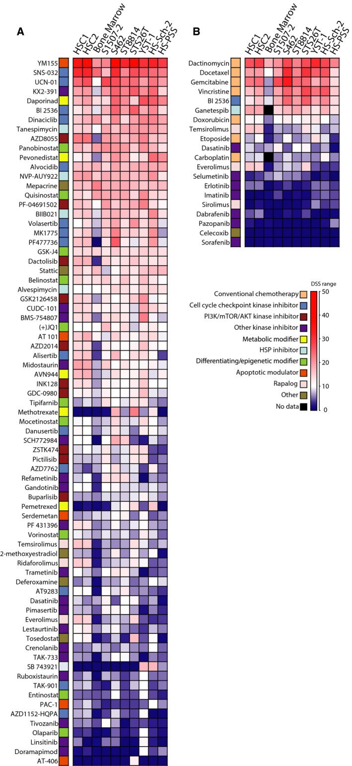
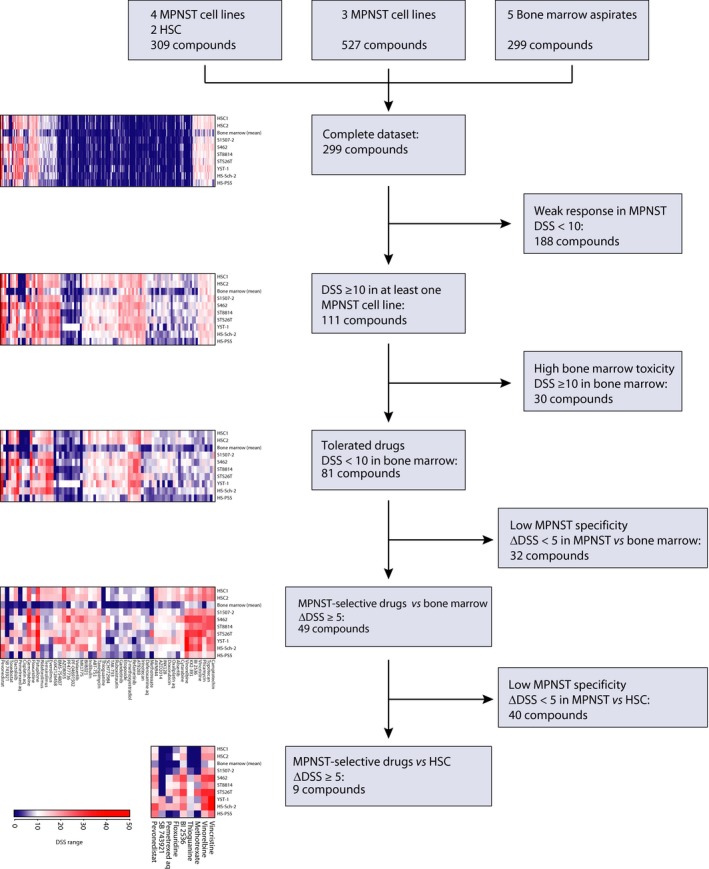
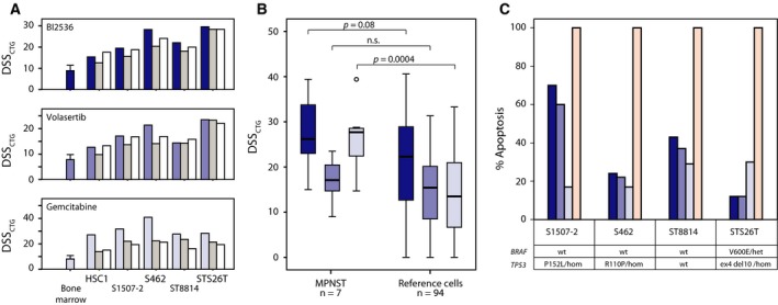
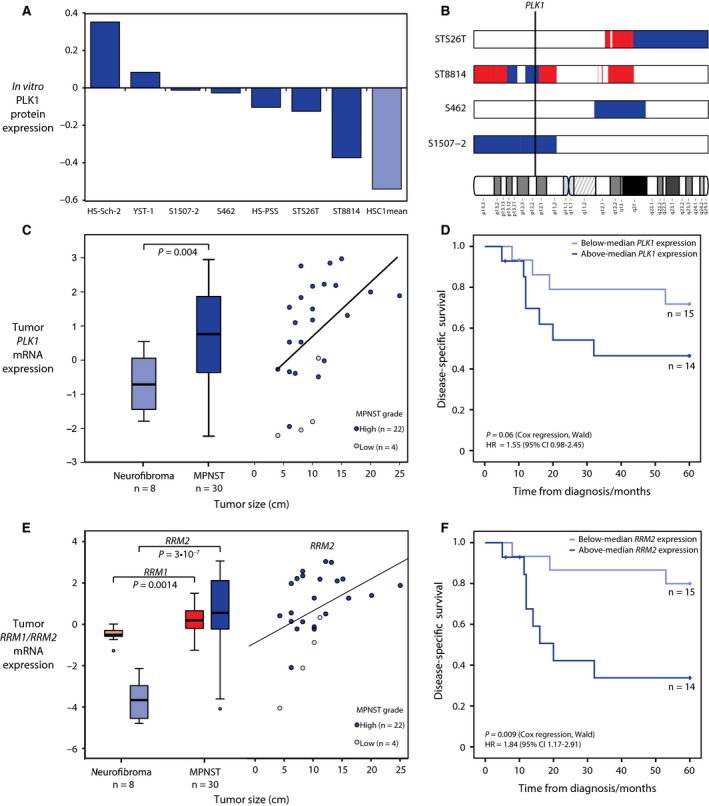
Patients with malignant peripheral nerve sheath tumor (MPNST), a rare soft tissue cancer associated with loss of the tumor suppressor neurofibromin (NF1), have poor prognosis and typically respond poorly to adjuvant therapy. We evaluated the effect of 299 clinical and investigational compounds on seven MPNST cell lines, two primary cultures of human Schwann cells, and five normal bone marrow aspirates, to identify potent drugs for MPNST treatment with few side effects. Top hits included Polo-like kinase 1 (PLK1) inhibitors (volasertib and BI2536) and the fluoronucleoside gemcitabine, which were validated in orthogonal assays measuring viability, cytotoxicity, and apoptosis. DNA copy number, gene expression, and protein expression were determined for the cell lines to assess pharmacogenomic relationships. MPNST cells were more sensitive to BI2536 and gemcitabine compared to a reference set of 94 cancer cell lines. PLK1, RRM1, and RRM2 mRNA levels were increased in MPNST compared to benign neurofibroma tissue, and the protein level of PLK1 was increased in the MPNST cell lines compared to normal Schwann cells, indicating an increased dependence on these drug targets in malignant cells. Furthermore, we observed an association between increased mRNA expression of PLK1, RRM1, and RRM2 in patient samples and worse disease outcome, suggesting a selective benefit from inhibition of these genes in the most aggressive tumors.

Keywords: MPNST; Schwann cell; drug screen; pharmacology.


Figures

Figure 1
Figure 1
Drug response heatmaps from CellTiter‐Glo (CTG) viability assay for seven MPNST cell lines, two normal human Schwann cell (HSC) cultures, and bone marrow (mean result from five healthy individuals). Drug responses for targeted drugs (excluding chemotherapeutic drugs) with a drug sensitivity score (DSSCTG) of 10, or more, in at least one MPNST cell line (A), and chemotherapeutic and other targeted drugs that have been used in clinical treatment of patients with MPNST (B). The same color coding and DSS gradient is used for both heatmaps.
Figure 2
Figure 2
Identification of candidate drugs for MPNST treatment. Filtering steps used to identify drugs with high specificity and selectivity toward MPNST as compared to bone marrow and normal human Schwann cells (HSC) based on drug sensitivity scores from CellTiter‐Glo viability assay (DSSCTG).
Figure 3
Figure 3
Independent validation and apoptosis assay of MPNST‐specific drugs. Comparison of drug sensitivity scores from initial (blue bars, including average data and standard deviation for the five bone marrow samples) CellTiter‐Glo viability assay (DSSCTG), and two subsequent validation rounds [manual (medium gray bars); custom plate (white bars)] (A). DSSCTG obtained for the three drugs BI2536 (dark blue), volasertib (medium blue), and gemcitabine (light blue) from MPNST cell lines in comparison with a reference set of 94 cancer cell lines (colon, ovarian, and leukemia); two‐tailed P‐values from independent samples t‐test, assuming unequal variance (B). The maximum level of apoptosis measured by a luminescence‐based caspase‐3/7 activation assay, induced by BI2536 (dark blue), volasertib (medium blue), and gemcitabine (light blue), in comparison with staurosporin (100% apoptosis, pink) and 0.1% DMSO (0% apoptosis) (C). The mutation status of TP53 and BRAF in each cell line is shown (het—heterozygous; hom—homozygous).
Figure 4
Figure 4
Expression of the drug targets PLK1, RRM1, and RRM2 in MPNST cell lines and prognostic relevance in patients with MPNST. Normalized RPPA protein expression of PLK1 in MPNST cell lines (median = 0 across 271 cancer‐relevant proteins) indicates an increased expression of PLK1 in seven MPNST cell lines as compared to the mean of duplicate runs of normal Schwann cell HSC1 (A). Regions of genomic gain (red) are not observed in the region on chromosome 16 harboring PLK1 (16p12.2) in the tested MPNST cell lines, while regions of loss (blue) are observed in the two cell lines S1507‐2 and ST8814 (B). Gene expression of PLK1 is significantly higher in MPNST tumor samples as compared to benign neurofibromas (C, left panel), and high expression in MPNST is associated with high tumor grade and large tumor size (C, right panel). MPNST patients with high expression of PLK1 in the tumor had worse outcome than patients with low expression, although not significantly at a 5% level (the P‐value and hazard ratio (HR) for PLK1 expression as a continuous variable in univariate Cox regression analysis for five‐year disease‐specific survival are shown) (D). The gene expression of gemcitabine target RRM1 and its activator RRM2 is significantly higher in MPNST tumor samples as compared to benign neurofibromas (E, left panel), and high expression of RRM2 in MPNST is associated with high tumor grade and large tumor size (E, right panel). MPNST patients with high expression of RRM2 in the tumor had significantly worse outcome than patients with low expression (P‐value and hazard ratio (HR) for gene expression as a continuous variable in univariate Cox regression analysis for 5‐year disease‐specific survival are shown) (F).

Similar articles

Cited by

References

    1. Abbou S, Lanvers‐Kaminsky C, Daudigeos‐Dubus E, LE Dret L, Laplace‐Builhe C, Molenaar J, Vassal G, Geoerger B, within the IB Preclinical Evaluation C (2016) Polo‐like kinase inhibitor volasertib exhibits antitumor activity and synergy with vincristine in pediatric malignancies. Anticancer Res 36, 599–609. - PubMed
    1. Ågesen TH, Flørenes VA, Molenaar WM, Lind GE, Berner JM, Plaat BE, Komdeur R, Myklebost O, van den Berg E and Lothe RA (2005) Expression patterns of cell cycle components in sporadic and neurofibromatosis type 1‐related malignant peripheral nerve sheath tumors. J Neuropathol Exp Neurol 64, 74–81. - PubMed
    1. Ahlquist T, Bottillo I, Danielsen SA, Meling GI, Rognum TO, Lind GE, Dallapiccola B and Lothe RA (2008) RAS signaling in colorectal carcinomas through alteration of RAS, RAF, NF1, and/or RASSF1A. Neoplasia 10(680–686), 682. - PMC - PubMed
    1. Albritton KH, Rankin C, Coffin CM, Ratner N, Budd GT, Schuetze SM, Randall RL, Declue JE and Borden EC (2006) Phase II study of erlotinib in metastatic or unresectable malignant peripheral nerve sheath tumors (MPNST). J Clin Oncol 24, S9518.
    1. Ambrosini G, Cheema HS, Seelman S, Teed A, Sambol EB, Singer S and Schwartz GK (2008) Sorafenib inhibits growth and mitogen‐activated protein kinase signaling in malignant peripheral nerve sheath cells. Mol Cancer Ther 7, 890–896. - PMC - PubMed

MeSH terms