. 2018 Jul;28(4):451-462.
doi: 10.1111/bpa.12532. Epub 2017 Jun 19.

Rapid amyloid-β oligomer and protofibril accumulation in traumatic brain injury

Affiliations

Rapid amyloid-β oligomer and protofibril accumulation in traumatic brain injury

Sami Abu Hamdeh et al. Brain Pathol. 2018 Jul.

Abstract

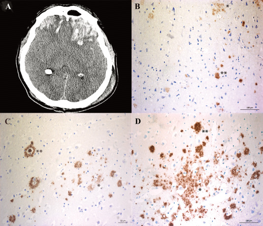
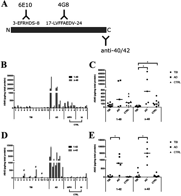
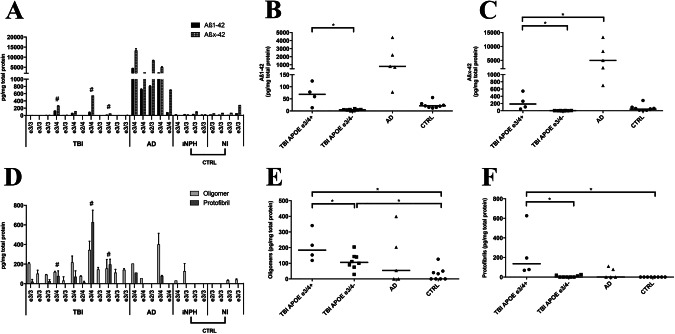
Deposition of amyloid-β (Aβ) is central to Alzheimer's disease (AD) pathogenesis and associated with progressive neurodegeneration in traumatic brain injury (TBI). We analyzed predisposing factors for Aβ deposition including monomeric Aβ40, Aβ42 and Aβ oligomers/protofibrils, Aβ species with pronounced neurotoxic properties, following human TBI. Highly selective ELISAs were used to analyze N-terminally intact and truncated Aβ40 and Aβ42, as well as Aβ oligomers/protofibrils, in human brain tissue, surgically resected from severe TBI patients (n = 12; mean age 49.5 ± 19 years) due to life-threatening brain swelling/hemorrhage within one week post-injury. The TBI tissues were compared to post-mortem AD brains (n = 5), to post-mortem tissue of neurologically intact (NI) subjects (n = 4) and to cortical biopsies obtained at surgery for idiopathic normal pressure hydrocephalus patients (iNPH; n = 4). The levels of Aβ40 and Aβ42 were not elevated by TBI. The levels of Aβ oligomers/protofibrils in TBI were similar to those in the significantly older AD patients and increased compared to NI and iNPH controls (P < 0.05). Moreover, TBI patients carrying the AD risk genotype Apolipoprotein E epsilon3/4 (APOE ε3/4; n = 4) had increased levels of Aβ oligomers/protofibrils (P < 0.05) and of both N-terminally intact and truncated Aβ42 (P < 0.05) compared to APOE ε3/4-negative TBI patients (n = 8). Neuropathological analysis showed insoluble Aβ aggregates (commonly referred to as Aβ plaques) in three TBI patients, all of whom were APOE ε3/4 carriers. We conclude that soluble intermediary Aβ aggregates form rapidly after TBI, especially among APOE ε3/4 carriers. Further research is needed to determine whether these aggregates aggravate the clinical short- and long-term outcome in TBI.

Keywords: Alzheimer's disease; amyloid β oligomers; amyloid-β; traumatic brain injury.


Conflict of interest statement

ERW and LS are employees of BioArctic AB. CM and HB are employees of BioArctic AB and own shares in the company. LL is a co‐founder and chairman of the board of BioArctic AB and owns shares in the company. The remaining authors declare no competing financial interests.

Figures

Figure 1
Figure 1
Amyloid‐β (Aβ) aggregates in surgically resected brain tissue in a subset of patients with severe TBI. A. An example of patients with a focal left frontal contusion injury following severe TBI causing midline shift, obliteration of the basal cisterns and life‐threatening mass effect to the surrounding brain tissue. Immediate surgical removal of the lesion was mandated, allowing immunohistochemical and biochemical analysis of the removed and injured brain tissue. B–D. Immunohistochemical image from the three TBI patients demonstrating accumulation of diffuse (*) and compact (**) insoluble Aβ aggregates.
Figure 2
Figure 2
TBI increases the levels of Aβ oligomers and protofibrils in surgically resected brain tissue. A. Individual oligomer Aβ levels in patient samples detected with the mAb82E1 sandwich ELISA. The absence of a bar indicates that the value was below the limit of detection. B. Dot‐plot of individual and median Aβ oligomer levels in TBI, Alzheimer's disease (AD) and in the control group (CTRL) consisting of idiopathic iNPH patients and non‐injured, NI individuals. Oligomers were significantly elevated (*) by TBI in comparison to the control group. C. Individual Aβ protofibril levels in patient samples detected with the mAb158 sandwich ELISA. D. Dot‐plot of individual and median Aβ protofibril levels in TBI, AD and in the control group. Aβ protofibril levels were significantly elevated (*) by TBI in comparison to the control group. (A,C) Each bar represents a mean value and standard deviation (SD) from two ELISA experiments performed on two different occasions. E,F. There was a significant positive correlation between Aβ1–42 (E), Aβx‐42 (F), and Aβ protofibril levels in TBI patients. Aβ = Amyloid β, # = TBI patients with Amyloid β aggregates, * = Significant difference (P < 0.05), Strep‐HRP = Streptavidin‐Horseradish Peroxidase; TMB = Tetramethylbenzidine.
Figure 3
Figure 3
No elevations of Aβ monomers in TBI brain tissue A. Detection of Aβ monomers with mAb6E10 and mAb4G8 antibodies. The mAb6E10 antibody detects more N‐terminally intact Aβ40 and 42 as compared to mAb4G8 that detects Aβ40 and Aβ42 by mid‐region binding. B. Individual full‐length (Aβ1‐40) and N‐truncated (Aβx‐40) Aβ40 monomer levels detected by mAb6E10 and mAb4G8, respectively, in TBI, Alzheimer's disease (AD) and in the control group (CTRL) consisting of idiopathic iNPH patients and NI individuals. C. Dot‐plot of individual and median Aβ1‐40 and Aβx‐40 levels. Levels of Aβx‐40 were significantly lower (*) in TBI patients than in both AD and the control group. D. Individual full‐length (Aβ1‐42) and truncated (Aβx‐42) Aβ42 monomer levels detected by mAb6E10 and mid‐region mAb4G8, respectively. Of the four TBI patients with markedly higher levels of Aβx‐42 than Aβ1‐42, the three patients with immunohistochemical evidence of Aβ aggregates are indicated with #. E. Dot‐plot of individual and median Aβ1‐42 and Aβx‐42 levels in TBI, AD and in the control group (CTRL). Levels of both Aβ1‐42 and Aβx‐42 were significantly lower (*) in brain tissue from TBI than from AD patients. (B,D); Data shown as the mean from one ELISA experiment with error bars showing variation between duplicate ELISA wells. Aβ = Amyloid β, * = Significant difference (P < 0.05).
Figure 4
Figure 4
The APOE ε3/4 genotype is associated with increased levels of Aβ42, Aβ oligomers and Aβ protofibrils following TBI. A. Individual full‐length (Aβ1‐42) and truncated (Aβx‐42) Aβ42 monomer levels detected by N‐terminal mAb6E10 and mid‐region mAb4G8, respectively, in TBI, Alzheimer's disease (AD) and in the control group (CTRL) consisting of idiopathic iNPH patients and NI individuals. The individual APOE genotype for each individual is shown. # indicates immunohistochemical evidence of Aβ aggregates. B. Dot‐plot of individual and median Aβ1‐42 in TBI patients with (APOE ε3/4+) and without (APOE ε3/4‐) the APOE ε3/4 genotype, in AD and in the control group. In TBI patients, the Aβ1‐42 levels were significantly elevated (*) in APOE ε3/4+ patients compared with APOE ε3/4‐ patients. C. Dot‐plot of individual and median Aβx‐42 levels in APOE ε3/4+ and APOE ε3/4‐ TBI patients, in AD and in the control group. In APOE ε3/4+ TBI patients, levels of Aβx‐42 were significantly lower than in Alzheimer's disease patients although elevated (*) in comparison to APOE ε3/4‐. D. Amyloid‐β (Aβ) oligomer and Aβ protofibril levels in TBI patients, in AD and in a control group consisting of iNPH and NI patients. The individual APOE genotype for each individual is shown. # indicates immunohistochemical evidence of Aβ aggregates. E. Dot‐plot of individual and median Aβ oligomer levels in APOE ε3/4+ and APOE ε3/4‐ TBI patients, in AD and in the control group. Compared to controls, Aβ oligomers were significantly elevated (*) in both APOE ε3/4+ and APOE ε3/4‐ TBI patients, while in TBI Aβ oligomers were significantly elevated (*) in APOE ε3/4+ when compared to APOE ε3/4‐ patients. F. Dot‐plot of individual and median Aβ protofibril levels in APOE ε3/4+ and APOE ε3/4‐ TBI patients, in AD and in the control group. Aβ protofibrils were significantly elevated (*) in APOE ε3/4+ TBI patients when compared both to APOE ε3/4‐ and controls. APOE = Apolipoprotein E, Aβ = Amyloid β, * = Significant difference (P < 0.05).

Comment in

References

    1. Benilova I, Karran E, De Strooper B (2012) The toxic Abeta oligomer and Alzheimer's disease: an emperor in need of clothes. Nat Neurosci 15:349–357. - PubMed
    1. Bouter Y, Dietrich K, Wittnam JL, Rezaei‐Ghaleh N, Pillot T, Papot‐Couturier S et al (2013) N‐truncated amyloid beta (Abeta) 4–42 forms stable aggregates and induces acute and long‐lasting behavioral deficits. Acta Neuropathol 126:189–205. - PMC - PubMed
    1. Braak H, Braak E (1991) Neuropathological stageing of Alzheimer‐related changes. Acta Neuropathol 82:239–259. - PubMed
    1. Brody DL, Magnoni S, Schwetye KE, Spinner ML, Esparza TJ, Stocchetti N et al (2008) Amyloid‐beta dynamics correlate with neurological status in the injured human brain. Science (New York, NY) 321:1221–1224. - PMC - PubMed
    1. Casoli T, Di Stefano G, Giorgetti B, Grossi Y, Balietti M, Fattoretti P, Bertoni‐Freddari C (2007) Release of beta‐amyloid from high‐density platelets: implications for Alzheimer's disease pathology. Ann New York Acad Sci 1096:170–178. - PubMed

Publication types