. 2017 Sep;11(9):1172-1188.
doi: 10.1002/1878-0261.12087. Epub 2017 Jun 26.

SK4 channels modulate Ca2+ signalling and cell cycle progression in murine breast cancer

Affiliations

SK4 channels modulate Ca2+ signalling and cell cycle progression in murine breast cancer

Friederike A Steudel et al. Mol Oncol. 2017 Sep.

Abstract

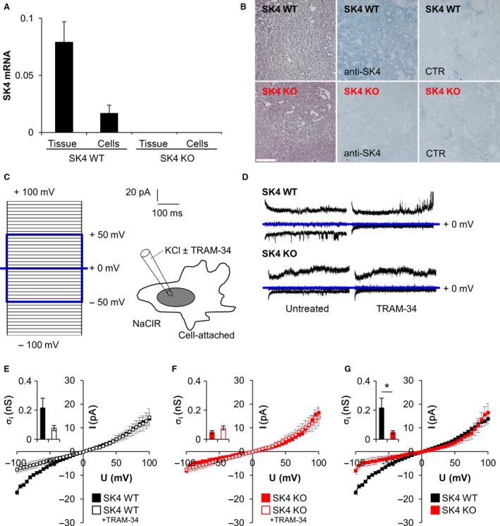
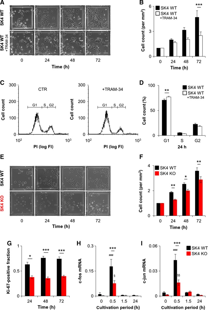
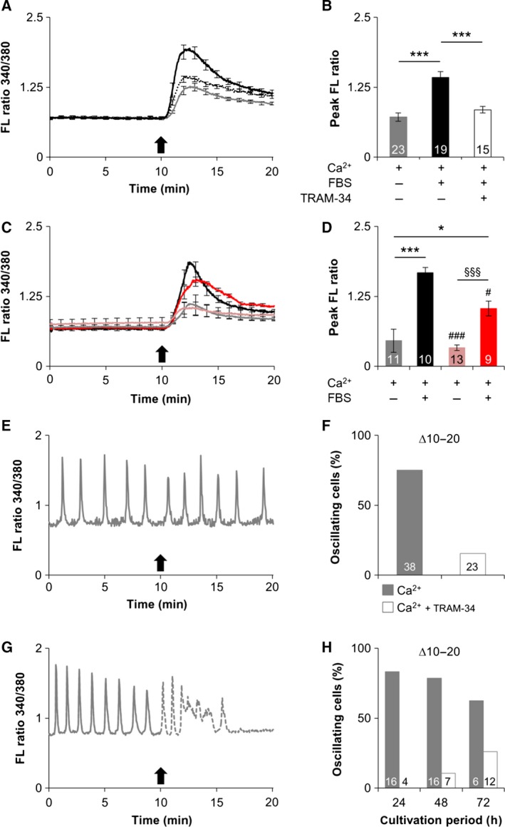
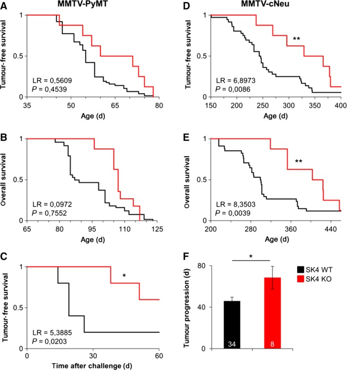
Oncogenic signalling via Ca2+ -activated K+ channels of intermediate conductance (SK4, also known as KCa 3.1 or IK) has been implicated in different cancer entities including breast cancer. Yet, the role of endogenous SK4 channels for tumorigenesis is unclear. Herein, we generated SK4-negative tumours by crossing SK4-deficient (SK4 KO) mice to the polyoma middle T-antigen (PyMT) and epidermal growth factor receptor 2 (cNeu) breast cancer models in which oncogene expression is driven by the retroviral promoter MMTV. Survival parameters and tumour progression were studied in cancer-prone SK4 KO in comparison with wild-type (WT) mice and in a syngeneic orthotopic mouse model following transplantation of SK4-negative or WT tumour cells. SK4 activity was modulated by genetic or pharmacological means using the SK4 inhibitor TRAM-34 in order to establish the role of breast tumour SK4 for cell growth, electrophysiological signalling, and [Ca2+ ]i oscillations. Ablation of SK4 and TRAM-34 treatment reduced the SK4-generated current fraction, growth factor-dependent Ca2+ entry, cell cycle progression and the proliferation rate of MMTV-PyMT tumour cells. In vivo, PyMT oncogene-driven tumorigenesis was only marginally affected by the global lack of SK4, whereas tumour progression was significantly delayed after orthotopic implantation of MMTV-PyMT SK4 KO breast tumour cells. However, overall survival and progression-free survival time in the MMTV-cNeu mouse model were significantly extended in the absence of SK4. Collectively, our data from murine breast cancer models indicate that SK4 activity is crucial for cell cycle control. Thus, the modulation of this channel should be further investigated towards a potential improvement of existing antitumour strategies in human breast cancer.

Keywords: Ca2+-activated K+ channels of intermediate conductance (SK4, KCa3.1, IK, KCNN4); breast cancer; epidermal growth factor receptor 2 (Her2/cNeu); mouse mammary tumour virus (MMTV); polyoma virus middle T-antigen (PyMT).


Figures

Figure 1
Figure 1
Expression and functional analysis of SK4 channels in MMTV‐PyMTtg/+ breast tumours and tumour‐derived cells. (A) MMTV‐PyMTtg/+ tumour tissue (n = 7) and cells (n = 4) express high levels of SK4 mRNA. β‐Actin was coamplified in each sample and used as a reference for normalization of mRNA abundance. As expected, no SK4 mRNA was detectable in SK4 KO tissue and cells (n = 2). (B) Haematoxylin and eosin staining of MMTV‐PyMTtg/+ SK4 WT and SK4 KO tumours (upper and lower left panels panel). Immunohistochemical detection of SK4 channels in PyMT transgenic SK4 WT tumours using a SK4‐specific primary antibody (upper middle panel). As expected, SK4‐negative tumours remained unstained (lower middle panel). The specificity of the immunohistochemical approach was further controlled by staining of SK WT and SK4 KO tumours in the absence of the primary antibody (upper and lower right panels) (n = 3 for each genotype; scale bar = 500 μm). (C) Macroscopic on‐cell currents were recorded from MMTV‐PyMTtg/+ tumour‐derived SK4 WT and SK4 KO cells using a KCl pipette and NaCl bath solution applying the depicted pulse protocol. (D) On‐cell current tracings recorded during voltage square pulses (C) to ‐50, 0 and +50 mV. Currents were recorded from MMTV‐PyMTtg/+ SK4 WT (top) and SK4 KO (bottom) cells with (right) and without (left) TRAM‐34 (10 μM) in the pipette solutions. (E–G) Dependence of the mean macroscopic on‐cell currents on clamp voltage in MMTV‐PyMTtg/+ SK4 WT (E, G) and SK4 KO (F, G) cells recorded in the absence (closed squares) and presence (open squares) of TRAM‐34 in the pipette. Inlays show inward conductance as calculated from the data in (E) and (F) by linear regression of MMTV‐PyMTtg/+ SK4 WT and SK4 KO cells in the absence (closed bars) and presence (open bars) of TRAM‐34 (*P < 0.05, unpaired two‐tailed Welch‐corrected Student's t‐test) (n = 11/7; 17/15; 11/15). SK4, calcium‐activated potassium channel with intermediate conductance; MMTV, mouse mammary tumour virus; PyMT, polyoma virus middle T‐antigen; WT, wild‐type; KO, knockout; CTR, control; mV, millivolt; pA, picoampere; KCl, potassium chloride; TRAM‐34, triarylmethan‐34; NaCl, sodium chloride; I, current; U, voltage.
Figure 2
Figure 2
Proliferation of MMTV‐PyMTtg/+breast tumour cells requires functional SK4 channels. (A) Effect of TRAM‐34 (10 μM) or vehicle (CTR) on the growth of MMTV‐PyMTtg/+ SK4 WT cells. Representative pictures were acquired at the different time points indicated in the mini‐grid assay (scale bar = 100 μm). (B) Cells depicted in (A) were counted with imagej software version 1.46 and cell numbers were normalized to t0 for each time point and treatment (n = 10). One‐way ANOVA followed by Bonferroni correction was used to test for statistical significance (***P < 0.001). (C) Representative flow cytometry of MMTV‐PyMTtg/+ SK4 WT cells treated for 24 h with vehicle (CTR) or TRAM‐34 (10 μM). DNA content was visualized with propidium iodide. (D) Flow cytometry analysis reveals a significantly higher amount of TRAM‐34‐treated MMTV‐PyMTtg/+ SK4 WT cells (open bars) in G1 and a decreased number of cells in S phase as compared to vehicle‐treated cells (black bars) indicating cell cycle arrest after SK4 blockade (n = 4). One‐way ANOVA followed by Bonferroni correction was used to test for statistical significance (*P < 0.05, **P < 0.01). (E) Representative mini‐grid pictures of MMTV‐PyMTtg/+ SK4 WT and SK4 KO cells at 0 and 72 h after serum restimulation (scale bar = 100 μm). (F) Mean cell number (n = 31) counted from the data in (E) with imagej software version 1.46. Cell numbers were normalized to t0 for each time point and genotype. Statistical analysis was performed by one‐way ANOVA followed by Bonferroni correction (**P < 0.01). (G) Ki‐67 immunofluorescence indicated the fraction of proliferative WT and SK4 KO cells after growth for 24, 48 or 72 h (n = 6). Statistical analysis was performed by one‐way ANOVA followed by Bonferroni correction (*P < 0.05, ***P < 0.001). (H–I) Analysis of G1‐phase cell cycle markers c‐fos and c‐jun in MMTV‐PyMTtg/+ SK4 WT (blacks bars) and SK4 KO (red bars) cells. Cells were arrested in G1 phase by serum withdrawal and were then restimulated with serum‐containing medium for the indicated cultivation periods (n = 4–5). Statistical analysis by one‐way ANOVA followed by Bonferroni correction (***P < 0.001). SK4, calcium‐activated potassium channel with intermediate conductance; WT, wild‐type; TRAM‐34, triarylmethan‐34; CTR, control; KO, knockout; PI, propidium iodide; FI, fluorescence intensity.
Figure 3
Figure 3
SK4 channels control [Ca2+]i signals in MMTV‐PyMTtg/+ breast tumour cells. (A‐B) Acute Ca2+ signals of MMTV‐PyMTtg/+ mammary tumour cells in the presence or absence of the SK4 inhibitor TRAM‐34. FURA‐2‐AM‐loaded cells were monitored in Ca2+‐free buffer for 10 min and then treated with incubation buffer containing Ca2+ (1.8 mM) only or Ca2+ (1.8 mM) plus FBS (5%) plus TRAM‐34 (0 or 10 μM) as indicated. Arrow indicates buffer replacement. (B) The mean maximal FL ratio of the 340/380‐nm wavelength was determined relatively to the respective baseline FL ratio [statistical analysis by one‐way ANOVA followed by Bonferroni correction (***P < 0.001)]. (C–D) Acute Ca2+ signals of MMTV‐PyMTtg/+ SK4 WT and SK4 KO mammary tumour cells. FURA‐2‐AM‐loaded cells were monitored in Ca2+‐free buffer for 10 min and then treated with incubation buffer containing Ca2+ (1.8 mM) only or Ca2+ (1.8 mM) plus FBS (5%) ±TRAM‐34 as indicated. Arrow indicates buffer change. (D) The mean maximal FL ratio of the 340/380‐nm wavelength was determined relatively to the respective baseline FL ratio. Statistical analysis by one‐way ANOVA followed by Bonferroni correction (**P < 0.01, ***P < 0.001 and # P < 0.05 or ###/§§§ P < 0.001 for the comparison to the SK4 WT group stimulated with Ca2+ and FBS ). (E–G) Spontaneous Ca2+ signals in MMTV‐PyMTtg/+ breast tumour cells in the absence or presence of the specific SK4 inhibitor TRAM‐34. Cells were cultivated for 24, 48 or 72 h and then loaded with FURA‐2‐AM in incubation buffer containing Ca2+ (1.8 mM). After monitoring Ca2+ oscillations for 10 min, cells were superfused with Ca2+‐containing incubation buffer in the (E) absence or (G) presence of TRAM‐34. Representative traces are shown for t = 48 h. Arrows indicate buffer change. (F) Percentage of all cells with Ca2+ oscillations during the first 10 min that continued to show alternations in [Ca2+]i in the next 10 min after adding TRAM‐34 or saline. (H) Percentage of initially oscillating cells at 24, 48 or 72 h after plating that continued to oscillate after the buffer change from 10 to 20 min ±TRAM‐34. FL, fluorescence; FBS, fetal bovine serum; TRAM‐34, triarylmethan‐34; MMTV, mouse mammary tumour virus; PyMT, polyoma virus middle T‐antigen; SK4, calcium‐activated potassium channel with intermediate conductance; FURA‐2‐AM, FURA‐2‐acetoxymethyl ester.
Figure 4
Figure 4
Kaplan–Meier analysis of MMTV‐PyMTtg/+ and MMTV‐cNeutg/+ SK4 WT and SK4 KO mice. (A–B) Tumour‐free survival (n = 25/8) and overall survival (n = 19/8) of MMTV‐PyMTtg/+ mice (FVB/N background) with a genetic ablation of SK4 compared to SK4 WT littermates. (C) Tumour‐free survival of FVB/N WT mice that were orthotopically transplanted with MMTV‐PyMTtg/+ SK4 WT (mean survival 20 ± 2 days) or SK4 KO (mean survival 45 ± 7 days) breast tumour cells, respectively (n = 5). (D–E) Tumour‐free and overall survival of MMTV‐cNeutg/+ mice (FVB/N background) with a genetic ablation of SK4 (322 ± 21 days and 391 ± 19 days, respectively) compared to SK4 WT littermates (247 ± 9 days and 291 ± 9 days, respectively) (n = 8/34). (Statistical analysis for A‐E was performed by the log‐rank test followed by P‐value calculation from χ2 with one degree of freedom (**P < 0.01, ***P < 0.001). (F) Tumour progression of MMTV‐cNeutg/+ SK4 WT (46 ± 4 days) and SK4 KO (68 ± 11 days) mice. Tumour progression was defined as the time between first palpation of a tumour until abort criterion (15‐mm‐diameter tumour size) was reached. Statistical analysis was performed by an unpaired two‐tailed Student's t‐test (*P < 0.05). SK4, calcium‐activated potassium channel with intermediate conductance; WT, wild‐type; KO, knockout; MMTV, mouse mammary tumour virus; PyMT, polyoma virus middle T‐antigen; LR, log‐rank value.

Similar articles

Cited by

References

    1. Agarwal JJ, Zhu Y, Zhang QY, Mongin AA and Hough LB (2013) TRAM‐34, a putatively selective blocker of intermediate‐conductance, calcium‐activated potassium channels, inhibits cytochrome P450 activity. PLoS ONE 8, e63028. - PMC - PubMed
    1. Azimi I, Roberts‐Thomson SJ and Monteith GR (2014) Calcium influx pathways in breast cancer: opportunities for pharmacological intervention. Br J Pharmacol 171, 945–960. - PMC - PubMed
    1. Bausch AE, Dieter R, Nann Y, Hausmann M, Meyerdierks N, Kaczmarek LK, Ruth P and Lukowski R (2015) The sodium‐activated potassium channel Slack is required for optimal cognitive flexibility in mice. Learn Mem 22, 323–335. - PMC - PubMed
    1. Begenisich T, Nakamoto T, Ovitt CE, Nehrke K, Brugnara C, Alper SL and Melvin JE (2004) Physiological roles of the intermediate conductance, Ca2 + ‐activated potassium channel Kcnn4. J Biol Chem 279, 47681–47687. - PubMed
    1. Bulk E, Ay AS, Hammadi M, Ouadid‐Ahidouch H, Schelhaas S, Hascher A, Rohde C, Thoennissen NH, Wiewrodt R, Schmidt E et al (2015) Epigenetic dysregulation of KCa 3.1 channels induces poor prognosis in lung cancer. Int J Cancer 137, 1306–1317. - PubMed

Publication types

Substances

LinkOut - more resources