

Lamin A and microtubules collaborate to maintain nuclear morphology
- PMID: 28557611
- PMCID: PMC5597299
- DOI: 10.1080/19491034.2017.1320460
Lamin A and microtubules collaborate to maintain nuclear morphology
Abstract
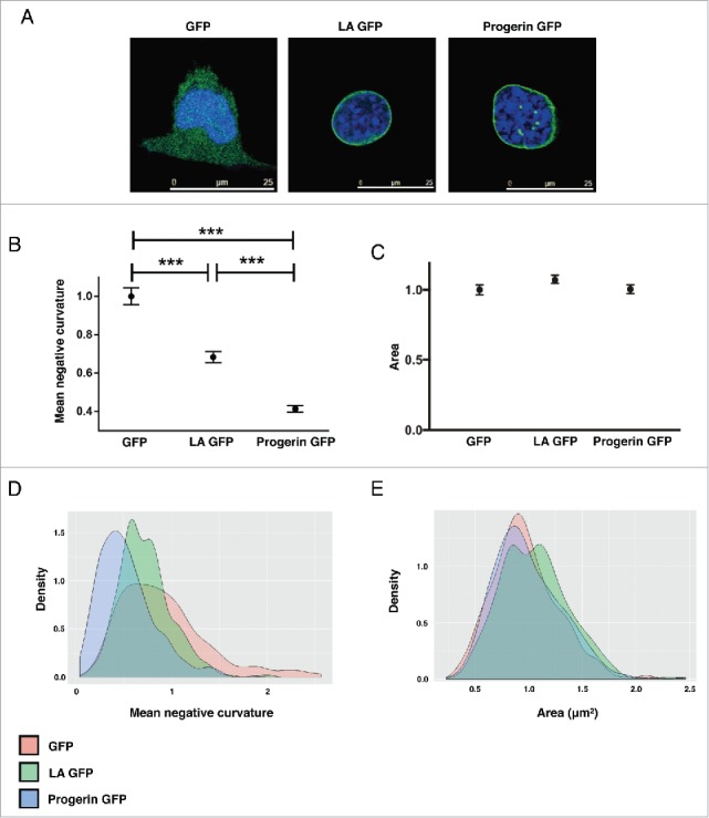
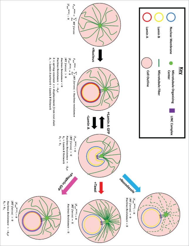
Lamin A (LA) is a critical structural component of the nuclear lamina. Mutations within the LA gene (LMNA) lead to several human disorders, most striking of which is Hutchinson-Gilford Progeria Syndrome (HGPS), a premature aging disorder. HGPS cells are best characterized by an abnormal nuclear morphology known as nuclear blebbing, which arises due to the accumulation of progerin, a dominant mutant form of LA. The microtubule (MT) network is known to mediate changes in nuclear morphology in the context of specific events such as mitosis, cell polarization, nucleus positioning and cellular migration. What is less understood is the role of the microtubule network in determining nuclear morphology during interphase. In this study, we elucidate the role of the cytoskeleton in regulation and misregulation of nuclear morphology through perturbations of both the lamina and the microtubule network. We found that LA knockout cells exhibit a crescent shape morphology associated with the microtubule-organizing center. Furthermore, this crescent shape ameliorates upon treatment with MT drugs, Nocodazole or Taxol. Expression of progerin, in LA knockout cells also rescues the crescent shape, although the response to Nocodazole or Taxol treatment is altered in comparison to cells expressing LA. Together these results describe a collaborative effort between LA and the MT network to maintain nuclear morphology.
Keywords: HGPS; lamin A; microbutuble; nuclear shape; progerin.
Figures


Similar articles
-
Inhibiting farnesylation of progerin prevents the characteristic nuclear blebbing of Hutchinson-Gilford progeria syndrome.Proc Natl Acad Sci U S A. 2005 Sep 6;102(36):12879-84. doi: 10.1073/pnas.0506001102. Epub 2005 Aug 29. Proc Natl Acad Sci U S A. 2005. PMID: 16129833 Free PMC article.
-
Incomplete processing of mutant lamin A in Hutchinson-Gilford progeria leads to nuclear abnormalities, which are reversed by farnesyltransferase inhibition.Hum Mol Genet. 2005 Oct 15;14(20):2959-69. doi: 10.1093/hmg/ddi326. Epub 2005 Aug 26. Hum Mol Genet. 2005. PMID: 16126733
-
Vitamin D receptor signaling improves Hutchinson-Gilford progeria syndrome cellular phenotypes.Oncotarget. 2016 May 24;7(21):30018-31. doi: 10.18632/oncotarget.9065. Oncotarget. 2016. PMID: 27145372 Free PMC article.
-
Epigenetic involvement in Hutchinson-Gilford progeria syndrome: a mini-review.Gerontology. 2014;60(3):197-203. doi: 10.1159/000357206. Epub 2014 Feb 28. Gerontology. 2014. PMID: 24603298 Review.
-
Hutchinson-Gilford progeria syndrome through the lens of transcription.Aging Cell. 2013 Aug;12(4):533-43. doi: 10.1111/acel.12070. Epub 2013 Apr 19. Aging Cell. 2013. PMID: 23496208 Review.
Cited by
-
Effect of Nuclear Stiffness on Cell Mechanics and Migration of Human Breast Cancer Cells.Front Cell Dev Biol. 2020 May 29;8:393. doi: 10.3389/fcell.2020.00393. eCollection 2020. Front Cell Dev Biol. 2020. PMID: 32548118 Free PMC article.
-
Transient nuclear lamin A/C accretion aids in recovery from vapor nanobubble-induced permeabilisation of the plasma membrane.Cell Mol Life Sci. 2022 Jan 4;79(1):23. doi: 10.1007/s00018-021-04099-9. Cell Mol Life Sci. 2022. PMID: 34984553 Free PMC article.
-
Cytoskeleton-mediated alterations of nuclear mechanics by extracellular mechanical signals.Biophys J. 2022 Jan 4;121(1):1-3. doi: 10.1016/j.bpj.2021.12.017. Epub 2021 Dec 22. Biophys J. 2022. PMID: 35000687 Free PMC article. No abstract available.
-
Nuclear F-actin and Lamin A antagonistically modulate nuclear shape.J Cell Sci. 2022 Jul 1;135(13):jcs259692. doi: 10.1242/jcs.259692. Epub 2022 Jul 4. J Cell Sci. 2022. PMID: 35665815 Free PMC article.
-
Altered microtubule structure, hemichannel localization and beating activity in cardiomyocytes expressing pathologic nuclear lamin A/C.Heliyon. 2020 Jan 23;6(1):e03175. doi: 10.1016/j.heliyon.2020.e03175. eCollection 2020 Jan. Heliyon. 2020. PMID: 32021920 Free PMC article.
References
-
- Eriksson M, Brown WT, Gordon LB, Glynn MW, Singer J, Scott L, Erdos MR, Robbins CM, Moses TY, Berglund P, et al.. Recurrent de novo point mutations in lamin A cause Hutchinson-Gilford progeria syndrome. Nature 2003; 423:293-8; PMID:12714972; https://doi.org/10.1038/nature01629 - DOI - PMC - PubMed
-
- Lammerding J, Schulze PC, Takahashi T, Kozlov S, Sullivan T, Kamm RD, Stewart CL, Lee RT. Lamin A/C deficiency causes defective nuclear mechanics and mechanotransduction. J Clin Invest 2004; 113:370-8; PMID:14755334; https://doi.org/10.1172/JCI200419670 - DOI - PMC - PubMed
-
- Worman HJ, Courvalin JC. How do mutations in lamins A and C cause disease? J Clin Invest 2004; 113:349-51; PMID:14755330; https://doi.org/10.1172/JCI20832 - DOI - PMC - PubMed
-
- Shumaker DK, Dechat T, Kohlmaier A, Adam SA, Bozovsky MR, Erdos MR, Eriksson M, Goldman AE, Khuon S, Collins FS, et al.. Mutant nuclear lamin A leads to progressive alterations of epigenetic control in premature aging. Proc Natl Acad Sci U S A 2006; 103:8703-8; PMID:16738054; https://doi.org/10.1073/pnas.0602569103 - DOI - PMC - PubMed
-
- Capell BC, Collins FS. Human laminopathies: nuclei gone genetically awry. Nat Rev Genet 2006; 7:940-52; PMID:17139325; https://doi.org/10.1038/nrg1906 - DOI - PubMed
MeSH terms
Substances
Grants and funding
LinkOut - more resources
Full Text Sources
Other Literature Sources
Miscellaneous