. 2017 May 30;18(1):110.
doi: 10.1186/s12931-017-0595-9.

Role of human rhinovirus in triggering human airway epithelial-mesenchymal transition

Affiliations

Role of human rhinovirus in triggering human airway epithelial-mesenchymal transition

Danielle M Minor et al. Respir Res. .

Abstract

Background: Structural changes in the airways, collectively referred to as airway remodeling, are a characteristic feature of asthma, and are now known to begin in early life. Human rhinovirus (HRV)-induced wheezing illnesses during early life are a potential inciting stimulus for remodeling. Increased deposition of matrix proteins causes thickening of the lamina reticularis, which is a well-recognized component of airway remodeling. Increased matrix protein deposition is believed to be due to the presence of increased numbers of activated mesenchymal cells (fibroblasts/myofibroblasts) in the subepithelial region of asthmatic airways. The origin of these increased mesenchymal cells is not clear, but one potential contributor is the process of epithelial-mesenchymal transition (EMT). We hypothesized that HRV infection may help to induce EMT.

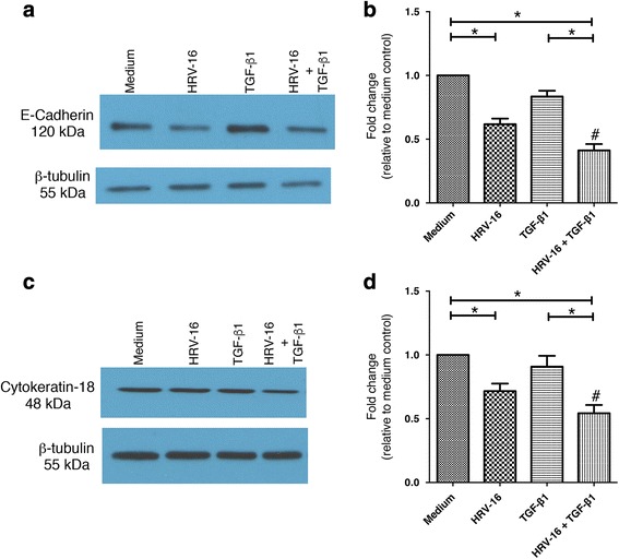
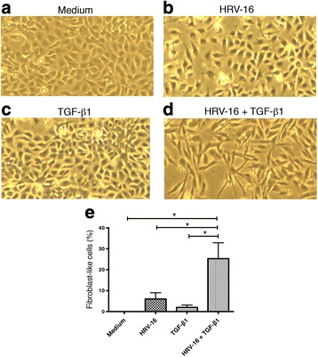
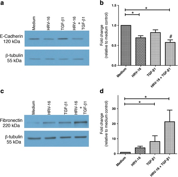
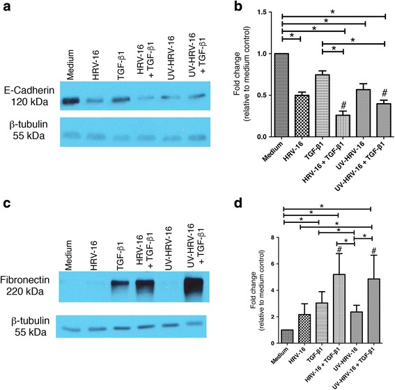
Methods: We used the BEAS-2B human bronchial epithelial cells line, which uniformly expresses the major group HRV receptor, to examine the effects of stimulation with HRV alone, transforming growth factor-β1 (TGF-β1), alone, and the combination, on induction of changes consistent with EMT. Western blotting was used to examine expression of epithelial and mesenchymal phenotypic marker proteins and selected signaling molecules. Cell morphology was also examined.

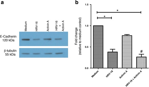
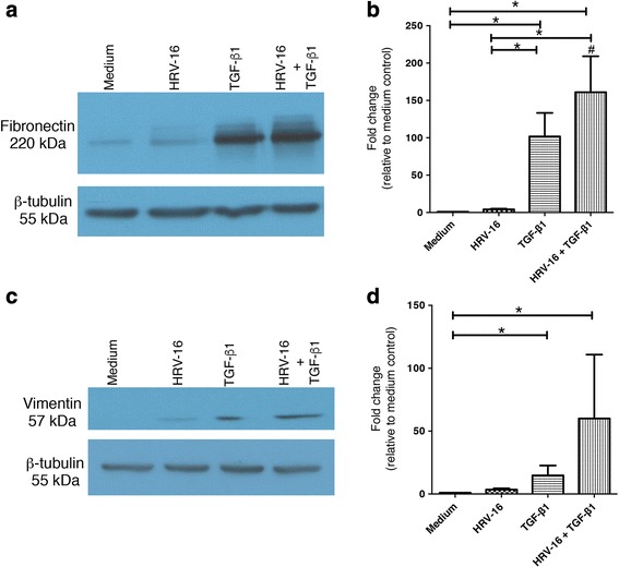
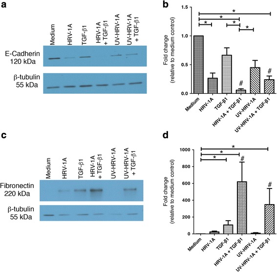
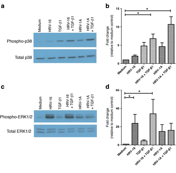
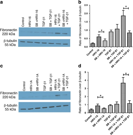
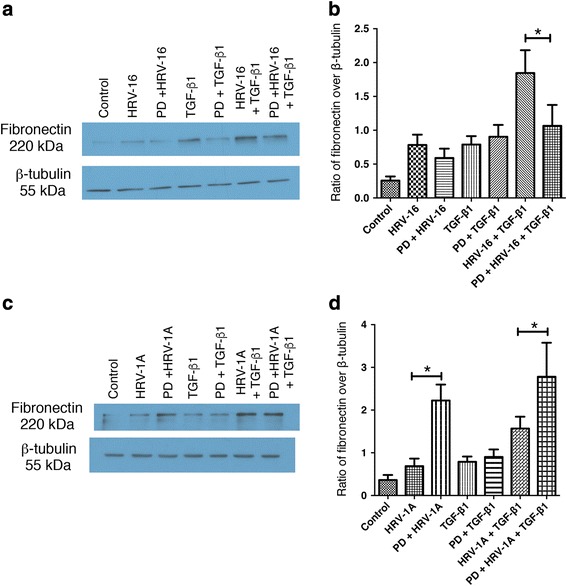
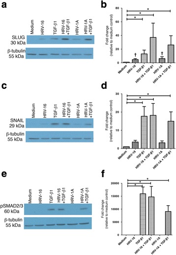
Results: In this study, we show that two different strains of HRV, which use two different cellular receptors, are each capable of triggering phenotypic changes consistent with EMT. Moreover, both HRV serotypes synergistically induced changes consistent with EMT when used in the presence of TGF-β1. Morphological changes were also most pronounced with the combination of HRV and TGF-β1. Viral replication was not essential for phenotypic changes. The synergistic interactions between HRV and TGF-β1 were mediated, at least in part, via activation of mitogen activated protein kinase pathways, and via induction of the transcription factor SLUG.

Conclusions: These data support a role for HRV in the induction of EMT, which may contribute to matrix protein deposition and thickening of the lamina reticularis in airways of patients with asthma.

Keywords: E-cadherin; Epithelial-mesenchymal transition; Fibronectin; Human rhinovirus; MAP kinases; SLUG; Transforming growth factor-β1.


Figures

Fig. 1
Fig. 1
Effects of exposure for 120 h to HRV-16 alone, TGF-β1 alone, and the combination on expression of epithelial markers in BEAS-2B cells. BEAS-2B cells were incubated with medium alone (labeled medium in Figures), HRV-16, TGF-β1, and HRV-16 + TGF-β1 for 120 h. a Representative western blot for E-cadherin. b Densitometry analysis for E-cadherin (n = 11). c Representative western blot for Cytokeratin-18. d Densitometry analysis for Cytokeratin-18 (n = 11). Asterisk indicates significance for comparisons shown by Dunn’s post-hoc analysis after Kruskal-Wallis ANOVA. Hatchmark indicates synergy compared to the sum of responses to individual stimuli by Wilcoxon matched-pairs signed ranks test
Fig. 2
Fig. 2
Effects of exposure for 120 h to HRV-16 alone, Activin A alone, and the combination on expression of E-cadherin in BEAS-2B cells. BEAS-2B cells were incubated with medium alone, HRV-16, Activin A, and HRV-16 + TGF-β1 for 120 h. a Representative western blot for E-cadherin. b Densitometry analysis for E-cadherin (n = 6). Asterisk indicates significance for comparisons shown by Dunn’s post-hoc analysis after Kruskal-Wallis ANOVA. Hatchmark indicates synergy compared to the sum of responses to individual stimuli by Wilcoxon matched-pairs signed ranks test
Fig. 3
Fig. 3
Effects of exposure for 120 h to HRV-16 alone, TGF-β1 alone, and the combination on expression of mesenchymal markers in BEAS-2B cells. BEAS-2B cells were stimulated with medium, HRV-16, TGF-β1, and HRV-16 + TGF-β1 for 120 h. a Representative western blot for fibronectin. b Densitometry analysis for fibronectin (n = 11). c Representative western blot for vimentin. d Densitometry analysis for vimentin (n = 11). Asterisk indicates significance for comparisons shown by Dunn’s post-hoc analysis after Kruskal-Wallis ANOVA. Hatchmark indicates synergy compared to the sum of responses to individual stimuli by Wilcoxon matched-pairs signed ranks test
Fig. 4
Fig. 4
Effects of exposure for 120 h to HRV-16 alone, TGF-β1 alone, and the combination on morphology of BEAS-2B cells. BEAS-2B cells were stimulated with medium, HRV-16, TGF-β1, and HRV-16 + TGF-β1 for 120 h. Light microscopy images (4X magnification) showing representative morphology of cells exposed to a medium. b HRV-16 alone. c TGF-β1 alone. d The combination of HRV-16 + TGF-β1. Data are from one of 3 experiments used for quantitative analysis. e Quantitative analysis of percentage of cells with fibroblast-like morphology (mean ± SEM from 3 experiments). Asterisk indicates significance for comparisons shown by Dunn’s post-hoc analysis after Kruskal-Wallis ANOVA
Fig. 5
Fig. 5
Exposure for 24 h to HRV-16 alone, TGF-β1 alone, and the combination alters expression of epithelial and mesenchymal markers in BEAS-2B cells. BEAS-2B cells were stimulated with medium, HRV-16, TGF-β1, and HRV-16 + TGF-β1 for 24 h. a Representative western blot for E-cadherin. b Densitometry analysis for E-cadherin (n = 10). c Representative western blot for fibronectin. d Densitometry analysis for fibronectin (n = 10). Asterisk indicates significance for comparisons shown by Dunn’s post-hoc analysis after Kruskal-Wallis ANOVA. Hatchmark indicates synergy compared to the sum of responses to individual stimuli by Wilcoxon matched-pairs signed ranks test
Fig. 6
Fig. 6
Viral replication is not essential for HRV-16 alone, or in combination with TGF-β1, to alter expression of epithelial and mesenchymal markers in BEAS-2B cells. BEAS-2B cells were stimulated with medium, HRV-16, TGF-β1, HRV-16 + TGF-β1, UV-HRV-16, or UV-HRV-16 + TGF-β1 for 120 h. a Representative western blot for E-cadherin. b Densitometry analysis for E-cadherin (n = 12). c Representative western blot for fibronectin. d Densitometry analysis for fibronectin (n = 12). Asterisk indicates significance for comparisons shown by Dunn’s post-hoc analysis after Kruskal-Wallis ANOVA. Hatchmark indicates synergy compared to the sum of responses to individual stimuli by Wilcoxon matched-pairs signed ranks test
Fig. 7
Fig. 7
The minor group HRV-1A, alone and in combination with TGF-β1, also alters expression of epithelial and mesenchymal markers in BEAS-2B cells and effects do not depend upon viral replication. BEAS-2B cells were stimulated with medium, HRV-1A, TGF-β1, HRV-1A + TGF-β1, UV-HRV-1A, or UV-HRV-1A + TGF-β1 for 120 h. a Representative western blot for E-cadherin. b Densitometry analysis for E-cadherin (n = 6). c Representative western blot for fibronectin. d Densitometry analysis for fibronectin (n = 6). Asterisk indicates significance for comparisons shown by Dunn’s post-hoc analysis after Kruskal-Wallis ANOVA. Hatchmark indicates synergy compared to the sum of responses to individual stimuli by Wilcoxon matched-pairs signed ranks test
Fig. 8
Fig. 8
Major and minor group HRV strains, and TGF-β1, differentially activate MAPK signaling pathways. BEAS-2B cells were stimulated with medium, HRV-16, HRV-1A, TGF-β1, HRV-16 + TGF-β1, or HRV-1A + TGF-β1 for 1 h. a Representative western blot for phospho- and total p38. b Densitometry analysis for p38 (n = 7). c Representative western blot for phospho and total ERK1/2. d Densitometry analysis for ERK1/2 (n = 5). Asterisk indicates significance for comparisons shown by Dunn’s post-hoc analysis after Kruskal-Wallis ANOVA
Fig. 9
Fig. 9
Inhibition of the p38 MAPK pathway significantly inhibits the induction of fibronectin by each HRV seroptype alone and in combination with TGF-β1. BEAS-2B cells were pre-treated for 1 h with the p38 MAPK pathway inhibitor, SB203580 (SB), and then stimulated with medium, HRV-16, HRV-1A, TGF-β1, HRV-16 + TGF-β1, or HRV-1A + TGF-β1 for 24 h. a Representative western blot for fibronectin using HRV-16 with and without SB. b Densitometry analysis for fibronectin using HRV-16 with and without SB. (n = 6). c Representative western blot for fibronectin using HRV-1A with and without SB. d Densitometry analysis for fibronectin using HRV-1A with and without SB. (n = 6). Asterisk indicates significance for comparisons shown by Dunn’s post-hoc analysis after Kruskal-Wallis ANOVA
Fig. 10
Fig. 10
Inhibition of the ERK/1/2 MAPK pathway differentially affects the induction of fibronectin by the combination of each HRV serotype with TGF-β1. BEAS-2B cells were pre-treated for 1 h with the ERK1/2 MAPK pathway inhibitor, PD98059 (PD), and then stimulated with medium, HRV-16, HRV-1A, TGF-β1, HRV-16 + TGF-β1, or HRV-1A + TGF-β1 for 24 h. a Representative western blot for fibronectin using HRV-16 with and without PD. b Densitometry analysis for fibronectin using HRV-16 with and without PD. (n = 6). c Representative western blot for fibronectin using HRV-1A with and without PD. d Densitometry analysis for fibronectin using HRV-1A with and without PD. (n = 6). Asterisk indicates significance for comparisons shown by Dunn’s post-hoc analysis after Kruskal-Wallis ANOVA
Fig. 11
Fig. 11
Activation of the transcriptional repressor SLUG, but not the SNAIL or Smad2/3 pathways may contribute to EMT induced by the combination of HRV and TGF-β1. BEAS-2B cells were stimulated with medium, HRV-16, HRV-1A, TGF-β1, HRV-16 + TGF-β1, or HRV-1A + TGF-β1 for 6 h. a Representative western blot for SLUG. b Densitometry analysis for SLUG (n = 8). c Representative western blot for SNAIL. d Densitometry analysis for SNAIL (n = 6). e Representative western blot for pSMAD2/3. f Densitometry analysis for pSMAD2/3 (n = 6). Asterisk indicates significance for comparisons shown by Dunn’s post-hoc analysis after Kruskal-Wallis ANOVA. † Indicates p < 0.02 compared to medium control stimuli by Wilcoxon matched-pairs signed ranks test

References

    1. Al-Muhsen S, Johnson JR, Hamid Q. Remodeling in asthma. J Allergy Clin Immunol. 2011;128:451–462. doi: 10.1016/j.jaci.2011.04.047. - DOI - PubMed
    1. Jeffery PK. Remodeling and inflammation of bronchi in asthma and chronic obstructive pulmonary disease. Proc Am Thorac Soc. 2004;1:176–183. doi: 10.1513/pats.200402-009MS. - DOI - PubMed
    1. Benayoun L, Druilhe A, Dombret M-C, Aubier M, Pretolani M. Airway structural alterations selectively associated with severe asthma. Am J Respir Crit Care Med. 2003;167:1360–1368. doi: 10.1164/rccm.200209-1030OC. - DOI - PubMed
    1. Brewster CEP, Howarth PH, Djukanovic R, Wilson J, Holgate ST, Roche wR. Myofibroblasts and subepithelial fibrosis in bronchial asthma. Am J Respir Cell Mol Biol. 1990;3:507–511. doi: 10.1165/ajrcmb/3.5.507. - DOI - PubMed
    1. Pohunek P, Warner JO, Turzíková J, Kudrmann J, Roche WR. Markers of eosinophilic inflammation and tissue re-modelling in children before clinically diagnosed bronchial asthma. Pediatr Allergy Immunol. 2005;16:43–51. doi: 10.1111/j.1399-3038.2005.00239.x. - DOI - PubMed

Publication types

MeSH terms

Substances