. 2017 May 30;36(1):72.
doi: 10.1186/s13046-017-0534-0.

FGF2/FGFR1 regulates autophagy in FGFR1-amplified non-small cell lung cancer cells

Affiliations

FGF2/FGFR1 regulates autophagy in FGFR1-amplified non-small cell lung cancer cells

Hong Yuan et al. J Exp Clin Cancer Res. .

Abstract

Background: Autophagy is a conserved catabolic process to degrade cellular organelles. The role of autophagy in cancer development is complex. Amplification of fibroblast growth factor receptor 1 (FGFR1) is one of the most frequent targets in lung squamous cell carcinoma (SQCC). Whether fibroblast growth factor 2 (FGF2)/FGFR1 contributes to the regulation of autophagy remains elusive.

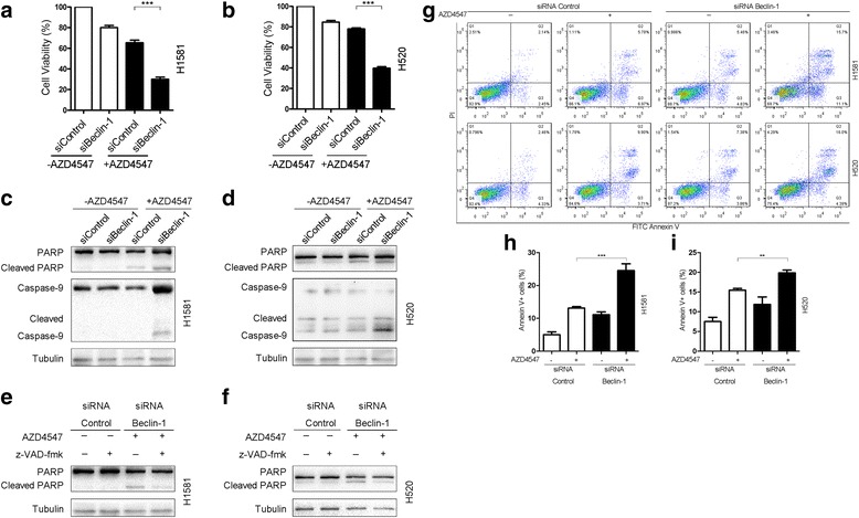
Methods: Autophagic activity was evaluated by immunoblotting for microtubule-associated protein 1 light chain 3 (LC3), formation of GFP-LC3 puncta, and monodansylcadaverine (MDC) staining. The effect of autophagy inhibition on cell survival was assessed by cell viability and apoptosis assays.

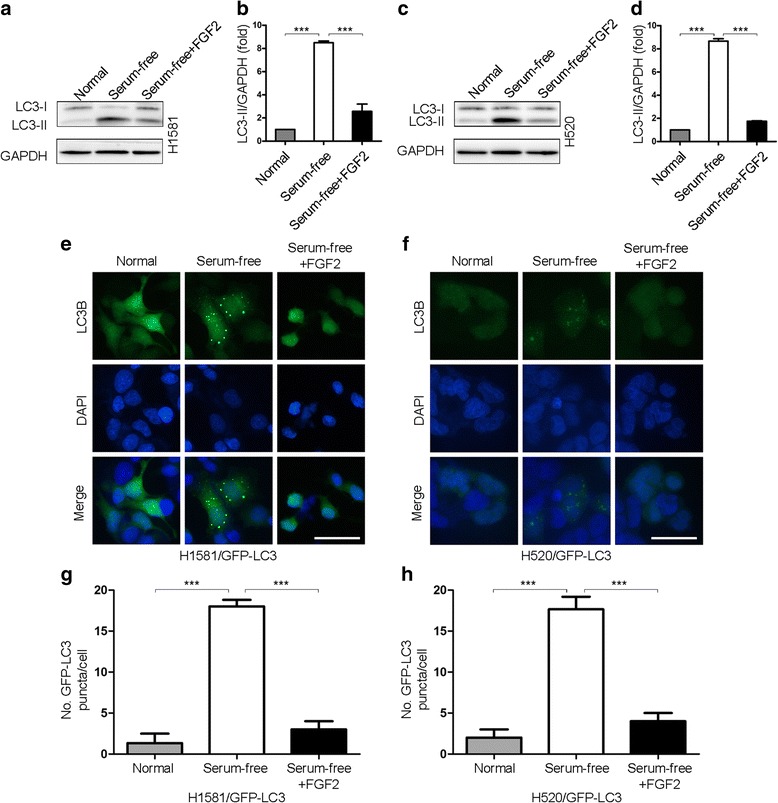
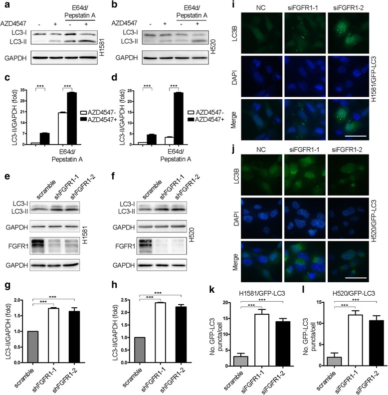
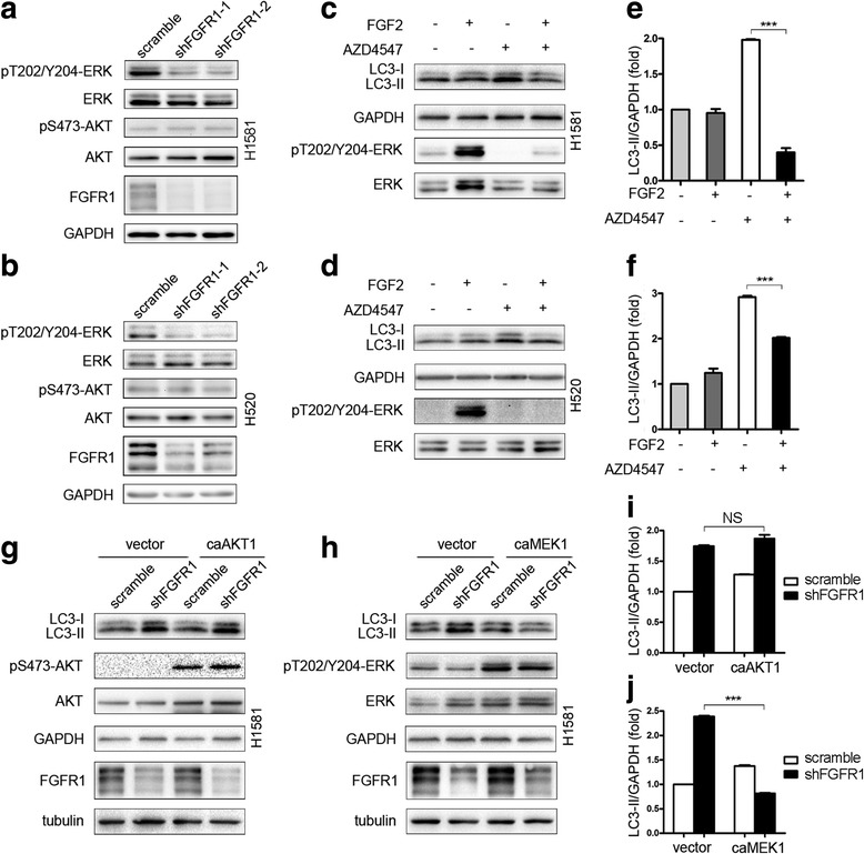
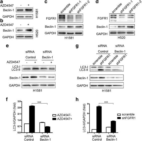
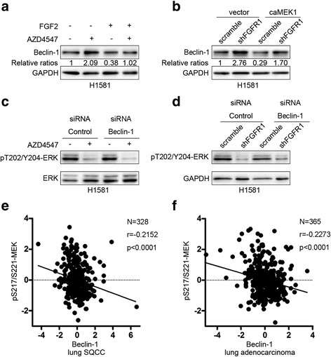
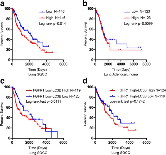
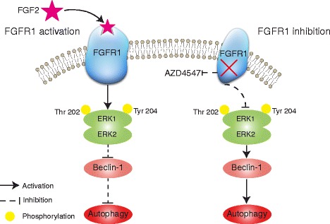
Results: We elucidated that FGFR1 activation suppressed autophagy. Pharmacological or genetic inhibition of FGFR1 by AZD4547 or FGFR1 short hairpin RNA (shRNA) induced autophagy in FGFR1-amplified non-small cell lung cancer (NSCLC) cells, H1581 and H520 cells. Mechanistic study revealed that the induction of autophagy by FGFR1 inhibition was mediated through inhibiting the ERK/MAPK pathway not by AKT pathway, accompanied by upregulation of beclin-1. Furthermore, activation of ERK/MAPK by transfection with a constitutively active MEK1 (caMEK1) construct or knockdown of beclin-1 by RNAi could attenuate autophagy induced by FGFR1 inhibition. Beclin-1 expression was inversely correlated with MEK1 phosphorylation. Inhibition of autophagy by beclin-1 silencing could enhance apoptosis after AZD4547 treatment in H1581 and H520 cells. High levels of LC3B mRNA was a marker of poor prognosis in NSCLC patients.

Conclusions: Simultaneously inhibiting FGFR1 and autophagy could enhance cell death which should be further explored in vivo.

Keywords: Autophagy; Beclin-1; ERK; FGFR1; NSCLC.


Figures

Fig. 1
Fig. 1
FGFR1 activation inhibits autophagy. a LC3-I/II western blot analysis in H1581 NSCLC cells cultured O/N in normal medium, serum-free medium, or serum-free medium plus FGF2 (25 ng/ml, 2 h). b Quantification of LC3-II levels in (a); mean ± SD, n = 3, ***p < 0.001. c LC3-I/II western blot analysis in H520 cells in conditions shown in (a). d Quantification of LC3-II levels in (c); mean ± SD, n = 3, ***p < 0.001. e Representative images of GFP-LC3 puncta in H1581/GFP-LC3 cells in conditions shown in (a). Scale bars represent 25 μm. f Representative images of GFP-LC3 puncta in H520/GFP-LC3 cells in conditions shown in (c). Scale bars represent 25 μm. g Quantitation of GFP-LC3 puncta in conditions shown in (e); mean ± SD, n = 3, ***p < 0.001. h Quantitation of GFP-LC3 puncta in conditions shown in (f); mean ± SD, n = 3, ***p < 0.001
Fig. 2
Fig. 2
FGFR1 inhibition induces autophagic activity. a H1581 cells were treated with 1 μM AZD4547 for 24 h. The last 8 h was in the presence or absence of E64d (10 μg/ml) and pepstatin A (10 μg/ml). Protein levels of LC3-I/II, and GAPDH were measured by western blot assays. b H520 cells were treated with 1 μM AZD4547 for 24 h. E64d and pepstatin A were added to the cells 8 h before harvesting. c Quantification of LC3-II levels in (a); mean ± SEM, n = 3, ***p < 0.001. d Quantification of LC3-II levels in (b); mean ± SEM, n = 3, ***p < 0.001. e The levels of LC3-II and GAPDH were determined by western blot in H1581 cells stably transduced by shRNA targeting FGFR1 and a control shRNA. FGFR1 and GAPDH protein expression levels by western blot were included as a control for the efficiency of shRNA knockdown. f Confirmation of changes in levels of autophagy after knockdown of FGFR1 by western blot with antibodies against LC3-I/II in H520 cells. g Quantification of LC3-II levels in (e); mean ± SD, n = 3, ***p < 0.001. h Quantification of LC3-II levels in (f); mean ± SD, n = 3, ***p < 0.001. i H1581/GFP-LC3 cells transfected with either siRNA targeting FGFR1 or negative control (NC) were analyzed by fluorescence microscopy to determine the distribution of punctate GFP-LC3. E64d and pepstatin A were added for 8 h before fixation. Scale bars represent 25 μm. j Punctate GFP-LC3 distribution of H520/GFP-LC3 cells transfected with two independent FGFR1 siRNA constructs or a negative control. E64d and pepstatin A were added for 8 h before fixation. Scale bars represent 25 μm. k Quantification of levels of autophagy in H1581/GFP-LC3 cells transfected with control siRNA or siRNA against FGFR1 in conditions shown in (i); mean ± SD, n = 3, ***p < 0.001. l Quantification of data from (j); mean ± SD, n = 3, ***p < 0.001
Fig. 3
Fig. 3
Constitutive activation of ERK/MAPK pathway, but not PI3K pathway, prevents autophagy induced by FGFR1 inhibition. a and b ERK and AKT phosphorylation by western blot in H1581 and H520 cells stably transduced by two independent FGFR1 shRNA constructs or a non-targeting control vector. FGFR1 protein expression levels were included as a control of knockdown efficiency. The filters were stripped and reprobed for total ERK and AKT to ensure equal loading of cell protein in each lane. c H1581 cells were treated with 1 μM AZD4547 for 24 h. FGF2 (25 ng/ml) was added for 2 h before harvesting. d H520 cells were treated with AZD4547 (1 μM) for 24 h. Cells were treated with FGF2 for 2 h before harvesting and then subjected to western blotting analysis. e Quantification of LC3-II levels in (c); mean ± SD, n = 3, ***p < 0.001. f Quantification of LC3-II levels in (d); mean ± SD, n = 3, ***p < 0.001. g Western blot showing that the expression of a caAKT1 construct failed to prevent increase in LC3-II induced by shFGFR1 in H1581 cells. h Western blot showing that the expression of a caMEK1 prevented increase in LC3-II induced by shFGFR1 in H1581 cells. i Quantification of LC3-II levels in (g); mean ± SEM, n = 3, NS, not significant. j Quantification of LC3-II levels in (h); mean ± SEM, n = 3, ***p < 0.001
Fig. 4
Fig. 4
Downregulation of beclin-1 prevents autophagy induced by FGFR1 inhibition. a and b H1581 and H520 cells were treated with 1 μM AZD4547 for 24 h. Then, total lysates were harvested and subjected to western blot analysis. c and d H1581 and H520 cells, stably transduced by the indicated lentiviruses (scramble or shFGFR1), were subjected to western blotting analysis. e H1581 cells were transfected with beclin-1 siRNA or control siRNA using Lipofectamine RNAiMAX transfection reagent as described in materials and methods. At 24 h after transfection, the cells were treated with or without AZD4547 (1 μM) for 24 h. f Quantification of LC3-II levels in (e); mean ± SEM, n = 3, ***p < 0.001. g H1581 cells, stably transduced by the indicated lentiviruses (scramble or shFGFR1), were transfected with beclin-1 siRNA or control siRNA, and then cells were subjected to western blotting analysis. h Quantification of LC3-II levels in (g); mean ± SEM, n = 3, ***p < 0.001
Fig. 5
Fig. 5
Upregulation of beclin-1 by FGFR1 inhibition contributes to induction of autophagy through inhibiting ERK pathway. a H1581 cells were treated with AZD4547 (1 μM) for 24 h prior to treatment with or without FGF2 (25 ng/ml) for 2 h. After treatment, cells were harvested and subjected to western blotting analysis. b H1581 cells, stably transduced by FGFR1 shRNA or control shRNA, were transfected with caMEK1 plasmid or control vector using jetPRIME transfection reagent as described in materials and methods. At 24–48 h after transfection, the cells were harvested and then subjected to western blotting analysis. c H1581 cells were transfected with beclin-1 siRNA or control siRNA using Lipofectamine RNAiMAX transfection reagent. At 24 h after transfection, the cells were treated with or without AZD4547 (1 μM) for 24 h. d H1581 cells, stably transduced by the indicated lentiviruses (scramble or shFGFR1), were transfected with beclin-1 siRNA or control siRNA, and then cells were subjected to western blotting analysis. e MEK phosphorylation was analyzed in a cohort of lung SQCC patients that had altered levels of beclin-1 protein expression using cBioPortal. f MEK phosphorylation was analyzed in a cohort of lung adenocarcinoma patients that had altered levels of beclin-1 expression using TCGA database
Fig. 6
Fig. 6
Inhibition of autophagy by beclin-1 silencing enhances apoptosis after AZD4547 treatment. a H1581 cells were transfected with beclin-1 siRNA or control siRNA using Lipofectamine RNAiMAX transfection reagent. At 24 h after transfection, the cells were treated with or without AZD4547 (1 μM) for 24 h. Cell viability was determined by CCK-8 assay; mean ± SD, n = 3, ***p < 0.001. b H520 cells were treated as described in (a), and cell viability was measured by CCK-8 assay; mean ± SD, n = 3, ***p < 0.001. c and d H1581 and H520 cells transfected with beclin-1 siRNA or control siRNA were treated with or without AZD4547 (1 μM) for another 24 h. Cleavage of both PARP and caspase-9 were analyzed by western blot assay. e and f H1581 and H520 cells transfected with beclin-1 siRNA or control siRNA were treated with or without AZD4547 (1 μM) for another 24 h. The last 5 h was in the presence or absence of z-VAD-fmk (20 μM). PARP cleavage and tubulin were analyzed by western blot assay. g H1581 and H520 cells transfected with beclin-1 siRNA or control siRNA were treated with or without AZD4547 (1 μM) for another 24 h. Cell apoptosis was determined by flow cytometry using FITC Annexin V/PI staining. The horizontal and vertical axes represent labeling with FITC Annexin V/PI, respectively. LR (Q3) represents early apoptotic cells (positive for Annexin V only), UR (Q2) represents late apoptotic cells (positive for both Annexin V and PI), and LL (Q4) represents live cells. h H1581 cells were treated as described in (g upper panel). The number of early/late apoptotic cells is shown as the sum of Annexin V positive and Annexin V/PI double positive cells; mean ± SD, n = 3, ***p < 0.001. i H520 cells were treated as described in (g lower panel); mean ± SD, n = 3, **p < 0.01
Fig. 7
Fig. 7
High levels of LC3B mRNA are associated with poor prognosis in NSCLC patients. a and b Kaplan-Meier curves of OS, with respect to LC3B mRNA level, in a lung SQCC (N = 146 for low expression and N = 146 for high expression, p = 0.014), and b lung adenocarcinoma (N = 123 for low expression and N = 123 for high expression, p = 0.5099). c Kaplan-Meier curves for OS in FGFR1 low - LC3B low expression (N = 125) and FGFR1 low - LC3B high expression (N = 119) patients, the latter had poorer OS (p = 0.0111). d Kaplan-Meier curves for OS in FGFR1 high - LC3B low expression (N = 119) and FGFR1 high - LC3B high expression (N = 124) patients, the latter conferred decreased OS (p = 0.1742). P-values are based on the log-rank test (a-d)
Fig. 8
Fig. 8
A schema depicting a mechanism by which FGF2/FGFR1 regulates autophagy. Left panel: FGFR1 activation by FGF2 upregulates ERK1/2 phosphorylation and then downregulates beclin-1, thereby suppresses autophagy. Right panel: FGFR1 inhibition (AZD4547 or FGFR1 knockdown) downregulates phosphorylation of ERK1/2 and subsequently upregulates beclin-1, thereby induces autophagy

Similar articles

Cited by

References

    1. Siegel RL, Miller KD, Jemal A. Cancer Statistics, 2016. Ca-a Cancer Journal for Clinicians. 2016;66:7–30. doi: 10.3322/caac.21332. - DOI - PubMed
    1. Drilon A, Rekhtman N, Ladanyi M, Paik P. Squamous-cell carcinomas of the lung: emerging biology, controversies, and the promise of targeted therapy. Lancet Oncol. 2012;13:E418–E426. doi: 10.1016/S1470-2045(12)70291-7. - DOI - PubMed
    1. Dieci MV, Arnedos M, Andre F, Soria JC. Fibroblast growth factor receptor inhibitors as a cancer treatment: from a biologic rationale to medical perspectives. Cancer Discov. 2013;3:264–279. doi: 10.1158/2159-8290.CD-12-0362. - DOI - PubMed
    1. Carter EP, Fearon AE, Grose RP. Careless talk costs lives: fibroblast growth factor receptor signalling and the consequences of pathway malfunction. Trends Cell Biol. 2015;25:221–233. doi: 10.1016/j.tcb.2014.11.003. - DOI - PubMed
    1. Weiss J, Sos ML, Seidel D, Peifer M, Zander T, Heuckmann JM, Ullrich RT, Menon R, Maier S, Soltermann A, et al. Frequent and focal FGFR1 amplification associates with therapeutically tractable FGFR1 dependency in squamous cell lung cancer. Sci Transl Med. 2010;2:62ra93. doi: 10.1126/scitranslmed.3001451. - DOI - PMC - PubMed

Publication types

MeSH terms

Substances