

Comparison of the fecal bacterial microbiota of healthy and diarrheic foals at two and four weeks of life
- PMID: 28558788
- PMCID: PMC5450145
- DOI: 10.1186/s12917-017-1064-x
Comparison of the fecal bacterial microbiota of healthy and diarrheic foals at two and four weeks of life
Abstract
Background: Diarrhea in foals affects up to 60% of foals during the first six months of life. The effect of diarrhea on the fecal bacterial microbiota in foals has not been investigated. Little is known on the fecal bacterial microbial richness and diversity of foals at a young age. The objective was to compare the fecal bacterial microbiota of healthy foals to foals with diarrhea at two and four weeks of life.
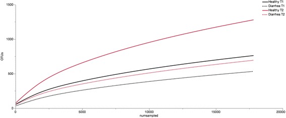
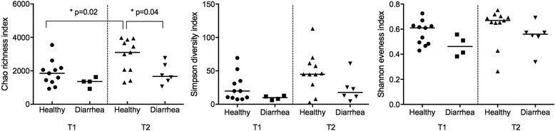
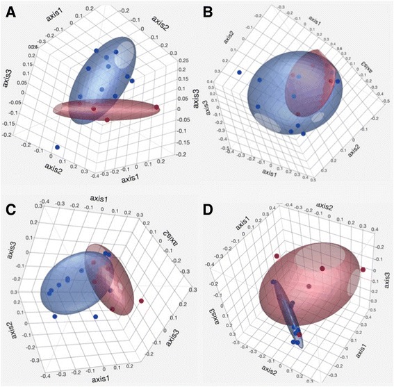
Methods: Fecal samples were collected from foals (n = 20) at 1-14 (T1) and 15-28 (T2) days of age and analyzed using high throughput sequencing. Differences in relative abundance of bacterial taxa, alpha diversity and beta diversity indices were assessed between age-matched foals with diarrhea (n = 9) and healthy foals (n = 11), and between time points.
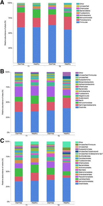
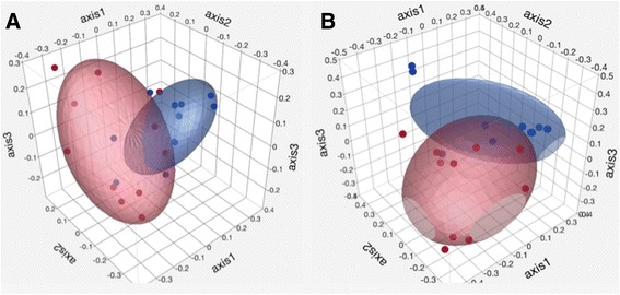
Results: Differences in microbial community composition based on time point and health status were observed on all taxonomic levels. Of 117 enriched species in healthy foals at T2, 50 (48%) were Lachnospiraceae or Ruminococcaceae. The Chao richness index was increased in healthy foals at T2 compared to T1 (p = 0.02). Foals with diarrhea had a significantly lower richness index than non-diarrheic foals at T2 (p = 0.04). Diarrhea had an inconsistent effect, while time point had a consistent effect on microbial community structure.
Conclusions: Preventative and therapeutic measures for diarrhea should focus on maintaining bacterial microbiota richness. Lachnospiraceae and Ruminococcaceae were underrepresented in foals with diarrhea. These should be evaluated further as potential therapeutic options.
Keywords: Clostridiales; Gastrointestinal microbiota; Horse; Lachnospiraceae; Metagenomic sequencing; Ruminococcaceae.
Figures
Similar articles
-
Dysbiosis is not present in horses with fecal water syndrome when compared to controls in spring and autumn.J Vet Intern Med. 2020 Jul;34(4):1614-1621. doi: 10.1111/jvim.15778. Epub 2020 Jun 26. J Vet Intern Med. 2020. PMID: 32588473 Free PMC article.
-
Effects of fecal microbiota transplantation on clinical outcomes and fecal microbiota of foals with diarrhea.J Vet Intern Med. 2024 Sep-Oct;38(5):2718-2728. doi: 10.1111/jvim.17185. Epub 2024 Sep 12. J Vet Intern Med. 2024. PMID: 39266472 Free PMC article.
-
Fecal microbiota changes associated with pathogenic and non-pathogenic diarrheas in foals.BMC Res Notes. 2025 Jan 23;18(1):34. doi: 10.1186/s13104-025-07110-9. BMC Res Notes. 2025. PMID: 39849534 Free PMC article.
-
Diagnostic procedures for isolation and characterization of Clostridium difficile associated with enterocolitis in foals.J Vet Diagn Invest. 1989 Jan;1(1):84-6. doi: 10.1177/104063878900100125. J Vet Diagn Invest. 1989. PMID: 2488656 Review. No abstract available.
-
Gastrointestinal diseases of foals.Vet Clin North Am Equine Pract. 1985 Apr;1(1):151-68. doi: 10.1016/s0749-0739(17)30774-5. Vet Clin North Am Equine Pract. 1985. PMID: 3907766 Free PMC article. Review.
Cited by
-
Dysbiosis is not present in horses with fecal water syndrome when compared to controls in spring and autumn.J Vet Intern Med. 2020 Jul;34(4):1614-1621. doi: 10.1111/jvim.15778. Epub 2020 Jun 26. J Vet Intern Med. 2020. PMID: 32588473 Free PMC article.
-
Expanded catalogue of metagenome-assembled genomes reveals resistome characteristics and athletic performance-associated microbes in horse.Microbiome. 2023 Jan 12;11(1):7. doi: 10.1186/s40168-022-01448-z. Microbiome. 2023. PMID: 36631912 Free PMC article.
-
Habitat environmental factors influence intestinal microbial diversity of the short-faced moles (Scaptochirus moschata).AMB Express. 2021 Jun 23;11(1):93. doi: 10.1186/s13568-021-01252-2. AMB Express. 2021. PMID: 34164757 Free PMC article.
-
Changes in the Fecal Microbiota Associated with a Broad-Spectrum Antimicrobial Administration in Hospitalized Neonatal Foals with Probiotics Supplementation.Animals (Basel). 2021 Aug 2;11(8):2283. doi: 10.3390/ani11082283. Animals (Basel). 2021. PMID: 34438741 Free PMC article.
-
Comparison of Gut Microbiota Diversity and Predicted Functions Between Healthy and Diseased Captive Rana dybowskii.Front Microbiol. 2020 Sep 1;11:2096. doi: 10.3389/fmicb.2020.02096. eCollection 2020. Front Microbiol. 2020. PMID: 32983063 Free PMC article.
References
-
- Costa MC, Weese JS. Animal health research reviews / conference of research Workers in Animal Diseases. 2012. The equine intestinal microbiome; pp. 1–8. - PubMed
-
- Milinovich GJ, Trott DJ, Burrell PC, Croser EL, Al Jassim RAM, Morton JM, et al. Fluorescence in situ hybridization analysis of hindgut bacteria associated with the development of equine laminitis. Environ Microbiol. 2007;9(8):2090–100. - PubMed
Publication types
MeSH terms
LinkOut - more resources
Full Text Sources
Other Literature Sources
Medical
