. 2017 Jun 13;114(24):6274-6279.
doi: 10.1073/pnas.1703391114. Epub 2017 May 30.

Characterization of the scrambling domain of the TMEM16 family

Affiliations

Characterization of the scrambling domain of the TMEM16 family

Sayuri Gyobu et al. Proc Natl Acad Sci U S A. .

Abstract

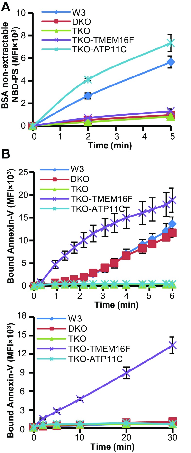
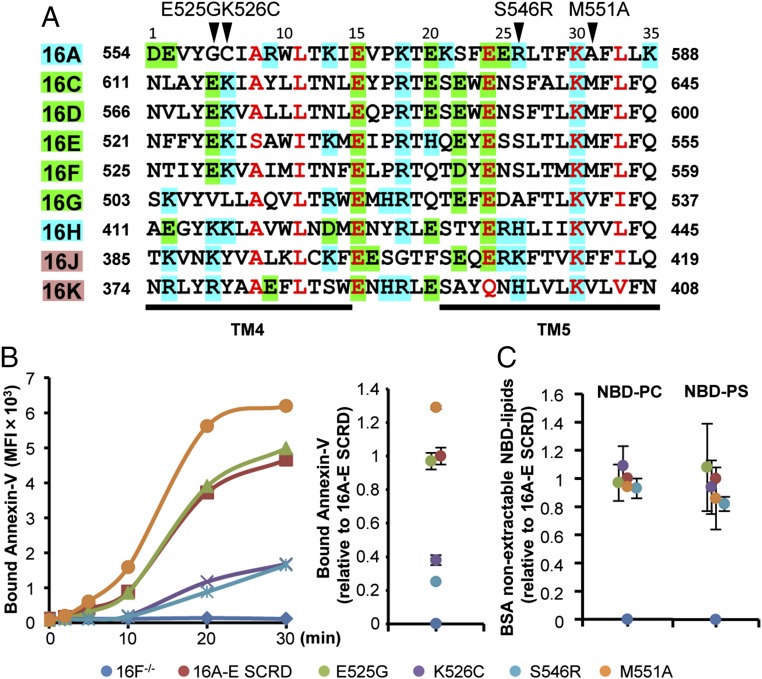
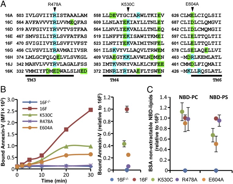
The TMEM16 protein family has 10 members, each of which carries 10 transmembrane segments. TMEM16A and 16B are Ca2+-activated Cl- channels. Several other members, including TMEM16F, promote phospholipid scrambling between the inner and outer leaflets of a cell membrane in response to intracellular Ca2+ However, the mechanism by which TMEM16 proteins translocate phospholipids in plasma membranes remains elusive. Here we show that Ca2+-activated, TMEM16F-supported phospholipid scrambling proceeds at 4 °C. Similar to TMEM16F and 16E, seven TMEM16 family members were found to carry a domain (SCRD; scrambling domain) spanning the fourth and fifth transmembrane segments that conferred scrambling ability to TMEM16A. By introducing point mutations into TMEM16F, we found that a lysine in the fourth transmembrane segment of the SCRD as well as an arginine in the third and a glutamic acid in the sixth transmembrane segment were important for exposing phosphatidylserine from the inner to the outer leaflet. However, their role in internalizing phospholipids was limited. Our results suggest that TMEM16 provides a cleft containing hydrophilic "stepping stones" for the outward translocation of phospholipids.

Keywords: TMEM16; phospholipids; point mutation; scramblase.


Conflict of interest statement

The authors declare no conflict of interest.

Figures

Fig. 1.
Fig. 1.
Ca2+-dependent phospholipid scrambling. (A) Expression of TMEM16F. Cell lysates were prepared from wild-type BaF3 cells (+/+), TMEM16F−/− BaF3 cells (−/−), and TMEM16F−/− BaF3 cells transformed with TMEM16F (16F). Proteins (5 µg for +/+ and −/−; 0.1 µg for 16F) were separated by 7.5% SDS/PAGE and analyzed by Western blotting with anti-TMEM16F (Upper) or anti–α-tubulin (Lower) antibodies. (B) Ca2+-dependent PtdSer exposure by TMEM16F. Wild-type BaF3 cells (BaF316F+/+), TMEM16F−/− BaF3 cells (BaF316F−/−), and TMEM16F transformants (BaF316F−/−16F) were incubated at 20 or 4 °C with 3 µM A23187 and 1 mM CaCl2 in the presence of Cy5-Annexin V and PI. Annexin V-staining profiles in the PI-negative population at the indicated time after A23187 addition are shown.
Fig. S1.
Fig. S1.
TMEM16F-dependent PtdSer exposure. Wild-type BaF3 cells (BaF316F+/+), TMEM16F−/− BaF3 cells (BaF316F−/−), and TMEM16F transformants (BaF316F−/−16F) were incubated at 20 or 4 °C with 3 µM A23187 and 1 mM CaCl2 in the presence of Cy5-Annexin V and PI. The PI- and Annexin V-staining profiles are shown before (0 min) and after the A23187 treatment (30 min at 4 °C or 5 min at 20 °C).
Fig. S2.
Fig. S2.
(A) Mutagenesis of ATP11A, ATP11C, and TMEM16F genes in W3 cells by the CRISPR-Cas9 system. The wild-type (Upper) and mutated sequences (Lower) of mouse ATP11A, ATP11C, and TMEM16F genes are shown. Protospacer-adjacent motifs are underlined in red. The deleted nucleotides are highlighted in green (for ATP11A, ATP11C, and the first allele of TMEM16F) or yellow (for the second allele of TMEM16F). Mutations caused homozygous truncations for ATP11A and ATP11C and heterozygous truncations for TMEM16F. Numbers at the top are amino acid positions. The red triplet asterisks indicate the termination codon. (B) Transformation of ATP11A−/−ATP11C−/−TMEM16F−/− W3 cells with mouse TMEM16F or human ATP11C. Western blotting was carried out with anti-mouse TMEM16F, anti-human ATP11C, or anti-GFP. CBB, Coomassie brilliant blue.
Fig. S3.
Fig. S3.
Flippase-independent phospholipid scrambling by TMEM16F. (A) ATP11A/ATP11C-dependent flipping of PtdSer. Wild-type (W3), ATP11A−/−ATP11C−/− (DKO), ATP11A−/−ATP11C−/−TMEM16F−/− (TKO), or ATP11A−/−ATP11C−/−TMEM16F−/− W3 cells transformed with human ATP11C (TKO-ATP11C) or mouse TMEM16F (TKO-TMEM16F) were incubated at 20 °C with NBD-PS, and the incorporated NBD-PS was analyzed by flow cytometry. (B) TMEM16F-dependent scrambling of PtdSer. Wild-type (W3), DKO, TKO, TKO-ATP11C, or TKO-TMEM16F was incubated at 20 °C (Upper) or 4 °C (Lower) with 3 μM A23187 and 1 mM CaCl2 in the presence of Cy5-labeled Annexin V and PI, and analyzed by flow cytometry. The experiments were carried out three times, and the average values of the bound Cy5-Annexin or the incorporated NBD-PS in the PI-negative population are expressed as mean fluorescence intensity (MFI) with SD (bars).
Fig. 2.
Fig. 2.
Expression of TMEM16A-SCRD chimeric proteins. (A) Schematic representation of the TMEM16A-SCRD chimeras. The numbers above each diagram indicate the amino acid positions in TMEM16A. In the chimeric constructions, the 35-amino acid segment in TMEM16A (positions 554 to 588) was replaced with the corresponding sequence of other TMEM16 family members (TMEM16C–H, J, or K). The chimeric proteins were fused with EGFP or DsRed at the C terminus. (B and C) Expression of EGFP-tagged TMEM16A-SCRD chimeras. TMEM16F−/− BaF3 cells were stably transformed with the indicated TMEM16A-SCRD chimera tagged with EGFP. (B) The cell lysates were analyzed by Western blotting with anti-GFP Ab. After blotting, the membrane was stained by Coomassie brilliant blue (Lower). Open arrowhead indicates TMEM16F, while closed arrowhead indicate TMEM16A or its chimera with SCRD. (C) The transformants were observed by fluorescence confocal microscopy. (Scale bars, 10 µm.)
Fig. 3.
Fig. 3.
Ca2+-dependent PtdSer exposure by TMEM16A-SCRD chimeras. TMEM16F−/− BaF3 transformants expressing the chimera of TMEM16A with 16H’s SCRD (H SCRD), 16J’s SCRD (J SCRD), or 16K’s SCRD (K SCRD) (A) or the chimera of TMEM16A with 16C’s SCRD (C SCRD), 16D’s SCRD (D SCRD), 16E’s SCRD (E SCRD), 16F’s SCRD (F SCRD), or 16G’s SCRD (G SCRD) (B) were treated at 4 °C with 3 µM A23187 in the presence of 1 mM CaCl2. At the indicated times, the exposed PtdSer, assessed by the binding of Cy5-labeled Annexin V in the PI-negative population, was determined by flow cytometry, and is expressed as the mean fluorescence intensity (MFI). The experiments were also carried out with the parental TMEM16F−/− BaF3 cell line (16F−/−) and its transformants expressing TMEM16A (16A) or TMEM16F (16F). (Right) The scramblase activity of each chimera determined at 30 min is plotted relative to that of the intact TMEM16F. The experiments were performed three times, and the average values are plotted with SD (bars).
Fig. S4.
Fig. S4.
Ca2+-dependent internalization of phospholipids by TMEM16A-SCRD chimeras. TMEM16F−/− BaF3 (16F−/−) transformants expressing the indicated TMEM16A-SCRD chimeras were preincubated at 4 °C for 3 min with 0.5 µM NBD-PC, 0.5 µM NBD-PS, 0.25 µM NBD-SM, or 0.5 µM NBD-GalCer. A23187 and CaCl2 were added to a final concentration of 3 µM and 1.0 mM, respectively, and the mixture was further incubated at 4 °C for 20 min. The BSA-nonextractable, internalized NBD-labeled lipids were quantified by FACSAria II, and are expressed as MFI. TMEM16F−/− BaF3 (16F−/−) transformants expressing the intact TMEM16A (16A) or TMEM16F (16F) were analyzed similarly.
Fig. 4.
Fig. 4.
Effect of SCRD point mutations on lipid scrambling activity. (A) Alignment of the SCRDs of mouse TMEM16 family members. The amino acid residues that are homologous among all members are in red. Basic and acidic amino acid residues are highlighted in blue and green, respectively. The fourth and fifth transmembrane segments (TM4 and TM5) are underlined. The four mutated residues are indicated above the top line. (B) TMEM16F−/− BaF3 (16F) transformants expressing the EGFP-tagged TMEM16A-chimera carrying 16E's SCRD or its mutants were treated at 4 °C with 3 µM A23187 and 1 mM CaCl2 in the presence of Cy5-Annexin V and PI. At the indicated times, the bound Annexin V in the PI-negative population was determined by flow cytometry, and is expressed as the MFI. (B, Right) The amount of bound Annexin V for each mutant was determined at 30 min, and is expressed relative to the value observed with the chimera of TMEM16A the wild-type 16E SCRD. (C) The cells were treated at 4 °C for 20 min with 3 µM A23187 and 1 mM CaCl2 in the presence of NBD-PC or NBD-PS. The incorporated NBD-phospholipids were determined by flow cytometry, and are expressed relative to the value obtained with the chimera of TMEM16A with 16E's SCRD. The experiments in B and C were carried out three times, and the average values are plotted with SD (bars).
Fig. S5.
Fig. S5.
TMEM16F−/− BaF3 cells (16F) were transformed with the EGFP-tagged TMEM16A-chimera carrying 16E's SCRD or its mutant, and analyzed for GFP by flow cytometry.
Fig. 5.
Fig. 5.
Effect of TMEM16F point mutations on phospholipid scrambling activity. (A) Alignment of mouse TMEM16 family members. Basic and acidic amino acid residues are highlighted in blue and green, respectively. The third, fourth, and sixth transmembrane segments are underlined, and the position of the first amino acid residue of each segment is indicated. The TMEM16F mutants analyzed are indicated above the top line. (B) TMEM16F−/− BaF3 (16F−/−) transformants expressing EGFP-tagged TMEM16F (16F) or its mutant (K530C, R478A, or E604A) were treated at 4 °C with 3 µM A23187 and 1 mM CaCl2 in the presence of Cy5-Annexin V and PI. The bound Annexin V in the PI-negative population was analyzed by flow cytometry at the indicated times, and is expressed as the MFI. (B, Right) The amount of bound Annexin V at 30 min is expressed relative to the value observed with TMEM16F. (C) The cells were treated with 3 µM A23187 and 1 mM CaCl2 in the presence of NBD-PC or NBD-PS, and incubated at 4 °C for 20 min. The incorporated NBD-phospholipids were determined by flow cytometry, and are expressed relative to the value obtained with TMEM16F. The experiments in B and C were carried out three times, and the average values are plotted with SD (bars).
Fig. S6.
Fig. S6.
TMEM16F−/− BaF3 cells (16F−/−) were transformed with the EGFP-tagged TMEM16F (16F) or its mutant (K530C, R478A, or E604A), and analyzed by flow cytometry for GFP.

Similar articles

Cited by

References

    1. Leventis PA, Grinstein S. The distribution and function of phosphatidylserine in cellular membranes. Annu Rev Biophys. 2010;39:407–427. - PubMed
    1. Balasubramanian K, Schroit AJ. Aminophospholipid asymmetry: A matter of life and death. Annu Rev Physiol. 2003;65:701–734. - PubMed
    1. Sebastian TT, Baldridge RD, Xu P, Graham TR. Phospholipid flippases: Building asymmetric membranes and transport vesicles. Biochim Biophys Acta. 2012;1821:1068–1077. - PMC - PubMed
    1. Coleman JA, Quazi F, Molday RS. Mammalian P4-ATPases and ABC transporters and their role in phospholipid transport. Mol Cell Biol. 2013;1831:555–574. - PMC - PubMed
    1. Segawa K, Kurata S, Nagata S. Human type IV P-type ATPases that work as plasma membrane phospholipid flippases, and their regulation by caspase and calcium. J Biol Chem. 2016;291:762–772. - PMC - PubMed

Publication types

LinkOut - more resources