. 2017 Jun 13;114(24):6292-6297.
doi: 10.1073/pnas.1705080114. Epub 2017 May 30.

A mechanism for lipid binding to apoE and the role of intrinsically disordered regions coupled to domain-domain interactions

Affiliations

A mechanism for lipid binding to apoE and the role of intrinsically disordered regions coupled to domain-domain interactions

Carl Frieden et al. Proc Natl Acad Sci U S A. .

Abstract

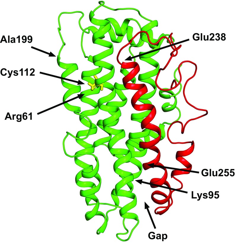
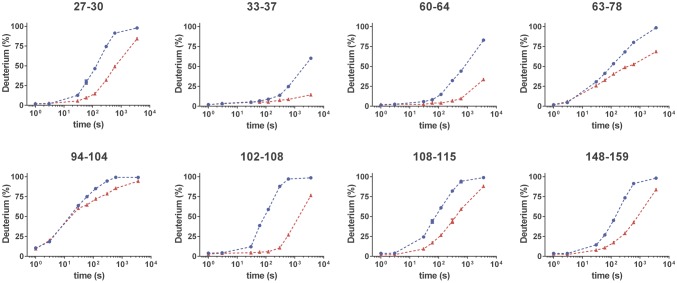
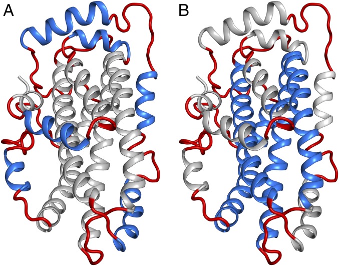
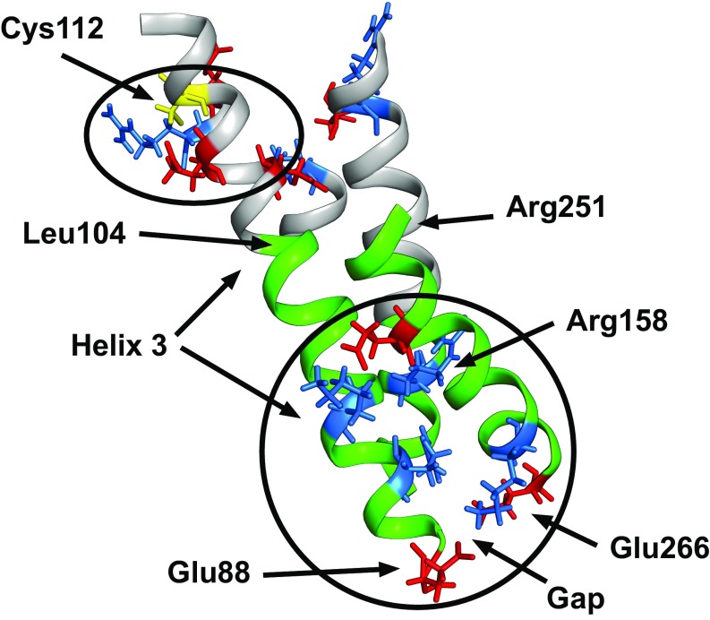
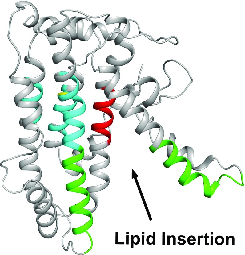
Relative to the apolipoprotein E (apoE) E3 allele of the APOE gene, apoE4 strongly increases the risk for the development of late-onset Alzheimer's disease. However, apoE4 differs from apoE3 by only a single amino acid at position 112, which is arginine in apoE4 and cysteine in apoE3. It remains unclear why apoE3 and apoE4 are functionally different. Described here is a proposal for understanding the functional differences between these two isoforms with respect to lipid binding. A mechanism is proposed that is based on the full-length monomeric structure of the protein, on hydrogen-deuterium exchange mass spectrometry data, and on the role of intrinsically disordered regions to control protein motions. It is proposed that lipid binds between the N-terminal and C-terminal domains and that separation of the two domains, along with the presence of intrinsically disordered regions, controls this process. The mechanism explains why apoE3 differs from apoE4 with respect to different lipid-binding specificities, why lipid increases the binding of apoE to its receptor, and why specific residues are conserved.

Keywords: apolipoprotein E; conserved residues; domain–domain interaction; hydrogen–deuterium exchange; protein structure.


Conflict of interest statement

The authors declare no conflict of interest.

Figures

Fig. 1.
Fig. 1.
An average NMR structure of full-length monomeric apoE3 as determined by Chen et al. (10) (PDB ID code 2L7B). In this depiction, the five residues that were mutated to prevent oligomer formation have been back mutated to their original amino acids using PyMol (The PyMOL Molecular Graphics System, Version 1.8 Schrödinger, LLC). The C-terminal domain is colored red. The gap between the N and C termini is indicated. The positions of Cys112, Lys95, Arg61, and Glu255 are shown, as are the end of the N-terminal domain (Ala199) and the beginning of the C-terminal domain (Glu238). As noted in the text, the authors proposed a salt bridge involving Glu255 to Lys95 rather than Arg61, as had been suggested earlier. This and other figures showing the apoE3 structure were generated using PyMol.
Fig. 2.
Fig. 2.
Structural differences between wild-type apoE3 and apoE4 as determined by HDX-MS studies plotted as a function of log time (s). The blue line represents data for apoE4, whereas the red line represents data for apoE3. Only those regions that show differences are shown here. The complete data are given in Fig. S1.
Fig. 3.
Fig. 3.
A representation of the full-length structure of apoE3 with the IDRs and flexible regions (in red) surrounding helical regions. (A) Short helices shown in blue. (B) Long helices shown in blue. The hinge connecting the N- and C-terminal domains is at the top of the figure. These regions comprise ∼30% of the structure.
Fig. 4.
Fig. 4.
Regions of apoE3, shown in green, that surround the gap between the N- and C-terminal domains. Shown within this gap are residues involved in salt bridges as proposed by Chen et al. (10) colored either blue (positively charged) or red (negatively charged). Arg158, which is a cysteine in apoE2, is labeled. Regions that may encompass the lipid-binding site are from Glu88 to Leu104 in the N-terminal domain and from Arg251 to Glu266 in the C-terminal domain. Also shown are charged residues around position 112 that, based on the data shown in Fig. 2, may be affected by the arginine residue at position 112 in apoE4. As discussed in the text, a portion of Helix 3 links the N-terminal portion of the lipid-binding site to the region of the cysteine to arginine difference at position 112. Residues that could be affected by this change, as indicated by Fig. 2, are shown.
Fig. 5.
Fig. 5.
Snapshot of domain–domain separation on lipid binding to apoE3. The structure was obtained using molecular dynamics to move apart the N- and C-terminal domains. Shown in green are portions of regions involved in lipid binding. In red is the apoE3 receptor-binding site and in blue are regions of structural differences based on HDX experiments. Cysteine112 (yellow) is within this region. Movie S1 provides a video moving from the closed to a completely open form.

Similar articles

Cited by

References

    1. Hauser PS, Narayanaswami V, Ryan RO. Apolipoprotein E: From lipid transport to neurobiology. Prog Lipid Res. 2011;50:62–74. - PMC - PubMed
    1. Corder EH, et al. Gene dose of apolipoprotein E type 4 allele and the risk of Alzheimer’s disease in late onset families. Science. 1993;261:921–923. - PubMed
    1. Saunders AM, et al. Association of apolipoprotein E allele epsilon 4 with late-onset familial and sporadic Alzheimer’s disease. Neurology. 1993;43:1467–1472. - PubMed
    1. Dong LM, et al. Novel mechanism for defective receptor binding of apolipoprotein E2 in type III hyperlipoproteinemia. Nat Struct Biol. 1996;3:718–722. - PubMed
    1. Phillips MC. Apolipoprotein E isoforms and lipoprotein metabolism. IUBMB Life. 2014;66:616–623. - PubMed

Publication types

MeSH terms