. 2017 Jun 13;114(24):6340-6345.
doi: 10.1073/pnas.1701644114. Epub 2017 May 30.

Bacteria exploit a polymorphic instability of the flagellar filament to escape from traps

Affiliations

Bacteria exploit a polymorphic instability of the flagellar filament to escape from traps

Marco J Kühn et al. Proc Natl Acad Sci U S A. .

Abstract

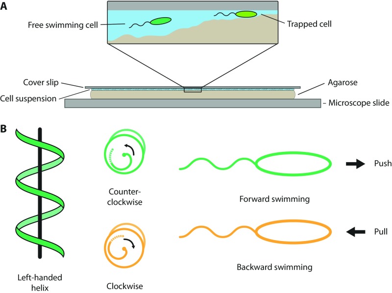
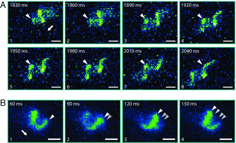
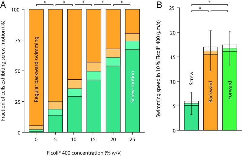
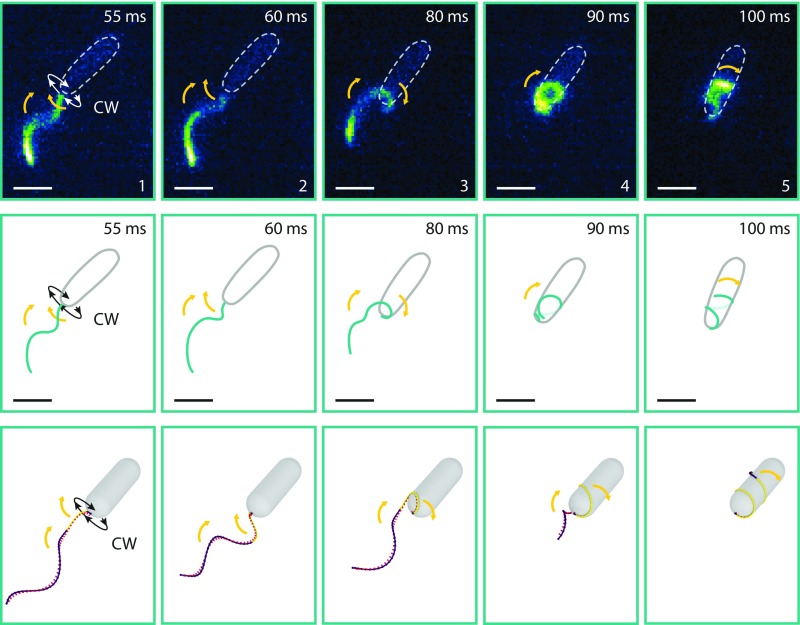
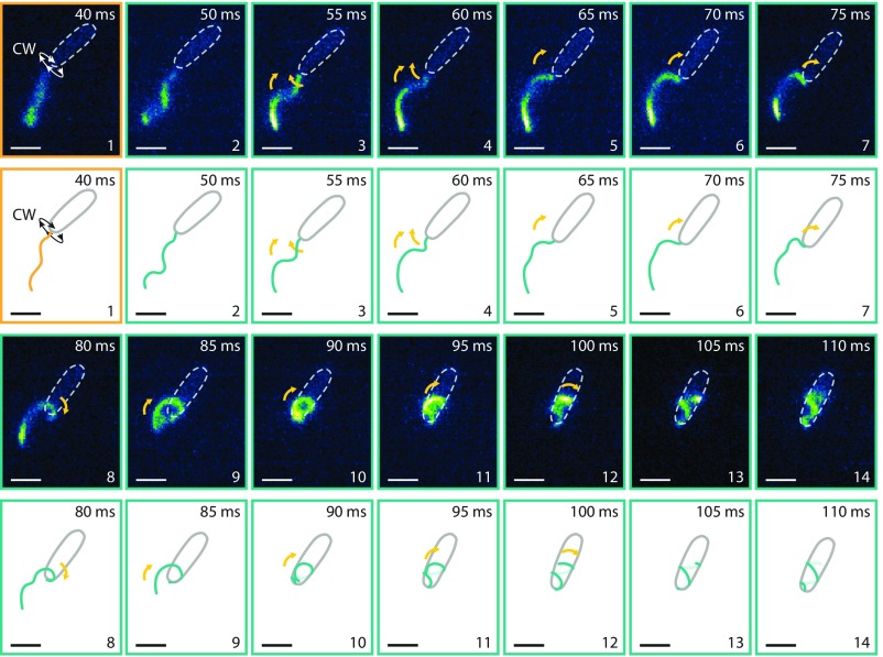
Many bacterial species swim by rotating single polar helical flagella. Depending on the direction of rotation, they can swim forward or backward and change directions to move along chemical gradients but also to navigate their obstructed natural environment in soils, sediments, or mucus. When they get stuck, they naturally try to back out, but they can also resort to a radically different flagellar mode, which we discovered here. Using high-speed microscopy, we monitored the swimming behavior of the monopolarly flagellated species Shewanella putrefaciens with fluorescently labeled flagellar filaments at an agarose-glass interface. We show that, when a cell gets stuck, the polar flagellar filament executes a polymorphic change into a spiral-like form that wraps around the cell body in a spiral-like fashion and enables the cell to escape by a screw-like backward motion. Microscopy and modeling suggest that this propagation mode is triggered by an instability of the flagellum under reversal of the rotation and the applied torque. The switch is reversible and bacteria that have escaped the trap can return to their normal swimming mode by another reversal of motor direction. The screw-type flagellar arrangement enables a unique mode of propagation and, given the large number of polarly flagellated bacteria, we expect it to be a common and widespread escape or motility mode in complex and structured environments.

Keywords: Shewanella; flagella; motility; structured environment.


Conflict of interest statement

The authors declare no conflict of interest.

Figures

Fig. S1.
Fig. S1.
(A) Experimental setup: Bacteria move in the small liquid pockets between the agarose and the coverslip where they can be monitored with a microscope from above. (B) Illustration of the regular swimming mechanism of S. putrefaciens. Changing the rotation of their flagellum while maintaining the handedness of the helix can switch the directional motion of the bacteria. CCW rotation (viewed from behind the cell) results in forward swimming, and CW rotation in backward swimming.
Fig. 1.
Fig. 1.
Approach, trapping, and escape of a bacterium between agarose and cover slide. (From Left to Right and Top to Bottom) The bright structures in the microscope image result from the fluorescently labeled flagellum. The cell body is indicated by a dotted ellipsoid. The yellow dashed line marks the position of the flagellated cell pole when the cell got stuck and maintained in subsequent frames to display the backward motion of the cell. The cartoon on the right summarizes the sequence of events and highlights the switch from counterclockwise (CCW) rotation of the motor (green frames) to clockwise (CW) rotation (orange frames; viewed from behind the cell). The cell approaches from the Lower Left (frame 1), moves upward, and then gets stuck (frames 2 and 3). While still pushing forward, the flagellum wiggles circularly in an instability known to occur for stronger applied torque. It then switches direction of rotation to CW (frame 4), and the flagellum wraps around the cell (frames 5 and 6). The strong forces during screw formation caused the cell to move, as indicated by the reorientation of the cell body between frames 5 and 6. The cell continues to the Lower Left, as indicated in frames 7–9. Exposure time was 30 ms; the number in the upper left corner of each micrograph displays the time each still image was taken from the movie (Movie S1 and Dataset S2). (Scale bars: 2 µm.) The freely rotating flagellum (frames 1–5) appears blurry because the rotation of the helical filament in normal medium is faster than the exposure time.
Fig. 2.
Fig. 2.
Screw-like propagation relies on interaction between the flagellar filament and asperities on the surface. The white arrow indicates the orientation of the cell body (flagellated pole). The white triangles indicate one peak of the flagellar waveform. (A) Still images taken from Movie S2 showing screw-like propagation of a cell that is stuck between agarose medium and the coverslip surface while the waveform of the flagellar helix does not move relative to the surrounding medium. (B) Still images taken from Movie S4 showing screw-like propagation of a free swimming cell (at least 50 µm away from the coverslip). Here, the flagellar helix waveform is moving relative to the surrounding medium, as indicated. (Image exposure time, 30 ms; scale bars, 1 µm.)
Fig. 3.
Fig. 3.
Inducing the transition to screw mode in media with higher viscosity (Left) and differences in swimming speed (Right). (A) Adding Ficoll 400 increases the viscosity and causes a larger torque when the cells switch from CCW to CW rotation. The fraction of cells in the “screw” state increases with Ficoll concentration (determined far away from surfaces). The viscosities are ∼2, 5, 10, 18, and 33 cP for 5%, 10%, 15%, 20%, and 25% Ficoll 400, respectively. Bars with an asterisk indicate that the differences are significant (Fisher’s exact test of independence; P ≤ 0.05, Bonferroni corrected). The exact P values from 0 to 25% Ficoll are 1.36 × 10⁻¹², 2.90 × 10⁻⁶, 3.96 × 10⁻⁴, 4.27 × 10⁻³, and 3.67 × 10⁻⁴, respectively. The number of counted backward swimming events are 309, 316, 313, 317, 324, and 313, respectively. Shaded boxes display 95% confidence intervals. (B) Measurements of the swimming speed show that, although forward and backward swimming have very similar speeds, the motion in the screw state is reduced. Bars with an asterisk indicate that the differences are significant (two-sample t test; P ≤ 0.01, Bonferroni corrected). The exact P values are <2.20 × 10⁻¹⁶ for screw vs. backward and screw vs. forward and 0.3 for backward vs. forward. For each swimming mode, 105 cell tracks were analyzed. Error bars represent SD; shaded boxes display 95% confidence intervals.
Fig. 4.
Fig. 4.
Comparison between observed and simulated flagellar states during screw formation. The upper panel shows flagellar states taken from Movie S5. These images are inverted horizontally to match the correct handedness of the flagellar helix (SI Materials and Methods). The flagellar filament was fluorescently labeled, and the medium was supplemented with 20% Ficoll 400 to slow down flagellar rotation (exposure, 5 ms; scale bars, 2 µm). The cell body is indicated by a dotted ellipsoid. Yellow arrows indicate the movement of the flagellar filament. The same states are depicted as cartoons in the middle panel. The lower panel displays the corresponding images from the simulation of screw formation (Movie S6). The stretched helix configuration is color-coded in purple, whereas the coiled state is marked in yellow. Details of the numerical simulation are described in SI Materials and Methods. Both imaging and simulations show that CW rotation results in an instability at the base of the flagellar filament, leading to a pull of the filament toward the cell body and, eventually, wrapping of the flagellum around the cell.
Fig. S2.
Fig. S2.
Fluorescence microscopy (Upper) and cartoon (Lower) showing the formation of the screw with high temporal resolution. Screw formation begins with the unwinding of the left-handed flagellar helix, which drives polymorphic transitions and leads to a movement of the filament toward the cell body. Ultimately, the filament is wrapped around the cell body and continues its CW rotation, now pushing instead of pulling the cell backward. The bright structures in the microscope image result from the fluorescently labeled flagellum. The cell body is indicated by a dotted ellipsoid. Yellow arrows indicate the movement of the flagellar filament. Still images were taken from Movie S5. These images are inverted horizontally to match the correct handedness of the flagellar helix (Materials and Methods). (Image exposure time, 5 ms; scale bars, 2 µm.)
Fig. S3.
Fig. S3.
Substitution of serine or threonine to cysteine in the variable domain of the polar flagellin monomers does not impair the swimming motility of S. putrefaciens. (A) Predicted structure of the polar flagellin FlaA1 of Shewanella putrefaciens CN-32 (calculated by Phyre V 2.0; illustration created with PyMOL 1.3). The threonine 174-to-cysteine substitution site is indicated by a blue sphere. The approximate corresponding substitution sites (T166C and S174C) are indicated by gray arrows. Thus, all substitution sites in both flagellins are located in the variable domain (approximately amino acids 163–183), which is exposed to the surrounding medium in the assembled flagellar filament. (B) Radial extension of FlaAB1-cys mutants and ∆flaAB2 as background strain (control) in 0.25% soft agar. The strains are labeled accordingly. The substitutions do not reduce the extension radius. (C) Swim speed and time between reversals for FlaAB1-cys are highly similar to its background strain ∆flaAB2 (control). For each strain, 598 cell tracks were evaluated (TrackMate plugin for ImageJ). The differences are not significant (two-sample t test; P = 0.087 and 0.891, respectively). Time between reversals was measured over the whole population, so the sample size is only three. Error bars represent SD; shaded boxes represent 95% confidence intervals. (D) FlaA1 and FlaB1 immunostaining subsequent to separation of the protein crude extract by SDS/PAGE and Western transfer to a membrane. Substitution of serine or threonine to cysteine does not cause a mass shift of the flagellins. This would only occur if glycosylation sites were substituted to cysteine.

References

    1. Kelly FX, Dapsis KJ, Lauffenburger DA. Effect of bacterial chemotaxis on dynamics of microbial competition. Microb Ecol. 1988;16:115–131. - PubMed
    1. Lauffenburger DA. Quantitative studies of bacterial chemotaxis and microbial population dynamics. Microb Ecol. 1991;22:175–185. - PubMed
    1. Fenchel T. Microbial behavior in a heterogeneous world. Science. 2002;296:1068–1071. - PubMed
    1. Alexandre G, Greer-Phillips S, Zhulin IB. Ecological role of energy taxis in microorganisms. FEMS Microbiol Rev. 2004;28:113–126. - PubMed
    1. Reichenbach T, Mobilia M, Frey E. Mobility promotes and jeopardizes biodiversity in rock-paper-scissors games. Nature. 2007;448:1046–1049. - PubMed

Publication types