. 2017 Jun 13;114(24):6310-6315.
doi: 10.1073/pnas.1610417114. Epub 2017 May 30.

Viral and cellular mRNA-specific activators harness PABP and eIF4G to promote translation initiation downstream of cap binding

Affiliations

Viral and cellular mRNA-specific activators harness PABP and eIF4G to promote translation initiation downstream of cap binding

Richard W P Smith et al. Proc Natl Acad Sci U S A. .

Abstract

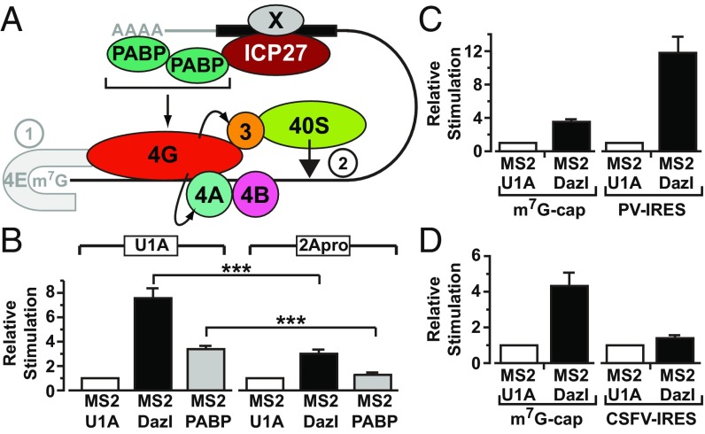
Regulation of mRNA translation is a major control point for gene expression and is critical for life. Of central importance is the complex between cap-bound eukaryotic initiation factor 4E (eIF4E), eIF4G, and poly(A) tail-binding protein (PABP) that circularizes mRNAs, promoting translation and stability. This complex is often targeted to regulate overall translation rates, and also by mRNA-specific translational repressors. However, the mechanisms of mRNA-specific translational activation by RNA-binding proteins remain poorly understood. Here, we address this deficit, focusing on a herpes simplex virus-1 protein, ICP27. We reveal a direct interaction with PABP that is sufficient to promote PABP recruitment and necessary for ICP27-mediated activation. PABP binds several translation factors but is primarily considered to activate translation initiation as part of the PABP-eIF4G-eIF4E complex that stimulates the initial cap-binding step. Importantly, we find that ICP27-PABP forms a complex with, and requires the activity of, eIF4G. Surprisingly, ICP27-PABP-eIF4G complexes act independently of the effects of PABP-eIF4G on cap binding to promote small ribosomal subunit recruitment. Moreover, we find that a cellular mRNA-specific regulator, Deleted in Azoospermia-like (Dazl), also employs the PABP-eIF4G interaction in a similar manner. We propose a mechanism whereby diverse RNA-binding proteins directly recruit PABP, in a non-poly(A) tail-dependent manner, to stimulate the small subunit recruitment step. This strategy may be particularly relevant to biological conditions associated with hypoadenylated mRNAs (e.g., germ cells/neurons) and/or limiting cytoplasmic PABP (e.g., viral infection, cell stress). This mechanism adds significant insight into our knowledge of mRNA-specific translational activation and the function of the PABP-eIF4G complex in translation initiation.

Keywords: DAZL; ICP27; mRNA-binding protein; mRNA-specific translational regulation; poly(A)-binding protein.


Conflict of interest statement

The authors declare no conflict of interest.

Figures

Fig. 1.
Fig. 1.
PABP interacts directly with ICP27 regions required for translational stimulation. (A) Immobilized purified GST-PABP or GST was incubated with purified His-ICP27. ICP27 was detected by immunoblotting; input is 10%. (B) Oocytes expressing ICP27 or U1A (negative control) were injected with VP16 mRNA and luciferase mRNA as an internal control. VP16 protein levels normalized to luciferase activity are plotted (±SEM; n = 5). (C) Y2H analysis of PABP interactions with indicated ICP27 regions. Iron regulatory protein (IRP) was used as a negative control. (D) Oocytes expressing MS2, MS2-ICP27, or MS2-ICP27 truncations (Fig. S3A) were coinjected with m7G-Luc-MS23 and β-gal mRNAs (Fig. S2A, [1] and [7]). Effects on translation were measured by luciferase assay normalized to β-gal activity. Translational stimulation relative to MS2 protein alone (=1) is plotted (±SEM; n = 3). (E) Immobilized purified GST or GST fusions of ICP27 or indicated truncations (Fig. S3A and Table S2) were incubated with purified His-PABP. PABP was detected by quantitative immunoblotting, and band intensities are shown, with the band intensity of lane 3 set to 100%. Input is 150%.
Fig. 2.
Fig. 2.
Translationally active ICP27 interacts with two domains of PABP and recruits PABP to mRNA. (A) Y2H analysis of ICP27 interactions with PABP domains. IRP was used as a negative control. (B) Immobilized purified GST or GST fusions of PABP or indicated truncations (Table S2) were incubated with HSV-1–infected cell extracts in the presence of RNase 1. ICP27 was detected by immunoblotting. (C) Y2H analysis of the PABP C-terminus (Ct, amino acids 396–633), RRM1-2Rd with ICP27 (amino acids 10–512), or ICP27 containing the M15 or M16 mutation. IRP and MS2 were used as negative controls. (D) Oocytes expressing MS2 or MS2-ICP27 were injected with ApG-Luc-MS23 (L) or ApG-Luc-ΔMS2 (LΔ) (Fig. S2A, [2] and [6]). PABP was immunoprecipitated (P) from lysates [control: nonspecific rabbit IgG (c)] and detected by immunoblotting (input is 27%). The presence of copurified luciferase or endogenous β-actin mRNAs was assessed by RT-PCR.
Fig. S1.
Fig. S1.
ICP27 associates with PABP in vivo, and PABP interacts directly with ICP27 in vitro. (A and B) PABP specifically coimmunoprecipitates with ICP27 from HSV-1–infected BHK cell extracts, prepared 5 h postinfection. (A) HSV-1 (KOS 1.1)–infected cell extracts were incubated with normal mouse serum (NMS) (lane 2) or anti-ICP27 monoclonal antibodies 1113 and 1119 (lanes 3–6) in the absence (lane 3) or presence of RNase A, 1, or T1 (lanes 4–6). IP, immunoprecipitation. ICP27 (Upper) or PABP (Lower) was detected by immunoblotting. (Upper) Lower band in the upper panel is IgG heavy chain. (B) Wild type (WT; KOS 1.1) HSV-1, ICP27-null (27lacZ) HSV-1 (where the gene encoding ICP27 is replaced by lacZ), or mock-infected (MI) BHK cell extracts were incubated with a mixture of anti-ICP27 monoclonal antibodies 1113 and 1119 (lanes 1–4) or NMS (lane 5). ICP27 (Upper) and coimmunoprecipitated PABP (Lower) were detected by immunoblotting. RNase A treatment is indicated by a + symbol. The lower protein band in each panel is the IgG heavy chain. PABP is only coimmunoprecipitated from infected cells expressing ICP27. (C) ICP27 and PABP interact directly on a subset of HSV-1 transcripts. The multifunctional ICP27 protein (red) binds to most viral mRNAs, which are polyadenylated and may therefore also bind PABP (green). This property allows for their fortuitous mRNA-mediated coisolation, which is RNase-sensitive. However, our data suggest that ICP27 stimulates the translation of a subset of transcripts (e.g., VP16) by directly recruiting additional PABP to these mRNAs (Fig. 5A). Consequently, on these mRNAs, ICP27 and PABP are present in a functional complex (mediated by protein–protein interactions) that is RNase-insensitive. (DG) Expression and purification of recombinant proteins. Recombinant proteins were expressed in and purified from E. coli BL21 (DE3) pLysS by affinity chromatography, resolved by SDS/PAGE, and visualized with Gelcode Blue. Molecular masses (kilodaltons) are indicated. (D) GST and GST-PABP. Full-length His-ICP27 (E) and GST-ICP27 (F) are indicated. The lower doublet in E and F was identified by immunoblotting as characteristic N-terminal ICP27 breakdown products. (G) His-PABP. (H) Immobilized purified GST-ICP27 or GST was incubated with purified His-PABP. Coisolated PABP was detected by immunoblotting; input represents 150%.
Fig. S2.
Fig. S2.
Tether-function reporter mRNAs are stable in oocytes. (A) Diagram of reporter and internal control mRNAs used in the tether-function assays throughout this report. ICP27 is expressed in X. laevis stage VI oocytes as a fusion with the phage MS2 coat protein (MS2) and tethered to luciferase mRNA via the interaction of MS2 with three cognate-binding sites within the 3′ UTR (Luc-MS23). Reporter mRNAs are unadenylated, unless otherwise stated ([3], [4]), and have a 5′ m7GpppG or ApppG cap (m7G-Luc-MS23 [1], [3] and ApG-Luc-MS23 [2], [4]) or an IRES as indicated (CSFV-Luc-MS23, PV-Luc-MS23, or HAV-Luc-MS23 [5]) in the main text. IRES-reporter constructs are ApppG-capped. A luciferase reporter mRNA lacking the MS2-binding sites is designated ApG-Luc-ΔMS2 [6]. An m7GpppG-capped polyadenylated lacZ mRNA containing no MS2-binding sites is used as an internal control (β-gal [7]). For the modified tether-function assay (Figs. 4C and 5B), an ApppG-capped, unadenylated, CSFV IRES-dependent internal control mRNA (CSFV–β-gal [8]) was used. (B and C) ICP27 alone is insufficient to activate VP16 mRNA translation efficiently. (B) X. laevis oocytes were injected with 50 ng of mRNA expressing ICP27, and ICP27 protein was detected by immunoblotting after overnight incubation. (C) In vitro-transcribed mRNA encoding VP16 (50, 250, or 500 pg) was injected into oocytes expressing either ICP27 or U1A (negative control), and VP16 protein was detected by immunoblotting after overnight incubation. (D) ICP27 does not affect reporter mRNA levels, consistent with the remarkable stability of mRNAs in oocytes (18). Oocytes expressing MS2 or MS2-ICP27 were injected with ApG-Luc-MS23, PV-Luc-MS23, HAV-Luc-MS23, or CSFV-Luc-MS23 reporter mRNAs. Total RNA was extracted either immediately (0h) or 14 h (14h) after injection and analyzed by quantitative RT-PCR with primers against the luciferase ORF, showing equivalent levels of mRNA. The number of amplification cycles required to obtain a detectable product in the linear range (threshold cycle) is shown. Error bars represent SEM.
Fig. S3.
Fig. S3.
ICP27 requires PABP binding to activate translation. (A) ICP27 deletion and point mutations used in this study. (Upper) Full-length ICP27 consists of 512 amino acids. RGG-box, RNA-binding domain; Zn, C-terminal zinc finger region. (Lower) Horizontal bars and gaps indicate amino acids present and absent, respectively. Vertical bars in M15 and M16 indicate the positions of point mutations. (B) Purification of GST fusions of ICP27 truncations. Recombinant proteins were expressed from the plasmids described in Table S2 and purified from E. coli BL21 (DE3) pLysS by affinity chromatography, resolved by SDS/PAGE, and visualized with Gelcode Blue. Molecular masses (kilodaltons) are indicated. (C) PABP contains four functionally nonequivalent RRM domains and a C-terminal region composed of a proline-rich linker (line) and the PABC domain. Minimal mapped binding regions for translation initiation factors are indicated (ref. and references therein). Interaction with eIF4B requires integrity of the PABP C-terminal region (dotted line). (D) GST fusions of different PABP domains purified from the plasmids listed in Table S2 as described in Fig. S1. (E) PABP coimmunoprecipitates with WT, but not M15/M16 mutant ICP27 from HSV-1–infected cell extracts. HSV-1–infected cell extracts were incubated anti-ICP27 monoclonal antibodies 1113 and 1119. ICP27 expression (Upper) and coimmunoprecipitated PABP (Lower) were detected by immunoblotting. The dashed vertical line indicates removal of irrelevant lanes from the gel.
Fig. S4.
Fig. S4.
ICP27-mediated translational activation is sensitive to reporter mRNA poly(A) tail length in a tether-function assay. (A) Oocytes expressing MS2 or MS2-ICP27 were coinjected with β-gal mRNA and m7G-Luc-MS23 or an identical reporter mRNA with a 3′ poly(A) tail (m7G-Luc-MS23-pA) (Fig. S2A, [7], [1], and [3], respectively). (Upper) Luciferase activity normalized to β-gal activity is plotted. (Lower) Relative stimulation compared with MS2 (set to 1) is plotted. (Upper) Polyadenylated reporter mRNA is translated more efficiently than its unadenylated counterpart, and translation of both reporters is stimulated by tethered ICP27. (Lower) Importantly, the relative increase in translational efficiency by ICP27 is greater with the unadenylated reporter than with the polyadenylated reporter. This result is consistent with ICP27 acting via recruitment of PABP, because this mode of action should result in a less pronounced stimulatory effect of MS2-ICP27 on polyadenylated reporter mRNAs, which will be bound by additional PABP molecules, compared with unadenylated reporter mRNAs, which can only recruit PABP via ICP27. (B) As in A, but substituting ApppG-capped for m7GpppG-capped reporter mRNAs (ApG-Luc-MS23 and ApG-Luc-MS23-pA; Fig. S2A, [2] and [4], respectively) and with the inclusion of MS2-PABP. The use of ApppG-capped reporters [with or without a poly(A) tail], which are translated at a substantially reduced rate (Fig. S5A), excludes the possibility that the reduced relative stimulation by ICP27 observed in A is a consequence of a maximal translation level being reached. A similar result was observed when PABP was directly tethered. Taken together, these results provide strong support for a mechanism whereby ICP27 functions by facilitating the recruitment of PABP. Error bars denote SEM from three repeats.
Fig. 3.
Fig. 3.
Tethered ICP27 stimulates small ribosomal subunit joining independently of cap binding. Oocytes expressing MS2 or MS2-ICP27 were coinjected with β-gal mRNA and m7G-Luc-MS23 or HAV-Luc-MS23 mRNA (A), PV-Luc-MS23 mRNA (B), or CSFV-Luc-MS23 mRNA (C) (Fig. S2A). Data (n = 3) are plotted as in Fig. 1D. (D) Oocytes expressing MS2 or MS2-PABP (RRM1-2) were coinjected with the reporter and control mRNAs as above. The plot represents luciferase activity (corrected for β-gal activity) of each reporter with MS2–RRM1-2 divided by the luciferase activity with MS2 alone. The m7G-Luc-MS23 ratio is set to 100%.
Fig. S5.
Fig. S5.
(A) Translation in X. laevis oocytes is highly sensitive to the presence of a functional cap, and the HAV IRES is active in X. laevis oocytes. Oocytes were coinjected with 375 pg of β-gal internal control mRNA and 750 pg of of m7G-Luc-MS23, ApG-Luc-MS23 (Fig. S2A, [1] and [2]), m7GpppG-capped HAV-Luc-MS23, or ApppG-capped HAV-Luc-MS23 mRNAs. The former two mRNAs show that replacing the physiological m7GpppG cap with ApppG results in a >70-fold reduction of translational efficiency. The latter two mRNAs contain the HAV IRES, although one is physiologically capped (m7GpppG) and the other is not (ApppG). The presence of the HAV IRES promotes efficient translation of the ApppG-capped mRNA (compare ApG-HAV-Luc-MS23 versus ApG-Luc-MS23 and m7G-Luc-MS23), showing that it is functional in oocytes. Luciferase activity normalized to β-gal activity is plotted. Error bars indicate SEM. (B) ICP27 activates a nonphysiologically (ApppG)-capped reporter mRNA in a tether-function assay. Oocytes expressing MS2 or MS2-ICP27 were coinjected with β-gal mRNA and m7G-Luc-MS23 or ApG-Luc-MS23 (Fig. S2A, [1], [2] and [7]). Effects on translation were measured by luciferase assay normalized for β-gal activity. Translational stimulation relative to MS2 protein alone (set to 1) is plotted. Error bars indicate SEM from three repeats. (C) Translational activation of different reporter mRNAs by tethered full-length PABP recapitulates translational stimulation by tethered ICP27. Oocytes expressing MS2, MS2-ICP27, or MS2-PABP were coinjected with β-gal mRNA and m7G-Luc-MS23 or CSFV-Luc-MS23 mRNA, HAV-Luc-MS23 mRNA, PV-Luc-MS23 mRNA, or ApG-Luc-MS23 mRNA (Fig. S2A, [7], [1], [5], and [2], respectively). Effects on translation were measured by luciferase assay normalized for minor changes in β-gal activity. Translational stimulation relative to translational stimulation of MS2 is plotted; error bars indicate SEM.
Fig. 4.
Fig. 4.
ICP27-PABP–mediated translational stimulation is dependent on eIF4G. (A) Y2H of PABP RRM1-2 (amino acids 1–182) or RRM1-2Nt (amino acids 3–137) with ICP27 (amino acids 10–512), eIF4G (amino acids 1–641), or Paip1. MS2 and IRP were used negative controls. (B) Immobilized purified GST-ICP27 or GST was incubated with purified FLAG-eIF4G and/or His-PABP as indicated. Proteins were detected by immunoblotting. (C) Oocytes expressing MS2-U1A (negative control), MS2-ICP27, MS2-PABP, or MS2-SLBP were injected with m7G-Luc-MS23 and CSFV–β-gal mRNAs as well as mRNAs expressing either 2Apro or U1A (n = 4; ±SEM). *P < 0.05; not significant (n.s.), P > 0.05.
Fig. S6.
Fig. S6.
(A) Purified eIF4G. FLAG fusion of eIF4G (amino acids 1–532) visualized with Gelcode Blue. (B) 2A protease does not disrupt CSFV IRES-dependent translation initiation. Oocytes were injected with m7G-Luc-MS23 (Left) or CSFV–β-gal (Right) mRNAs (Fig. S2A, [3] and [8]) and increasing amounts of mRNA expressing 2Apro. Luciferase and β-gal activities were determined, showing that 2Apro inhibits cap-dependent (Left) but not eIF4G-independent (Right) CSFV IRES-driven translation, as expected. (C) Oocytes were uninjected (−) or injected with mRNA expressing 2Apro or U1A (a proteolytically and translationally inert control protein). After 3 h, extracts were prepared and endogenous eIF4G, PABP, and Paip1 were analyzed by immunoblotting. (D) Dazl activates a nonphysiologically (ApppG)-capped reporter mRNA in a tether-function assay. Oocytes expressing MS2-U1A or MS2-Dazl were coinjected with β-gal mRNA and m7G-Luc-MS23 or ApG-Luc-MS23 (Fig. S2A, [1], [2], and [7]). Effects on translation were measured by luciferase assay normalized for β-gal activity. Translational stimulation relative to MS2-U1A (set to 1) is plotted. Error bars indicate SEM from six repeats.
Fig. 5.
Fig. 5.
(A) Model for ICP27-mediated translational stimulation of HSV-1 mRNA. ICP27 (red) binds to the translational target mRNA (solid black bar, putative ICP27-binding site) and recruits PABP (green). ICP27 target binding may be aided by an additional viral factor (Fig. 1B), denoted by X. Herpesvirus mRNAs have a 5′ cap (m7G) and 3′ poly(A) tail; however, these structures and eIF4E (gray) are dispensable for ICP27-mediated translational activation. Rather, the ICP27–PABP complex promotes small ribosomal subunit recruitment through eIF4G, which interacts with eIF3 and eIF4A (curved arrows): eIF4A removes the secondary structure to allow small subunit binding, which is facilitated by direct eIF4G–eIF3 interactions. The eIFs are represented by their numbers. Steps of translation initiation: , cap binding; , 43S recruitment. (B) Oocytes expressing MS2-U1A (negative control), MS2-Dazl, or MS2-PABP were injected with m7G-Luc-MS23 and CSFV–β-gal mRNAs as well as mRNAs expressing either 2Apro or U1A. (n = 6; ±SEM). ***P < 0.001. Oocytes expressing MS2-U1A or MS2-Dazl were coinjected with β-gal mRNA and m7G-Luc-MS23 or with PV-Luc-MS23 mRNA (C) or CSFV-Luc-MS23 mRNA (D) (n = 3).

Similar articles

Cited by

References

    1. Hentze MW, Gebauer F, Preiss T. cis-Acting regulatory sequences and trans-acting factors in translational control. In: Mathews MB, Sonenberg N, Hershey JWB, editors. Translational Control in Biology and Medicine. Cold Spring Harbor Laboratory Press; Cold Spring Harbor, NY: 2007. pp. 269–295.
    1. Jackson RJ, Hellen CU, Pestova TV. The mechanism of eukaryotic translation initiation and principles of its regulation. Nat Rev Mol Cell Biol. 2010;11:113–127. - PMC - PubMed
    1. Michel YM, Poncet D, Piron M, Kean KM, Borman AM. Cap-Poly(A) synergy in mammalian cell-free extracts. Investigation of the requirements for poly(A)-mediated stimulation of translation initiation. J Biol Chem. 2000;275:32268–32276. - PubMed
    1. Kahvejian A, Svitkin YV, Sukarieh R, M’Boutchou MN, Sonenberg N. Mammalian poly(A)-binding protein is a eukaryotic translation initiation factor, which acts via multiple mechanisms. Genes Dev. 2005;19:104–113. - PMC - PubMed
    1. Yanagiya A, et al. Requirement of RNA binding of mammalian eukaryotic translation initiation factor 4GI (eIF4GI) for efficient interaction of eIF4E with the mRNA cap. Mol Cell Biol. 2009;29:1661–1669. - PMC - PubMed

Publication types

MeSH terms