. 2017 Jun 13;114(24):E4753-E4761.
doi: 10.1073/pnas.1702560114. Epub 2017 May 30.

RNA-seq reveals conservation of function among the yolk sacs of human, mouse, and chicken

Affiliations

RNA-seq reveals conservation of function among the yolk sacs of human, mouse, and chicken

Tereza Cindrova-Davies et al. Proc Natl Acad Sci U S A. .

Abstract

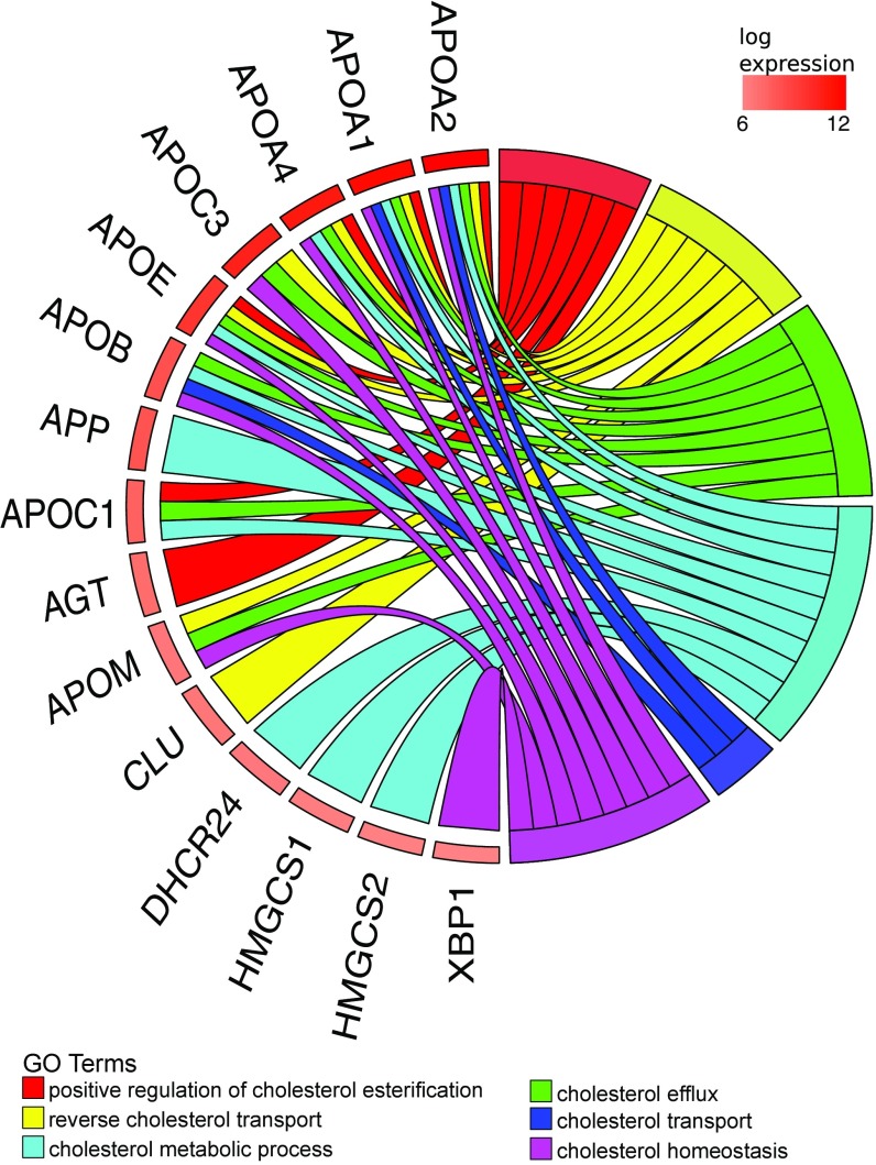
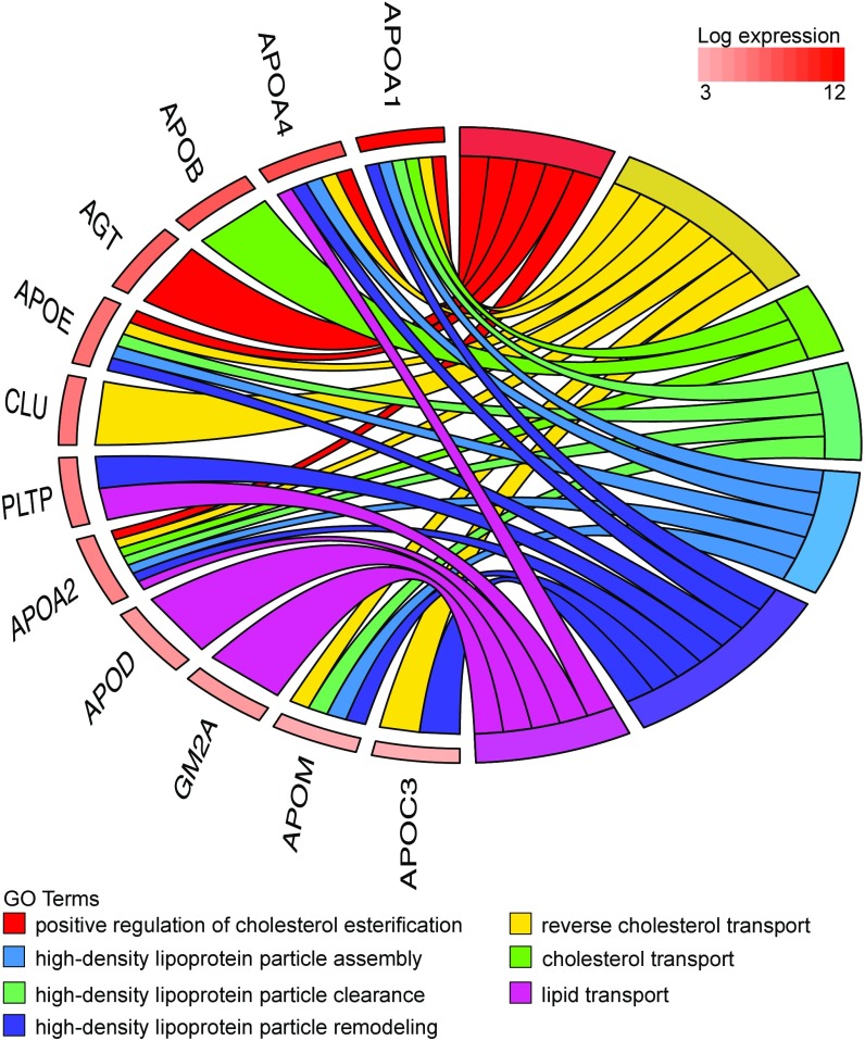
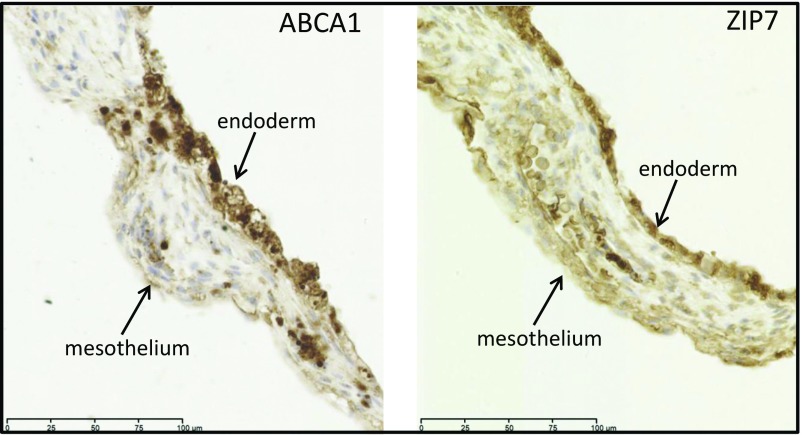
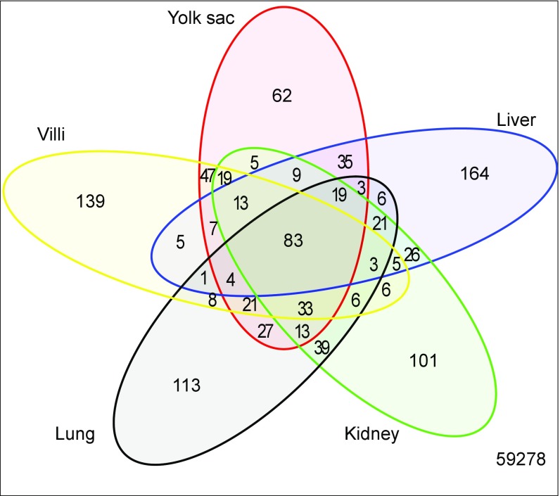
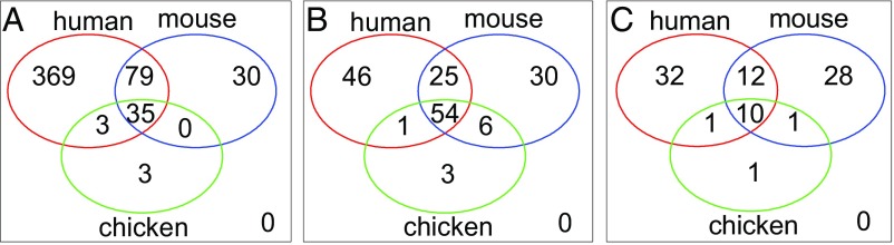
The yolk sac is phylogenetically the oldest of the extraembryonic membranes. The human embryo retains a yolk sac, which goes through primary and secondary phases of development, but its importance is controversial. Although it is known to synthesize proteins, its transport functions are widely considered vestigial. Here, we report RNA-sequencing (RNA-seq) data for the human and murine yolk sacs and compare those data with data for the chicken. We also relate the human RNA-seq data to proteomic data for the coelomic fluid bathing the yolk sac. Conservation of transcriptomes across the species indicates that the human secondary yolk sac likely performs key functions early in development, particularly uptake and processing of macro- and micronutrients, many of which are found in coelomic fluid. More generally, our findings shed light on evolutionary mechanisms that give rise to complex structures such as the placenta. We identify genetic modules that are conserved across mammals and birds, suggesting these modules are part of the core amniote genetic repertoire and are the building blocks for both oviparous and viviparous reproductive modes. We propose that although a choriovitelline placenta is never established physically in the human, the placental villi, the exocoelomic cavity, and the secondary yolk sac function together as a physiological equivalent.

Keywords: evolution; placenta; yolk sac.


Conflict of interest statement

The authors declare no conflict of interest.

Figures

Fig. 1.
Fig. 1.
Chord plot illustrating the GO biological process terms that include “cholesterol” and that are overrepresented in the 400 most abundant yolk sac transcripts (Right) and the genes contributing to that enrichment (Left) arranged in order of their expression level.
Fig. 2.
Fig. 2.
Chord plot illustrating proteins present in the coelomic fluid that relate to GO biological process terms involving “cholesterol” and “lipid transport.” The presence of these proteins is consistent with the high level of their transcripts in the human yolk sac illustrated in Fig. 1.
Fig. 3.
Fig. 3.
Immunolocalization of ABCA1 and SLC39A7/ZIP7 transporter proteins in the human yolk sac at gestational age 11 wk. Sections were immunostained with anti-ABCA1 or anti-ZIP7 antibodies. In both cases, staining was present in the inner endodermal and outer mesothelial layers, although it was stronger in the former.
Fig. 4.
Fig. 4.
Venn diagram comparing the most abundant 400 transcripts in the human yolk sac with first-trimester placental villi and adult liver, lung, and kidney. Transcripts shared by all five tissues (n = 83) principally encoded housekeeping proteins, whereas those shared uniquely with liver (n = 35) encoded proteins involved in cholesterol and lipid metabolism, suggesting that the yolk sac may perform these functions while the fetal liver develops. By contrast, there are few transcripts shared uniquely with the kidney (n = 5), suggesting that the yolk sac plays little role in excretion.
Fig. 5.
Fig. 5.
Chord plot connecting GO biological process terms associated with “lipid metabolism” and genes encoding transcripts that are shared by the human yolk sac and adult liver. The overlap suggests the yolk sac may perform hepatic functions while the fetal liver differentiates.
Fig. 6.
Fig. 6.
Venn diagrams illustrating the overlap among overrepresented GO terms associated with the 400 most abundant transcripts in each of the human, mouse, and chicken yolk sacs. (A) Biological process terms. (B) Cellular component terms. (C) Molecular function terms. The considerable overlap among the species in all three categories suggests conservation of functions.
Fig. 7.
Fig. 7.
Diagrammatic comparison of the nutrient pathway during early pregnancy in the mouse (A) and the speculated pathway in the human (B). In the mouse, histotrophic secretions (green) released from the endometrial glands are phagocytosed (step 1) by the endodermal cells of the visceral layer of the inverted yolk sac. Following fusion with lysosomes (step 2), digestion of maternal proteins leads to release of amino acids that are transported (step 3) to the fetal circulation (FC). In the human, histotrophic secretions are released from the endometrial glands through the developing basal plate of the placenta into the intervillous space (IVS) and are phagocytosed (step 1) by the syncytiotrophoblast (STB) (42). We speculate that following digestion by lysosomal enzymes (step 2), free amino acids are transported (step 3) by efflux transporters to the coelomic fluid (CF), where they accumulate. Nutrients in the CF may be taken up by the mesothelial cells (M) of the yolk sac and transported (step 4) into the fetal circulation (FC). Alternatively, they may diffuse into the cavity of the yolk sac and be taken up by the endodermal cells (step 5). Some intact maternal proteins may also be released into the CF by exocytosis of residual bodies (step 6) and may be engulfed by the mesothelial cells (step 7). CTB, cytotrophoblast cells.

References

    1. Mossman H. Vertebrate Fetal Membranes: Comparative Ontogeny and Morphology: Evolution; Phylogenetic Significance; Basic Functions, Research Opportunities. Macmillan; London: 1987.
    1. Brent RL, Fawcett LB. Nutritional studies of the embryo during early organogenesis with normal embryos and embryos exhibiting yolk sac dysfunction. J Pediatr. 1998;132:S6–S16. - PubMed
    1. Woollett LA. Where does fetal and embryonic cholesterol originate and what does it do? Annu Rev Nutr. 2008;28:97–114. - PubMed
    1. Baardman ME, et al. The origin of fetal sterols in second-trimester amniotic fluid: Endogenous synthesis or maternal-fetal transport? Am J Obstet Gynecol. 2012;207:202.e19–202.e25. - PubMed
    1. Baardman ME, et al. The role of maternal-fetal cholesterol transport in early fetal life: Current insights. Biol Reprod. 2013;88:1–9. - PubMed

Publication types