. 2017 Jul 5;37(27):6423-6441.
doi: 10.1523/JNEUROSCI.2732-16.2017. Epub 2017 May 30.

Parallel Regulation of Memory and Emotion Supports the Suppression of Intrusive Memories

Affiliations

Parallel Regulation of Memory and Emotion Supports the Suppression of Intrusive Memories

Pierre Gagnepain et al. J Neurosci. .

Abstract

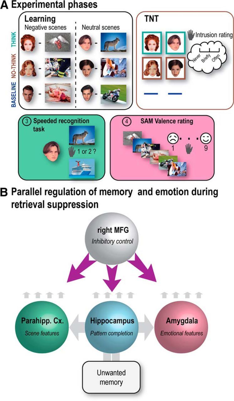
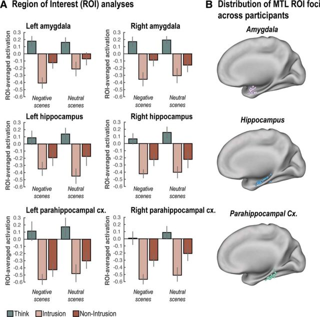
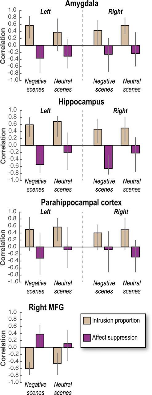
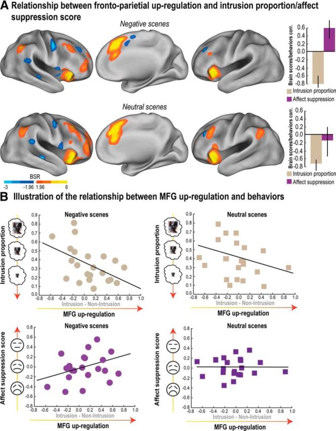
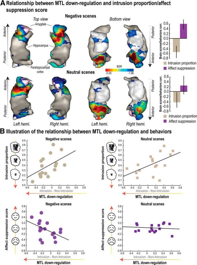
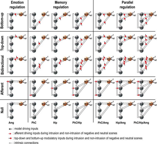
Intrusive memories often take the form of distressing images that emerge into a person's awareness, unbidden. A fundamental goal of clinical neuroscience is to understand the mechanisms allowing people to control these memory intrusions and reduce their emotional impact. Mnemonic control engages a right frontoparietal network that interrupts episodic retrieval by modulating hippocampal activity; less is known, however, about how this mechanism contributes to affect regulation. Here we report evidence in humans (males and females) that stopping episodic retrieval to suppress an unpleasant image triggers parallel inhibition of mnemonic and emotional content. Using fMRI, we found that regulation of both mnemonic and emotional content was driven by a shared frontoparietal inhibitory network and was predicted by a common profile of medial temporal lobe downregulation involving the anterior hippocampus and the amygdala. Critically, effective connectivity analysis confirmed that reduced amygdala activity was not merely an indirect consequence of hippocampal suppression; rather, both the hippocampus and the amygdala were targeted by a top-down inhibitory control signal originating from the dorsolateral prefrontal cortex. This negative coupling was greater when unwanted memories intruded into awareness and needed to be purged. Together, these findings support the broad principle that retrieval suppression is achieved by regulating hippocampal processes in tandem with domain-specific brain regions involved in reinstating specific content, in an activity-dependent fashion.SIGNIFICANCE STATEMENT Upsetting events sometimes trigger intrusive images that cause distress and that may contribute to psychiatric disorders. People often respond to intrusions by suppressing their retrieval, excluding them from awareness. Here we examined whether suppressing aversive images might also alter emotional responses to them, and the mechanisms underlying such changes. We found that the better people were at suppressing intrusions, the more it reduced their emotional responses to suppressed images. These dual effects on memory and emotion originated from a common right prefrontal cortical mechanism that downregulated the hippocampus and amygdala in parallel. Thus, suppressing intrusions affected emotional content. Importantly, participants who did not suppress intrusions well showed increased negative affect, suggesting that suppression deficits render people vulnerable to psychiatric disorders.

Keywords: affect regulation; dynamic causal modeling; emotion; forgetting; inhibitory control; memory suppression.


Figures

Figure 1.
Figure 1.
Experimental phases and hypothesized dynamics. A, After learning face-scene pairs (Negative or Neutral), participants were scanned during the TNT task. For Think items (bounded by green box), participants recalled the associated scene; for No-Think items (bounded by red box), they tried to stop the memory of the scene from entering awareness. Baseline cues were not presented during this TNT phase. Next, participants performed a speeded associative recognition task followed by an SAM valence rating task on all picture categories (Think, No-Think, and Baseline) to evaluate how suppression affected memory and emotional perceptions, respectively. On these final tests, baseline items provided an estimate of memory or affect, given that neither retrieval nor suppression has been performed in the interim. The pictures displayed here are not issued from IAPS database but are free-use pictures taken from the internet for illustrative purpose. B, The dynamics predicted to bring about parallel inhibition of memory and emotion for intrusive negative scenes. Cue input to the hippocampus is predicted to drive pattern completion, followed by recurrent reactivation of scene and emotional features in parahippocampal cortex and amygdala, respectively; intrusion-related reactivation in these regions is predicted to trigger parallel inhibition by the right MFG. We do not propose that MFG directly inhibits these structures given the weak anatomical projections between MFG and amygdala (Anderson et al., 2016). However, the MFG is proposed to modulate these regions polysynaptically via pathways yet to be fully understood.
Figure 2.
Figure 2.
Behavioral and neural indices of mnemonic and affective regulation. A, Intrusion proportions (i.e., the proportion of trials in which the associated memory entered into awareness on No-Think trials as measured by our trial-by-trial intrusion report measure; see Procedure) over the five scanning blocks of the TNT phase. Shaded error bands represent within-participant SDs. B, Left, The relationship between intrusion proportion and affect suppression score (No-Think − Baseline) for Negative scenes. Right, Participants who were better at controlling intrusions of unpleasant scenes also showed reduced negative feelings toward them afterward. ★p < 0.05. C, Brain areas more engaged by retrieval suppression than by retrieval (No-Think > Think; hot colors) and vice versa (No-Think < Think; cold colors), thresholded at the uncorrected level of p < 0.001 for visualization purposes. Pink spheres represent right MFG ROI foci across participants used in subsequent activation, correlation, and DCM analyses (see Materials and Methods). These ROIs were derived from individual local maxima centered around the supramodal control system described by Depue et al. (2015) and are projected onto a common standard space for visualization purposes. Statistical parametric maps were rendered on the top of the PALS human surface using Caret software (Van Essen et al., 2001) (RRID:SCR_006260).
Figure 3.
Figure 3.
MTL downregulation. A, Suppressing scene memories reduced activity across the whole MTL overall. Additionally, we observed more pronounced downregulation in these MTL regions during suppression attempts that were accompanied by intrusions. B, Distribution of MTL ROI foci across participants once projected back to MNI space. Error bars indicate SEM.
Figure 4.
Figure 4.
Brain/behavior correlations. Pearson correlation (skipping bivariate outliers) between affect suppression/intrusion proportion and neural marker of memory suppression (Intrusion − Non-Intrusion) for each scene type. For neural markers of memory suppression in the MTL, negative (lower) scores are assumed to indicate more successful suppression of activity (Intrusion − Non-Intrusion is a downregulation in most cases, suggesting control); in contrast, neural markers for memory suppression in MFG are upregulations of activity (again, Intrusion − Non-Intrusion), and so positive scores are assumed to indicate greater engagement of control. Higher behavioral scores for affect suppression scores indicate greater reduction in negative valence for No-Think compared with Baseline items (i.e., No-Think − Baseline); in contrast, higher intrusion scores indicate worse control over intrusions. Together, these considerations indicate that, in MTL, positive correlations for intrusions signify that downregulations predict fewer intrusions, whereas negative correlations for affect suppression signify that downregulations predict reduced affect. In MFG, the direction of correlations is expected to invert because greater control is indicated by upregulation. Error bars indicate 99.3% bootstrapped CI corrected for multiple comparisons across ROIs. Significant correlations occur when the CI does not encompass zero.
Figure 5.
Figure 5.
Relationship between neural markers of inhibitory control and the reduction of mnemonic awareness/affective response. Outcome of the PLS analysis for both Negative and Neutral scenes (conducted within the retrieval suppression network; see Materials and Methods) between intrusion-related upregulation (Intrusion − Non-Intrusion) and behavioral measures (intrusion proportion and affect suppression score). A, Voxels showing a significant pattern of brain/behavior correlations as revealed by the first (significant) LV were identified using a BSR threshold higher/lower than 1.96/−1.96, respectively (i.e., p < 0.05). Correlations between participants' brain scores and behavioral measures for the first significant LV are also reported in A. Error bars indicate bootstrapped 95% CI. Brain scores reflect the contribution of each participant to a given LV. The correlation between brain scores and behaviors thus reveals the meaning of the LV. B, Scatter plots observed in the right MFG illustrating the relationship captured by PLS analysis between the upregulation (Intrusion − Non-Intrusion) and behavioral scores for Negative and Neutral scenes. These findings reveal voxels whose upregulation is associated with reduced intrusion frequency for both Negative and Neutral scenes and also with increased affect suppression score only in the case of Negative scenes (reduced negative affect for suppressed images). BSR maps were rendered on the top of the PALS human surface using Caret software (Van Essen et al., 2001) (RRID:SCR_006260).
Figure 6.
Figure 6.
Relationship between MTL downregulation and reductions of mnemonic awareness/affective response. Outcome of the behavioral PLS analysis for both Negative and Neutral scenes conducted within the MTL, including bilateral amygdala, hippocampus, and parahippocampal cortex. PLS correlations were tested with the MTL contrast map for the differences between Intrusion and Non-Intrusion conditions on one hand (i.e., downregulation), and behavioral scores (intrusion proportion and affect suppression, i.e., the reduction of negative feelings for No-Think items relative to Baseline) on the other hand. A, Voxels significantly associated to the first significant LV and whose downregulation significantly correlated with intrusion proportion for both Negative and Neutral scenes, as well as with affect suppression exclusively for Negative scenes. Error bars indicate bootstrapped 95% CI. Voxels were identified using a BSR higher or lower than 1.96/−1.96, respectively (i.e., p < 0.05). Correlations between participants' brain scores and behaviors for the first significant LV are also reported in A. Clusters of BSR exceeding threshold were rendered onto a 3D reconstruction of a standard MTL template. The 3D representation of the MTL was obtained by transforming MTL binary masks into 3D meshes using “Anatomist/BrainVISA” software (http://www.brainvisa.info/; RRID:SCR_007354). B, Scatter plots illustrating the relationship captured by PLS analysis between the downregulation observed in the MTL cluster (A) and behavioral scores for both Negative and Neutral scenes.
Figure 7.
Figure 7.
DCM model space. DCM models were organized into two families. The first (the Modulatory family) divided the model space into five subgroups differing according to whether the intrinsic connection from the right MFG was modulated or not by No-Think trials (modeled as 3 s short epochs separately for Intrusion and Non-Intrusion of each emotion type). Subgroups 1–5 of this family (rows 1–5) either included modulation on bottom up (e.g., hippocampus to MFG, row 1), top-down (row 2), or bidirectional (row 3) connections; or no modulation, but variable afferent input to MTL regions from a source independent of MFG (row 4); or no modulation at all (row 5). The second family (the Regulation family) divided the model space into families according to modulatory targets. Subgroups 1–3 of this family (columns 1–3) include the Emotion Regulation family (left, amygdala modulation only), the Memory Regulation family (middle, modulation of hippocampus, parahippocampal cortex, or both), and the Parallel regulation family (right, modulation of amygdala, and other memory-related regions). After estimating all 35 models for each participant, we performed the group BMS as implemented in SPM12 (version DCM12 revision 4750; RRID:SCR_007037). This produces the exceedance probability (i.e., the extent to which each model is more likely than any other considered model) and expected posterior probability (i.e., the probability of a model generating the observed data). By positing connectivity relationships between MFG and these MTL structures in our DCM models, we do not presuppose direct anatomical connections (an assumption that DCM's analytical method does not require); rather, we are modeling the data to evaluate the existence of a (potentially) polysynaptic and directional causal influence of each region on the activity of others to which it is connected.

Comment in

References

    1. Abdi H, Williams LJ, Beaton D, Posamentier MT, Harris TS, Krishnan A, Devous MD Sr (2012) Analysis of regional cerebral blood flow data to discriminate among Alzheimer's disease frontotemporal dementia and elderly controls: multi-block barycentric discriminant (MUBADA) methodology. J Alzheimer Dis 31:S189–S201. 10.3233/JAD-2012-112111 - DOI - PMC - PubMed
    1. Anderson MC, Green C (2001) Suppressing unwanted memories by executive control. Nature 410:366–369. 10.1038/35066572 - DOI - PubMed
    1. Anderson MC, Hanslmayr S (2014) Neural mechanisms of motivated forgetting. Trends Cogn Sci 18:279–292. 10.1016/j.tics.2014.03.002 - DOI - PMC - PubMed
    1. Anderson MC, Ochsner KN, Kuhl B, Cooper J, Robertson E, Gabrieli SW, Glover GH, Gabrieli JD (2004) Neural systems underlying the suppression of unwanted memories. Science 303:232–235. 10.1126/science.1089504 - DOI - PubMed
    1. Anderson MC, Bunce JG, Barbas H (2016) Prefrontal-hippocampal pathways underlying inhibitory control over memory. Neurobiol Learn Mem 134:145–161. 10.1016/j.nlm.2015.11.008 - DOI - PMC - PubMed

LinkOut - more resources