

Is Abdominal Fetal Electrocardiography an Alternative to Doppler Ultrasound for FHR Variability Evaluation?
- PMID: 28559852
- PMCID: PMC5432618
- DOI: 10.3389/fphys.2017.00305
Is Abdominal Fetal Electrocardiography an Alternative to Doppler Ultrasound for FHR Variability Evaluation?
Abstract
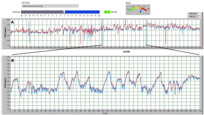
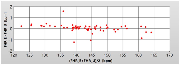
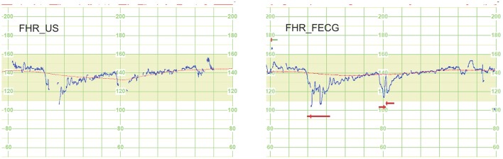
Great expectations are connected with application of indirect fetal electrocardiography (FECG), especially for home telemonitoring of pregnancy. Evaluation of fetal heart rate (FHR) variability, when determined from FECG, uses the same criteria as for FHR signal acquired classically-through ultrasound Doppler method (US). Therefore, the equivalence of those two methods has to be confirmed, both in terms of recognizing classical FHR patterns: baseline, accelerations/decelerations (A/D), long-term variability (LTV), as well as evaluating the FHR variability with beat-to-beat accuracy-short-term variability (STV). The research material consisted of recordings collected from 60 patients in physiological and complicated pregnancy. The FHR signals of at least 30 min duration were acquired dually, using two systems for fetal and maternal monitoring, based on US and FECG methods. Recordings were retrospectively divided into normal (41) and abnormal (19) fetal outcome. The complex process of data synchronization and validation was performed. Obtained low level of the signal loss (4.5% for US and 1.8% for FECG method) enabled to perform both direct comparison of FHR signals, as well as indirect one-by using clinically relevant parameters. Direct comparison showed that there is no measurement bias between the acquisition methods, whereas the mean absolute difference, important for both visual and computer-aided signal analysis, was equal to 1.2 bpm. Such low differences do not affect the visual assessment of the FHR signal. However, in the indirect comparison the inconsistencies of several percent were noted. This mainly affects the acceleration (7.8%) and particularly deceleration (54%) patterns. In the signals acquired using the electrocardiography the obtained STV and LTV indices have shown significant overestimation by 10 and 50% respectively. It also turned out, that ability of clinical parameters to distinguish between normal and abnormal groups do not depend on the acquisition method. The obtained results prove that the abdominal FECG, considered as an alternative to the ultrasound approach, does not change the interpretation of the FHR signal, which was confirmed during both visual assessment and automated analysis.
Keywords: Doppler ultrasound; fetal electrocardiogram; fetal heart rate analysis; fetal outcome; fetal state assessment.
Figures
References
-
- Agostinelli A., Fioretti S., Di Nardo F., Burattini L. (2015a). Clinical Application of the Segmented-Beat Modulation Method for Fetal ECG Extraction, in Proceedings of the 12th International Workshop on Intelligent Solutions in Embedded Systems (WISES) eds Conti M., Orcioni S. (Ancona: IEEE Press; ), 35–40.
-
- Agostinelli A., Grillo M., Biagini A., Giuliani C., Burattini L., Fioretti S. A. (2015b). Noninvasive fetal electrocardiography: an overview of the signal electrophysiological meaning, recording procedures, and processing techniques. Ann. Noninvas. Electro. 20, 303–313. 10.1111/anec.12259 - DOI - PMC - PubMed
-
- Almeida R., Goncalves H., Rocha A. P., Bernardes J. (2013). A wavelet-based method for assessing fetal cardiac rhythms from abdominal ECGs. Comput. Cardiol. 40, 289–292.
LinkOut - more resources
Full Text Sources
Other Literature Sources