. 2017 May 25:10:135.
doi: 10.1186/s13068-017-0822-0. eCollection 2017.

Genome-wide characterization of cellulases from the hemi-biotrophic plant pathogen, Bipolaris sorokiniana, reveals the presence of a highly stable GH7 endoglucanase

Affiliations

Genome-wide characterization of cellulases from the hemi-biotrophic plant pathogen, Bipolaris sorokiniana, reveals the presence of a highly stable GH7 endoglucanase

Shritama Aich et al. Biotechnol Biofuels. .

Abstract

Background: Bipolaris sorokiniana is a filamentous fungus that causes spot blotch disease in cereals like wheat and has severe economic consequences. However, information on the identities and role of the cell wall-degrading enzymes (CWDE) in B. sorokiniana is very limited. Several fungi produce CWDE like glycosyl hydrolases (GHs) that help in host cell invasion. To understand the role of these CWDE in B. sorokiniana, the first step is to identify and annotate all possible genes of the GH families like GH3, GH6, GH7, GH45 and AA9 and then characterize them biochemically.

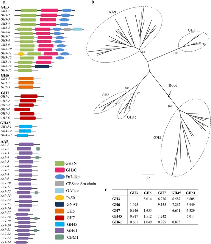
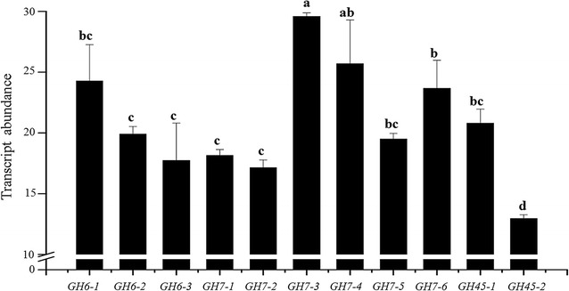
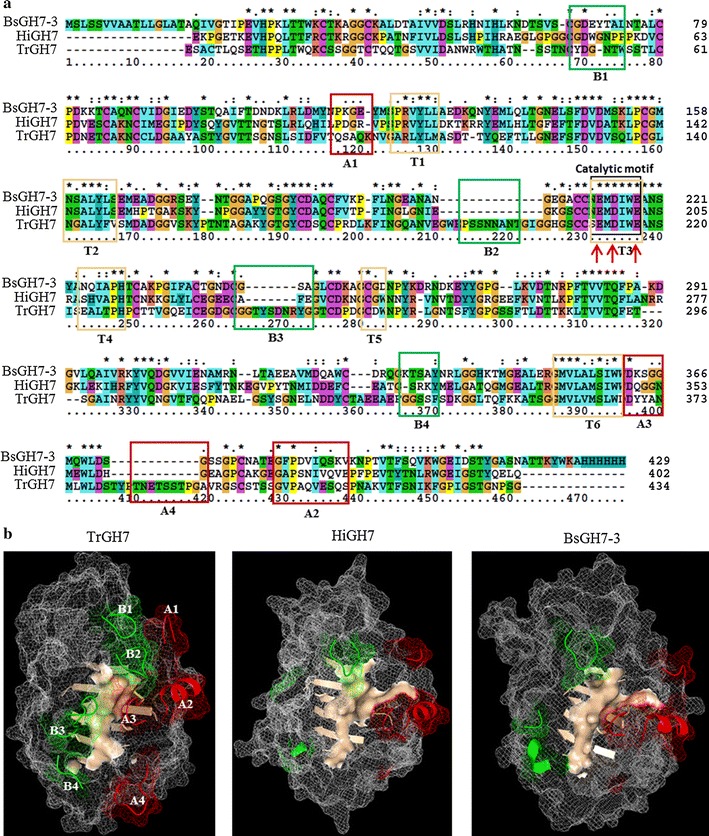
Results: We confirmed and annotated the homologs of GH3, GH6, GH7, GH45 and AA9 enzymes in the B. sorokiniana genome using the sequence and domain features of these families. Quantitative real-time PCR analyses of these homologs revealed that the transcripts of the BsGH7-3 (3rd homolog of the GH 7 family in B. sorokiniana) were most abundant. BsGH7-3, the gene of BsGH7-3, was thus cloned into pPICZαC Pichia pastoris vector and expressed in X33 P. pastoris host to be characterized. BsGH7-3 enzyme showed a temperature optimum of 60 °C and a pHopt of 8.1. BsGH7-3 was identified to be an endoglucanase based on its broad substrate specificity and structural comparisons with other such endoglucanases. BsGH7-3 has a very long half-life and retains 100% activity even in the presence of 4 M NaCl, 4 M KCl and 20% (v/v) ionic liquids. The enzyme activity is stimulated up to fivefold in the presence of Mn+2 and Fe+2 without any deleterious effects on enzyme thermostability.

Conclusions: Here we reanalysed the B. sorokiniana genome and selected one GH7 enzyme for further characterization. The present work demonstrates that BsGH7-3 is an endoglucanase with a long half-life and no loss in activity in the presence of denaturants like salt and ionic liquids, and lays the foundation towards exploring the Bipolaris genome for other cell wall-degrading enzymes.

Keywords: Alkaliphilic; Bipolaris sorokiniana; Cell wall-degrading enzymes; GH7 endoglucanases; Glycosyl hydrolase; Ionic liquids; Salt tolerant; Thermostable.


Figures

Fig. 1
Fig. 1
Characterization of GHs in B. sorokiniana genome. a Domain distributions in the homologs of B. sorokiniana GHs and redox enzymes (AA9). The figure shows the schematic of arrangement of domains in each of the BsGH homologs (comparative lengths are unscaled). b Phylogeny and evolution of B. sorokiniana GHs. Maximum likelihood rooted phylogeny of B. sorokiniana GHs and redox enzymes (AA9). The values on branches show the bootstrap (%). The dotted terminal branches in the clades of GH3, GH45, GH6 and GH7 families are the corresponding GHs from the bacterial species used as an out-group in the phylogenetic analysis. c Evolutionary divergence between GH family members. The upper diagonal represents the number of substitutions per site between the respective GH families, while the lower diagonal represents its standard deviation
Fig. 2
Fig. 2
Transcript abundance of the GH family genes in B. sorokiniana. A standard curve using known amounts of cDNA was prepared for the three homologs of BsGH6, six belonging to BsGH7 and two of BsGH45; (further details in “Results”) in four replicates. Based on the standard curve, cDNA corresponding to 150 ng of total RNA was used to evaluate the absolute transcript amount based on the respective CT values. Three independent experiments were conducted, each comprising four replicates, and the mean values were used to plot the graph. Data are represented as mean ± SE. Assistat 7.6 beta was used for statistical analysis (DMRT-Duncan multiple range test). The Duncan test at a level of 5% of probability was applied. Bars with the same letter do not differ statistically between themselves (p ≤ 0.05)
Fig. 3
Fig. 3
Biochemical characterization of BsGH7-3. a 10% SDS-PAGE of purified BsGH7-3. Lane M PageRuler plus pre-stained protein ladder (ThermoFisher, Waltham, USA) and BsGH7-3 stained by Coomassie Brilliant Blue R250. b Specific activity and stability of BsGH7-3 reported as percent residual activity at 4 and 60 °C, respectively. c Effect of temperature on GH7-3 measured over a range of 48 to 68 °C with 0.8% CMC as a substrate and McIlvaine buffer, pH 8.1. The residual activity was measured by the standard DNS assay and reported as % specific activity. d Effect of pH was determined by incubating the enzyme in buffer of pH range 5.2–8.6 for 6 h at 4 °C and then the kinetic assay was performed to determine the % specific activity
Fig. 4
Fig. 4
BsGH7-3 catalysed hydrolysis of carboxymethyl cellulose (CMC) as determined by visible spectrophotometry. The solid line indicates the best fit of the Michaelis–Menten equation. The values determined were K m = 0.7461 mg mL−1, V max = 21.64 µM min−1 and k cat = 288 min−1
Fig. 5
Fig. 5
Comparative analysis of B. sorokiniana GH7-3 with H. insolens endoglucanase GH7 (PDB ID: 1OJJ-B) and T. reesei cellobiohydrolase GH7 (PDB ID: 7CEL). (a) Multiple sequence alignment of BsGH7-3, HiGH7 and TrGH7. The black rectangular box contains a stretch of six residues of the catalytic motif that are conserved across the three proteins. The catalytic residues are marked with red arrows. The coloured rectangular boxes denote the residues of motifs B (green), A (red) and tunnel T (light yellow). b Homology model-based structure of BsGH7-3 shows variation in the secondary structure of motifs A, B and T (same colour as in a, with HiGH7 and TrGH7). The motifs A (red colour) and B (green colour) are in cartoon view on the mesh background. The surface view of the inner lining of the tunnel is shown in salmon colour

References

    1. Durand H, Clanet M, Tiraby G. Genetic improvement of Trichoderma reesei for large scale cellulase production. Enzyme Microbiol Technol. 1988;10(6):341–346. doi: 10.1016/0141-0229(88)90012-9. - DOI
    1. Martinez D, Berka RM, Henrissat B, Saloheimo M, Arvas M, Baker SE, Chapman J, Chertkov O, Coutinho PM, Cullen D, et al. Genome sequencing and analysis of the biomass-degrading fungus Trichoderma reesei (syn. Hypocrea jecorina) Nat Biotechnol. 2008;26(5):553–560. doi: 10.1038/nbt1403. - DOI - PubMed
    1. Harris PV, Welner D, McFarland KC, Re E, Navarro Poulsen JC, Brown K, Salbo R, Ding H, Vlasenko E, Merino S, et al. Stimulation of lignocellulosic biomass hydrolysis by proteins of glycoside hydrolase family 61: structure and function of a large, enigmatic family. Biochemistry. 2010;49(15):3305–3316. doi: 10.1021/bi100009p. - DOI - PubMed
    1. Datta S. Recent strategies to overexpress and engineer cellulases for biomass degradation. Curr Metabol. 2016;4(1):14–22. doi: 10.2174/2213235X03666150702155845. - DOI
    1. Walton JD. Deconstructing the cell wall. Plant Physiol. 1994;104(4):1113–1118. doi: 10.1104/pp.104.4.1113. - DOI - PMC - PubMed

LinkOut - more resources