

Quantitative photoacoustic imaging for early detection of muscle ischemia injury
- PMID: 28559976
- PMCID: PMC5446508
Quantitative photoacoustic imaging for early detection of muscle ischemia injury
Abstract
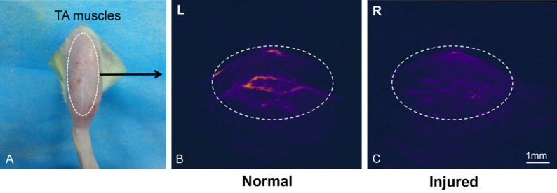
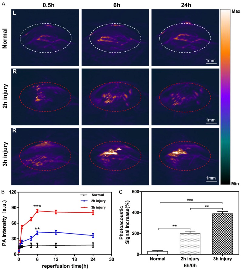
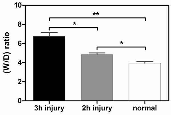
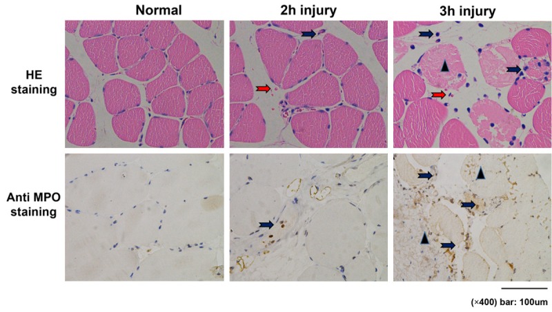
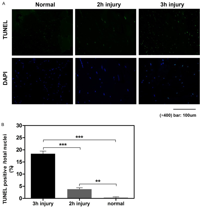
Acute lower extremity ischemia is a limb-and life-threatening problem. The timing of clinical intervention is critical to achieving optimal outcomes. However, there has been a lack of effective techniques capable of evaluating muscle and limb damage. Microcirculatory injury is the initial pathological change during ischemic muscle injury. Here, we performed photoacoustic imaging (PAI) in real time to quantitatively detect the degree of microcirculatory injury of ischemic muscles in a rat model in which Evans blue (EB), which strongly binds to albumin in blood, was used as a nontoxic molecular PA probe. The right lower hind limbs of Sprague-Dawley (SD) rats were subjected to 2 or 3 hours of tourniquet-induced ischemia. Then, PA imaging of the tibialis anterior (TA) muscles in the anterior compartment was performed for 0-24 h after the release of compression. Twenty-four hours after reperfusion, rats were euthanized and examined for pathology, edema and muscle viability. Imaging at 680 nm on rats revealed that there was significant signal enhancement in the TA muscles of the two injury groups compared to the control group, and the 3-h injury group had significantly higher PA signal intensity than the 2-h injury group at each time point. Histopathology results obtained from both the normal and the damaged muscles correlated well with the PAI findings. In conclusion, PA imaging is a promising modality for quantitatively detecting limb and muscle ischemic injury and may pave the road for further clinical application.
Keywords: Muscle; extremity; ischemic injury; microcirculatory injury; photoacoustic imaging.
Conflict of interest statement
None.
Figures
References
-
- Malinoski DJ, Slater MS, Mullins RJ. Crush injury and rhabdomyolysis. Crit Care Clin. 2004;20:171–192. - PubMed
-
- Walker TG. Acute limb ischemia. Tech Vasc Interv Radiol. 2009;12:117–129. - PubMed
-
- Henke PK. Contemporary management of acute limb ischemia: factors associated with amputation and in-hospital mortality. Semin Vasc Surg. 2009;22:34–40. - PubMed
-
- Aftabuddin M, Islam N, Jafar MA, Haque I. The status of lower-limb amputation in Bangladesh: a 6-year review. Surg Today. 1997;27:130–134. - PubMed
-
- Nakayama T, Fujita M, Ishihara M, Ishihara M, Ogata S, Yamamoto Y, Shimizu M, Maehara T, Kanatani Y, Tachibana S. Improved survival rate by temperature control at compression sites in rat model of crush syndrome. J Surg Res. 2014;188:250–259. - PubMed
LinkOut - more resources
Full Text Sources
Other Literature Sources