. 2017 May 15;9(5):2352-2362.
eCollection 2017.

Peptide-modified chitosan hydrogels promote skin wound healing by enhancing wound angiogenesis and inhibiting inflammation

Affiliations

Peptide-modified chitosan hydrogels promote skin wound healing by enhancing wound angiogenesis and inhibiting inflammation

Xionglin Chen et al. Am J Transl Res. .

Abstract

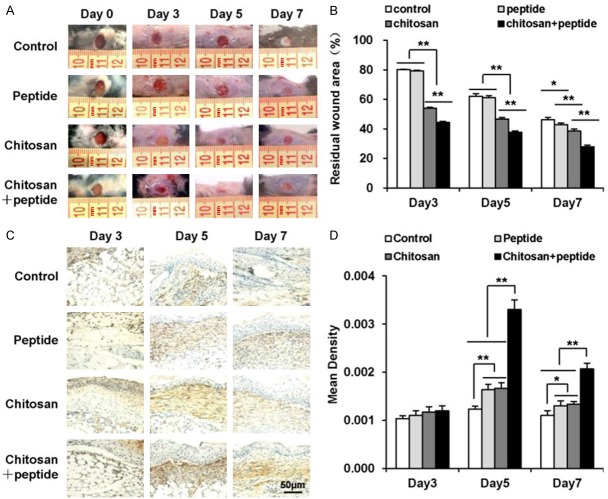
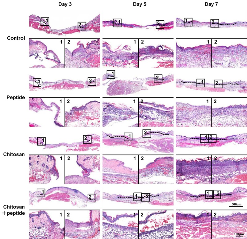
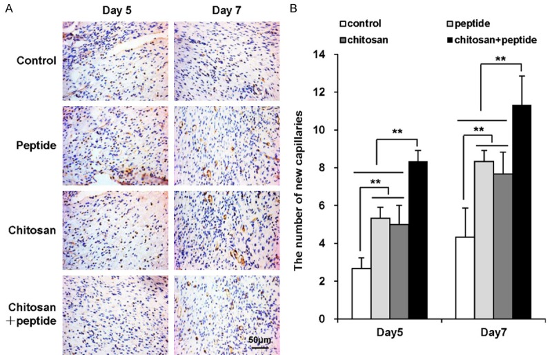
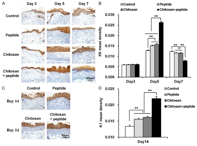
Cutaneous wound healing following trauma is a complex and dynamic process involving multiple overlapping events following trauma. Two critical elements affecting skin wound healing are neovascularization and inflammation. A nascent vessel can provide nutrition and oxygen to a healing wound. Therefore, treatments strategies that enhance angiogenesis and inhibit inflammation can promote skin wound healing. Previous studies have shown that the SIKVAV peptide (Ser-Ile-Lys-Val-Ala-Val) from laminin can promote angiogenesis in vitro. This study evaluated the effects of peptide SIKVAV-modified chitosan hydrogels on skin wound healing. We established skin wounds established in mice and treated them with SIKVAV-modified chitosan hydrogels. H&E staining showed that peptide-modified chitosan hydrogels accelerated the reepithelialization of wounds compared with the negative and positive controls. Immunohistochemistry analysis demonstrated that more myofibroblasts were deposited at wounds treated with peptide-modified chitosan hydrogels that at those treated with negative and positive controls. In addition, peptide-modified chitosan hydrogels promoted angiogenesis as well as keratinocyte proliferation and differentiation, but inhibited inflammation in skin wounds. Taken together, these results suggest that SIKVAV-modified chitosan hydrogels are a promising treatment component for healing-impaired wounds.

Keywords: SIKVAV; angiogenesis; chitosan hydrogel; inflammation; re-epithelialization; wound healing.


Conflict of interest statement

None.

Figures

Figure 1
Figure 1
Peptide-modified chitosan hydrogels significantly promote skin wound healing in mice. A: Representative photographs of wounds in the control, peptide, chitosan hydrogel, and peptide + chitosan hydrogel groups taken 3, 5, and 7 days after the initial treatment in mice. B: The residual wound percentages in the control, peptide, chitosan hydrogel, and peptide + chitosan hydrogel groups (n = 3). C: α-SMA expression in the control, peptide, chitosan hydrogel, and peptide + chitosan hydrogel groups detected on days 3, 5, and 7 after the initial treatments as detected by immunohistochemical staining (scale bar: 50 μm). D: Mean α-SMA expression in the control, peptide, chitosan hydrogel, and peptide + chitosan hydrogel groups measured 3, 5, and 7 days after the initial treatments (n = 3).
Figure 2
Figure 2
Peptide-modified chitosan hydrogels promote skin re-epithelialization in mice. H&E staining showed the re-epithelialization process in the control, peptide, chitosan hydrogel, and peptide + chitosan hydrogel groups on days 3, 5, and 7 after the initial treatments (scale bar: 500 μm).
Figure 3
Figure 3
Peptide-modified chitosan hydrogels significantly promote angiogenesis in mouse skin wounds. A: Immunohistochemical staining for CD31 expression in vascular endothelial cells of wounded mouse skin 5 and 7 days after the initial treatments (scale bar: 50 μm). B: Mean numbers of new blood capillaries per field of view in mouse skin wounds in the control, peptide, chitosan hydrogel, and peptide + chitosan hydrogel groups (n = 3).
Figure 4
Figure 4
Peptide-modified chitosan hydrogels promote the proliferation and differentiation of keratinocytes in skin wounds. A: Immunohistochemical staining to show K6 expression in keratinocytes in the control, peptide, chitosan hydrogel, and peptide + chitosan hydrogel groups 3, 5, and 7 days after the initial treatments (scale bar: 50 μm). B: K6 expression in the control, peptide, chitosan hydrogel, and peptide + chitosan hydrogel groups on days 3, 5, and 7 after the initial treatments (n = 3). C: Immunohistochemical staining of K1 expression in keratinocytes in the control, peptide, chitosan hydrogel, and peptide + chitosan hydrogel groups on day 14 after the initial treatments (scale bar: 50 μm). D: K1 expression in the control, peptide, chitosan hydrogel, and peptide + chitosan hydrogel groups on day 14 after the initial treatments (n = 3).
Figure 5
Figure 5
Peptide-modified chitosan hydrogels inhibit inflammation in skin wounds. A: Immunohistochemical staining of CD86-positive cells in the control, peptide, chitosan hydrogel, and peptide + chitosan hydrogel groups on days 3, 5, and 7 after the initial treatments (scale bar: 50 μm). B: Mean numbers of CD86-positive cells per field of view in the control, peptide, chitosan hydrogel, and peptide + chitosan hydrogel groups on days 3, 5, and 7 after the initial treatments (n = 3). C: Immunohistochemical staining of CD163-positive cells in the control, peptide, chitosan hydrogel, and peptide + chitosan hydrogel groups on days 3, 5, and 7 after the initial treatments (scale bar: 50 μm). D: Mean numbers of CD163-positive cells in the control, peptide, chitosan hydrogel, and peptide + chitosan hydrogel groups on days 3, 5, and 7 after the initial treatments (n = 3).

Similar articles

Cited by

References

    1. Zoller N, Valesky E, Butting M, Hofmann M, Kippenberger S, Bereiter-Hahn J, Bernd A, Kaufmann R. Clinical application of a tissue-cultured skin autograft: an alternative for the treatment of non-healing or slowly healing wounds? Dermatology. 2014;229:190–198. - PubMed
    1. Takayama Y, Aoki R. Roles of lactoferrin on skin wound healing. Biochem Cell Biol. 2012;90:497–503. - PubMed
    1. Curtis K, Lam M, Mitchell R, Black D, Taylor C, Dickson C, Jan S, Palmer CS, Langcake M, Myburgh J. Acute costs and predictors of higher treatment costs of trauma in New South Wales, Australia. Injury. 2014;45:279–284. - PubMed
    1. Santema TB, Poyck PP, Ubbink DT. Skin grafting and tissue replacement for treating foot ulcers in people with diabetes. Cochrane Database Syst Rev. 2016;2:CD011255. - PMC - PubMed
    1. Tadokoro S, Ide S, Tokuyama R, Umeki H, Tatehara S, Kataoka S, Satomura K. Leptin promotes wound healing in the skin. PLoS One. 2015;10:e0121242. - PMC - PubMed

LinkOut - more resources