

microRNA-383 mediates high glucose-induced oxidative stress and apoptosis in retinal pigment epithelial cells by repressing peroxiredoxin 3
- PMID: 28559987
- PMCID: PMC5446519
microRNA-383 mediates high glucose-induced oxidative stress and apoptosis in retinal pigment epithelial cells by repressing peroxiredoxin 3
Abstract
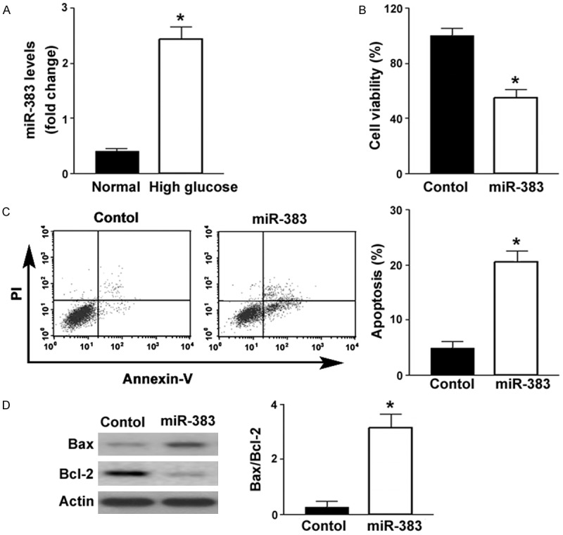
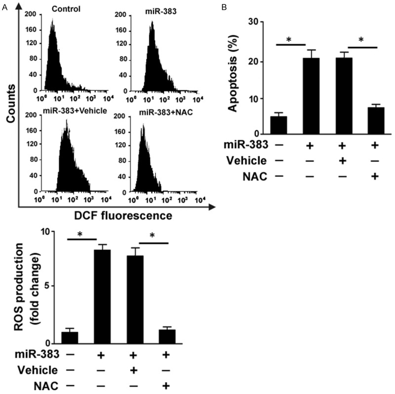
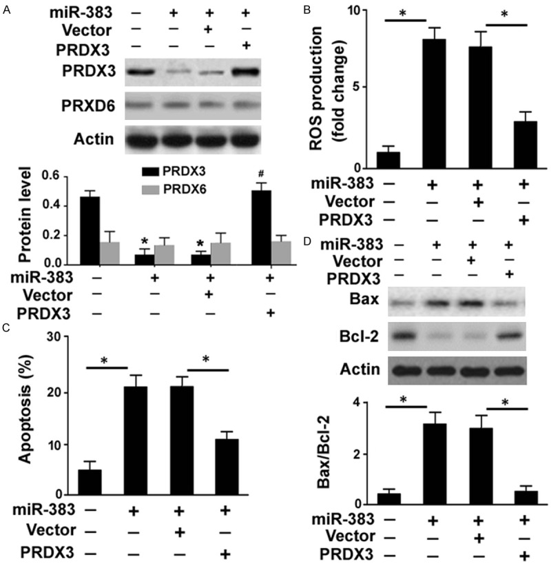
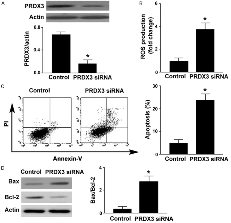
Hyperglycemia-mediated damage to retinal pigment epithelial (RPE) cells plays a central role in the pathogenesis of diabetic retinopathy. Dysregulation of microRNA (miR)-383 modulates pancreatic beta cell survival in diabetes; however, its role in diabetic retinopathy remains unclear. In this study, we examined the expression of miR-383 in ARPE-19 human RPE cell lines after high glucose treatment and investigated its functions in high glucose-induced reactive oxygen species (ROS) generation and apoptotic responses. The downstream target gene that mediated the action of miR-383 was functionally characterized. It was found that high glucose induced a 2.4-fold increase in miR-383 abundance, compared to ARPE-19 cells treated with normal glucose. Overexpression of miR-383 inhibited cell viability and promoted apoptosis and ROS formation in ARPE-19 cells, which was coupled with deregulation of Bcl-2 and Bax. Peroxiredoxin 3 (PRDX3) expression was repressed by miR-383 in ARPE-19 cells. Restoration of PRDX3 counteracted miR-383-induced ROS generation and apoptosis, while silencing of PRDX3 phenocopied the detrimental effects of miR-383 on ARPE-19 cells. Delivery of anti-miR-383 inhibitors led to an increase of PRDX3 expression and prevented high glucose-elicited ROS formation and apoptosis in ARPE-19 cells. Overall, miR-383 upregulation accounts for high glucose-induced oxidative stress and apoptosis in RPE cells by repressing PRDX3 expression. Targeting miR-383 may have therapeutic potential in the treatment of diabetic retinopathy.
Keywords: Apoptosis diabetic retinopathy; microRNA; oxidative stress; target gene.
Conflict of interest statement
None.
Figures

Similar articles
-
MiR-455-5p ameliorates HG-induced apoptosis, oxidative stress and inflammatory via targeting SOCS3 in retinal pigment epithelial cells.J Cell Physiol. 2019 Dec;234(12):21915-21924. doi: 10.1002/jcp.28755. Epub 2019 Apr 30. J Cell Physiol. 2019. PMID: 31041827
-
Hyperglycemia-induced GLP-1R downregulation causes RPE cell apoptosis.Int J Biochem Cell Biol. 2015 Feb;59:41-51. doi: 10.1016/j.biocel.2014.11.018. Epub 2014 Dec 5. Int J Biochem Cell Biol. 2015. PMID: 25483438
-
Long non-coding RNA XIST regulates hyperglycemia-associated apoptosis and migration in human retinal pigment epithelial cells.Biomed Pharmacother. 2020 May;125:109959. doi: 10.1016/j.biopha.2020.109959. Epub 2020 Feb 25. Biomed Pharmacother. 2020. PMID: 32106367
-
Dihydromyricetin Alleviates High Glucose-Induced Oxidative Stress and Apoptosis in Human Retinal Pigment Epithelial Cells by Downregulating miR-34a Expression.Diabetes Metab Syndr Obes. 2021 Jan 27;14:387-397. doi: 10.2147/DMSO.S290633. eCollection 2021. Diabetes Metab Syndr Obes. 2021. PMID: 33536772 Free PMC article.
-
microRNA-125b contributes to high glucose-induced reactive oxygen species generation and apoptosis in HK-2 renal tubular epithelial cells by targeting angiotensin-converting enzyme 2.Eur Rev Med Pharmacol Sci. 2016 Oct;20(19):4055-4062. Eur Rev Med Pharmacol Sci. 2016. PMID: 27775793
Cited by
-
Down-regulation of microRNA-216a confers protection against yttrium aluminium garnet laser-induced retinal injury via the GDNF-mediated GDNF/GFRα1/RET signalling pathway.J Biosci. 2018 Dec;43(5):985-1000. J Biosci. 2018. PMID: 30541958
-
Elevation of the vitreous body concentrations of oxidative stress-responsive apoptosis-inducing protein (ORAIP) in proliferative diabetic retinopathy.Graefes Arch Clin Exp Ophthalmol. 2019 Jul;257(7):1519-1525. doi: 10.1007/s00417-019-04343-w. Epub 2019 May 6. Graefes Arch Clin Exp Ophthalmol. 2019. PMID: 31062144
-
Control-released Alpha-lipoic acid-loaded PLGA microspheres enhance bone formation in type 2 diabetic rat model.Int J Clin Exp Pathol. 2017 Sep 1;10(9):10019-10031. eCollection 2017. Int J Clin Exp Pathol. 2017. PMID: 31966892 Free PMC article.
-
MicroRNA-140-5p ameliorates the high glucose-induced apoptosis and inflammation through suppressing TLR4/NF-κB signaling pathway in human renal tubular epithelial cells.Biosci Rep. 2020 Mar 27;40(3):BSR20192384. doi: 10.1042/BSR20192384. Biosci Rep. 2020. PMID: 32073611 Free PMC article.
-
SIRT3 deficiency increases mitochondrial oxidative stress and promotes migration of retinal pigment epithelial cells.Exp Biol Med (Maywood). 2021 Apr;246(8):877-887. doi: 10.1177/1535370220976073. Epub 2021 Jan 10. Exp Biol Med (Maywood). 2021. PMID: 33423553 Free PMC article.
References
-
- Cunha-Vaz J, Bernardes R, Lobo C. Blood-retinal barrier. Eur J Ophthalmol. 2011;21(Suppl 6):S3–S9. - PubMed
-
- Kim DI, Park MJ, Lim SK, Choi JH, Kim JC, Han HJ, Kundu TK, Park JI, Yoon KC, Park SW, Park JS, Heo YR, Park SH. High-glucose-induced CARM1 expression regulates apoptosis of human retinal pigment epithelial cells via histone 3 arginine 17 dimethylation: role in diabetic retinopathy. Arch Biochem Biophys. 2014;560:36–43. - PubMed
-
- Kim DI, Park MJ, Choi JH, Lim SK, Choi HJ, Park SH. Hyperglycemia-induced GLP-1R downregulation causes RPE cell apoptosis. Int J Biochem Cell Biol. 2015;59:41–51. - PubMed
LinkOut - more resources
Full Text Sources
Research Materials
Miscellaneous