Review
. 2017 May 15:8:2041731417707339.
doi: 10.1177/2041731417707339. eCollection 2017 Jan-Dec.

A mini review focused on the proangiogenic role of silicate ions released from silicon-containing biomaterials

Affiliations
Review

A mini review focused on the proangiogenic role of silicate ions released from silicon-containing biomaterials

Khandmaa Dashnyam et al. J Tissue Eng. .

Abstract

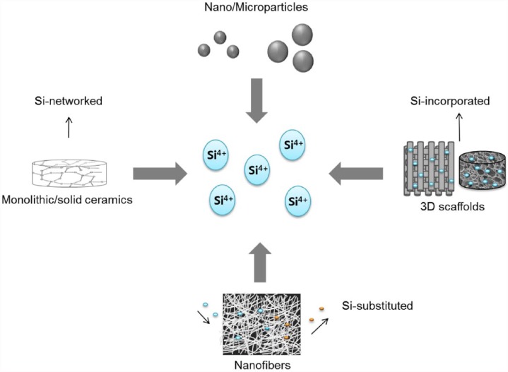
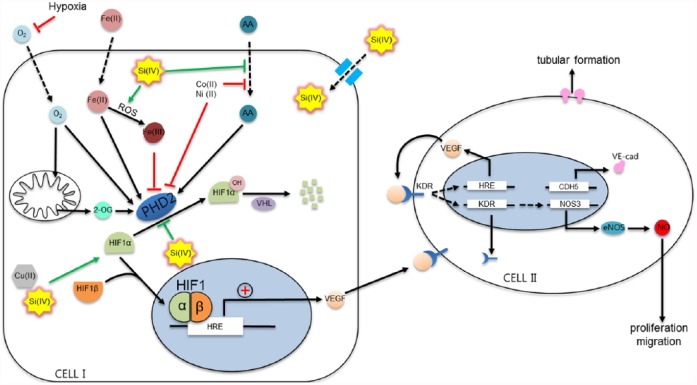
Angiogenesis is considered an important issue in the development of biomaterials for the successful regeneration of tissues including bone. While growth factors are commonly used with biomaterials to promote angiogenesis, some ions released from biomaterials can also contribute to angiogenic events. Many silica-based biomaterials have been widely used for the repair and regeneration of tissues, mainly hard tissues such as bone and tooth structure. They have shown excellent performance in bone formation by stimulating angiogenesis. The release of silicate and others (Co and Cu ions) has therefore been implicated to play critical roles in the angiogenesis process. In this short review, we highlight the in vitro and in vivo findings of angiogenesis (and the related bone formation) stimulated by the various types of silicon-containing biomaterials where silicate ions released might play essential roles. We discuss further the possible molecular mechanisms underlying in the ion-induced angiogenic events.

Keywords: Angiogenesis; bone stimulation; silicate ions; silicon-containing biomaterials.


Conflict of interest statement

Declaration of conflicting interests: The author(s) declared no potential conflicts of interest with respect to the research, authorship, and/or publication of this article.

Figures

Figure 1.
Figure 1.
Various forms of silicon-containing biomaterials that can release silicate ions at proper therapeutic doses (silicate ions are symbolized as Si4+ for simplicity).
Figure 2.
Figure 2.
Silica-based biomaterials that release silicate ions, stimulating angiogenic events in vitro and in vivo, including endothelial (progenitor) cell homing, polarization, cell migration, angiogenesis, tubule-like formation, and neo-vessel sprouting.
Figure 3.
Figure 3.
In vitro angiogenesis of HAECs cultured on ECMatrix in the presence of Ake and TCP extracts at 1/256 and 1/64 dilutions. (a) Optical images of HAECs cultured on ECMatrix in the presence of Ake and TCP extracts at 1/256 and 1/64 dilutions for 2.5 h (bar = 100 µm), 5.5 h (bar = 100 µm), and 17 h (bar = 200 µm). (b–d) The statistics of the number of nodes, circles, and tubes formed in the culture after 2.5, 5.5, and 17 h, respectively. Data represent mean values ± SD (n = 4). The symbols /, #, and $ represent p < 0.05 of node number, p < 0.01 of circle number, and p < 0.01 of tube-like number, respectively, when compared with the control. Reprint permission was obtained from Zhai et al.
Figure 4.
Figure 4.
In vivo angiogenesis stimulated by silica-based biomaterials in (a) wound healing model: wound closure at different time points and CD31 staining for the new blood vessels (arrows) at day 14 and in (b) bone defect model: β-TCP and PLDLA/β-CS implantation after 4 weeks, radiographs and Van Gieson’s picrofuchsin staining performed (BV: blood vessel; MC: multinucleate cell, and OB: osteoblast cell). Reprint permission was obtained from Wang et al. and Yu et al.
Figure 5.
Figure 5.
Synergistic effect of the silicate ion and VEGF releasing from mesoporous silica microspheres presented excellent neovascularization from existing vessels in the CAM model. Quantification of the total length, total size, and total junctions showed significantly enhancement of each parameter. Reprint permission was obtained from Dashnyam et al.
Figure 6.
Figure 6.
Schematic illustration of the possible mechanisms of silicate ion in the angiogenic pathway. The key step in this diagram is the activation or inactivation of PHD2. PHD2 is in charge of the hydroxylation of HIF-1α for further degradation in the proteasome. If any cofactor (ascorbic acid, Fe2+) or substrate (oxygen or 2-oxyglutarate) is reduced or substituted by other ions, PHD2 cannot hydroxylate HIF-1α. Silicate ion may in part share the mechanisms in common with other metallic ions such as copper, cobalt, or nickel by interacting directly with PHD2 or with any of the substrates or cofactors. Besides, silicon may enhance the production of ROS and thus affecting Fe2+ role in PHD2 blocking. Modified from original article with reprint permission from Fong and Takeda.

Similar articles

Cited by

References

    1. Laschke MW, Harder Y, Amon M, et al. Angiogenesis in tissue engineering: breathing life into constructed tissue substitutes. Tissu Eng 2006; 12: 60913044658052. - PubMed
    1. Shi M, Zhou Y, Shao J, et al. Stimulation of osteogenesis and angiogenesis of hBMSCs by delivering Si ions and functional drug from mesoporous silica nanospheres. Acta Biomater 2015; 21: 178–189. - PubMed
    1. Hoppe A, Güldal NS, Boccaccini AR. A review of the biological response to ionic dissolution products from bioactive glasses and glass-ceramics. Biomaterials 2011; 32: 2757–2774. - PubMed
    1. Vallet-Regí M, Ruiz-Hernández E. Bioceramics: from bone regeneration to cancer nanomedicine. Adv Mater 2011; 23: 5177–5218. - PubMed
    1. Andrades JA, Narváez-ledesma L, Cerón-torres L, et al. Bone engineering: a matter of cells, growth factors and biomaterials.

LinkOut - more resources