. 2017 May 17:15:333-342.
doi: 10.1016/j.nicl.2017.05.010. eCollection 2017.

Application and evaluation of NODDI in the cervical spinal cord of multiple sclerosis patients

Affiliations

Application and evaluation of NODDI in the cervical spinal cord of multiple sclerosis patients

Samantha By et al. Neuroimage Clin. .

Abstract

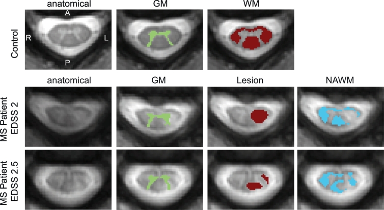
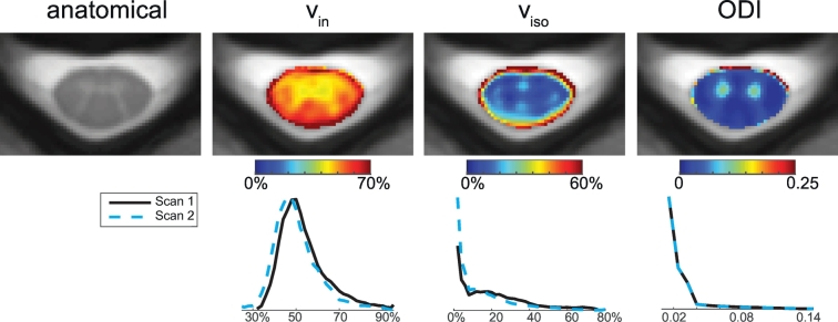
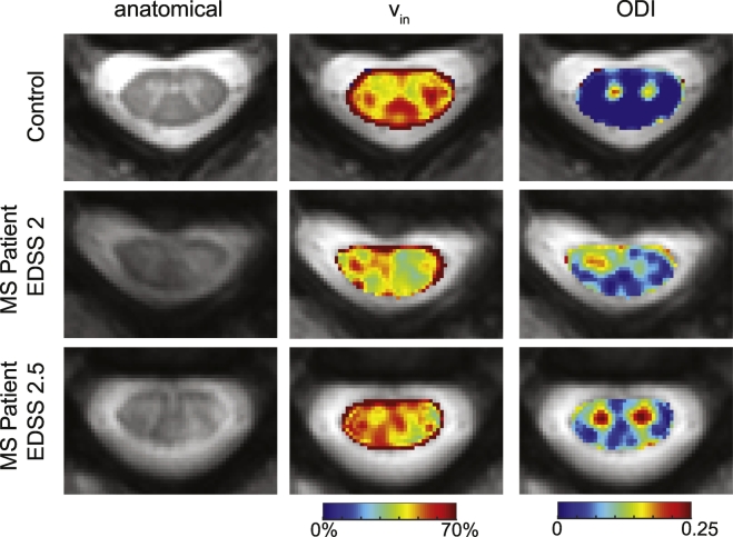
Introduction: There is a need to develop imaging methods sensitive to axonal injury in multiple sclerosis (MS), given the prominent impact of axonal pathology on disability and outcome. Advanced multi-compartmental diffusion models offer novel indices sensitive to white matter microstructure. One such model, neurite orientation dispersion and density imaging (NODDI), is sensitive to neurite morphology, providing indices of apparent volume fractions of axons (vin), isotropic water (viso) and the dispersion of fibers about a central axis (orientation dispersion index, ODI). NODDI has yet to be studied for its sensitivity to spinal cord pathology. Here, we investigate the feasibility and utility of NODDI in the cervical spinal cord of MS patients.

Methods: NODDI was applied in the cervical spinal cord in a cohort of 8 controls and 6 MS patients. Statistical analyses were performed to test the sensitivity of NODDI-derived indices to pathology in MS (both lesion and normal appearing white matter NAWM). Diffusion kurtosis imaging (DKI) and diffusion tensor imaging (DTI) analysis were also performed to compare with NODDI.

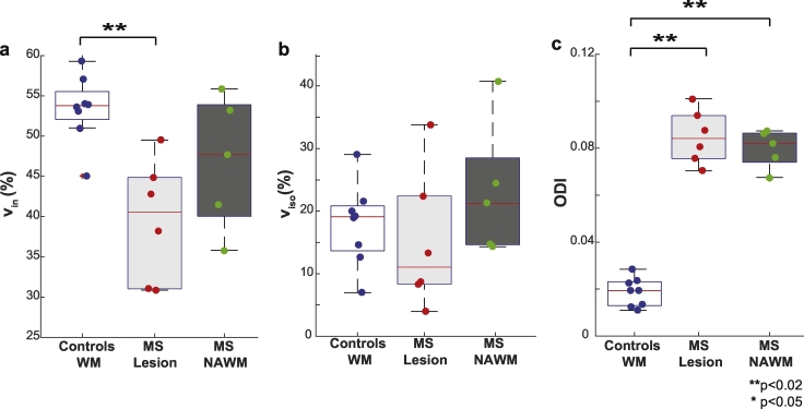
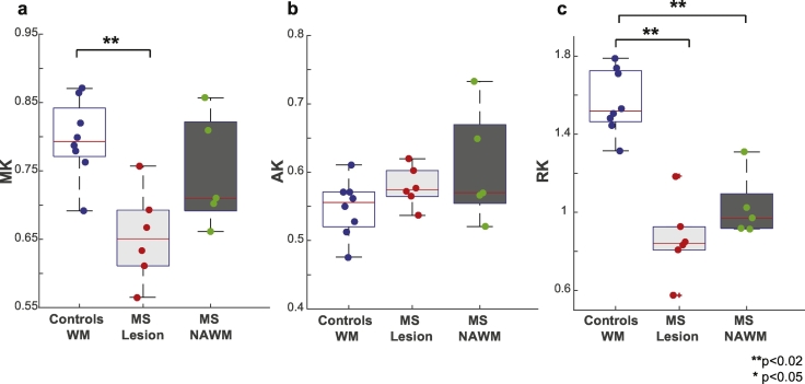
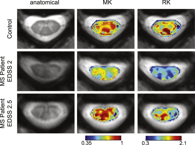
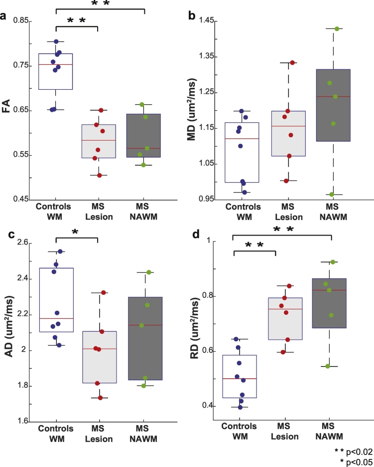
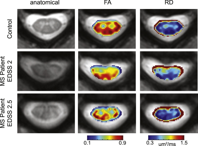
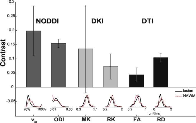
Results: A decrease in NODDI-derived vin was observed at the site of the lesion (p < 0.01), whereas a global increase in ODI was seen throughout white matter (p < 0.001). DKI-derived mean kurtosis (MK) and radial kurtosis (RK) and DTI-derived fractional anisotropy (FA) and radial diffusivity (RD) were all significantly different in MS patients (p < 0.02), however NODDI provided higher contrast between NAWM and lesion in all MS patients.

Conclusion: NODDI provides unique contrast that is not available with DKI or DTI, enabling improved characterization of the spinal cord in MS.

Keywords: Axon; Diffusion; Multiple sclerosis; NODDI; Spinal cord; Volume fraction.


Figures

Fig. 1
Fig. 1
Examples of ROI identification. Representative control (top row) includes the anatomical (left) from which the GM (second column) and WM (third column) were automatically segmented. For MS patients (middle and bottom row), the GM (second column) and WM are automatically segmented the same way as controls. WM, however, is separated into manually delineated lesions (third column) and any WM voxels containing no lesion was considered NAWM (fourth column). Note for all images the radiological coordinate system is used.
Fig. 2
Fig. 2
Maps and histograms of fitted parameters using NODDI in controls. Top row: Mean maps over all of the controls were calculated for the anatomical (left), vin, viso, and ODI (right). Bottom row: Histograms over all white matter voxels for scan 1 and scan 2 for vin, viso, and ODI (right).
Fig. 3
Fig. 3
Comparison of vin, viso, and ODI. Boxplots highlighting the median, 25th and 75th percentiles over controls (WM) and MS patients (lesions and NAWM) for (a) vin, (b) viso and (c) ODI. Mean values from each subject plotted and asterisks indicate significant differences between the groups, where double asterisks indicate significance after Bonferroni correction.
Fig. 4
Fig. 4
Example images from NODDI. Representative control is shown in the first row, followed by examples from two different MS patients (same as Fig. 1). From left to right, the anatomical, vin, viso, and ODI are shown.
Fig. 5
Fig. 5
Comparison of MK, AK and RK. Boxplots highlighting the median, 25th and 75th percentiles over controls (WM) and MS patients (lesions and NAWM) for (a) MK, (b) AK and (c) RK. Mean values from each subject plotted and asterisks indicate significant differences between the groups. Kurtosis metrics are unitless.
Fig. 6
Fig. 6
Example images from DKI. Representative control is shown in the first row, followed by examples from two MS patients. From left to right, the anatomical, MK and RK are shown. The same control and patients from Fig. 4 are included.
Fig. 7
Fig. 7
Comparison of FA, MD, AD and RD. Boxplots highlighting the median, 25th and 75th percentiles over controls (WM) and MS patients (lesions and NAWM) for (a) FA, (b) MD, (c) AD, and (d) RD. Mean values from each subject plotted and asterisks indicate significant differences between the groups.
Fig. 8
Fig. 8
Example images from DTI. Representative control is shown in the first row, followed by examples from two MS patients. From left to right, the anatomical, FA and RD are shown. The same control and patients from Fig. 4 are included.
Fig. 9
Fig. 9
Contrast in diffusion maps. Mean contrast between lesions and NAWM over all patients is shown, with error bars indicating the standard deviation over all of the patients.

References

    1. Alexander D.C. A general framework for experiment design in diffusion MRI and its application in measuring direct tissue-microstructure features. Magn. Reson. Med. 2008;60:439–448. - PubMed
    1. Asman A.J., Bryan F.W., Smith S.A., Reich D.S., Landman B.A. Groupwise multi-atlas segmentation of the spinal cord's internal structure. Med. Image Anal. 2014;18:460–471. - PMC - PubMed
    1. Assaf Y., Basser P.J. Composite hindered and restricted model of diffusion (CHARMED) MR imaging of the human brain. NeuroImage. 2005;27:48–58. - PubMed
    1. Assaf Y., Cohen Y. Assignment of the water slow-diffusing component in the central nervous system using q-space diffusion MRS: implications for fiber tract imaging. Magn. Reson. Med. 2000;43:191–199. - PubMed
    1. Assaf Y., Blumenfeld-Katzir T., Yovel Y., Basser P.J. AxCaliber: a method for measuring axon diameter distribution from diffusion MRI. Magn. Reson. Med. 2008;59:1347–1354. - PMC - PubMed

Publication types

MeSH terms

LinkOut - more resources