

Application of calibrated fMRI in Alzheimer's disease
- PMID: 28560160
- PMCID: PMC5443910
- DOI: 10.1016/j.nicl.2017.05.009
Application of calibrated fMRI in Alzheimer's disease
Abstract
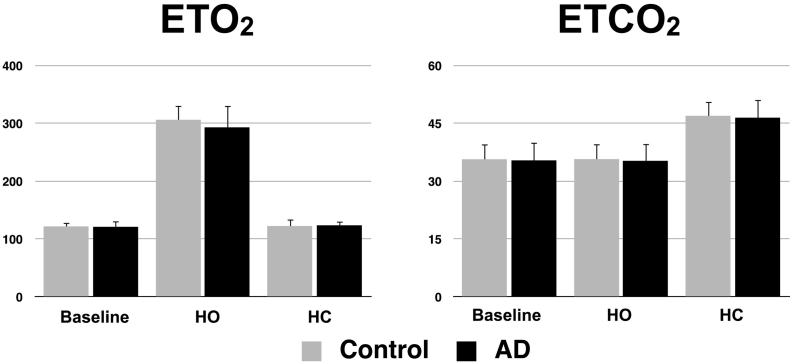
Calibrated fMRI based on arterial spin-labeling (ASL) and blood oxygen-dependent contrast (BOLD), combined with periods of hypercapnia and hyperoxia, can provide information on cerebrovascular reactivity (CVR), resting blood flow (CBF), oxygen extraction fraction (OEF), and resting oxidative metabolism (CMRO2). Vascular and metabolic integrity are believed to be affected in Alzheimer's disease (AD), thus, the use of calibrated fMRI in AD may help understand the disease and monitor therapeutic responses in future clinical trials. In the present work, we applied a calibrated fMRI approach referred to as Quantitative O2 (QUO2) in a cohort of probable AD dementia and age-matched control participants. The resulting CBF, OEF and CMRO2 values fell within the range from previous studies using positron emission tomography (PET) with 15O labeling. Moreover, the typical parietotemporal pattern of hypoperfusion and hypometabolism in AD was observed, especially in the precuneus, a particularly vulnerable region. We detected no deficit in frontal CBF, nor in whole grey matter CVR, which supports the hypothesis that the effects observed were associated specifically with AD rather than generalized vascular disease. Some key pitfalls affecting both ASL and BOLD methods were encountered, such as prolonged arterial transit times (particularly in the occipital lobe), the presence of susceptibility artifacts obscuring medial temporal regions, and the challenges associated with the hypercapnic manipulation in AD patients and elderly participants. The present results are encouraging and demonstrate the promise of calibrated fMRI measurements as potential biomarkers in AD. Although CMRO2 can be imaged with 15O PET, the QUO2 method uses more widely available imaging infrastructure, avoids exposure to ionizing radiation, and integrates with other MRI-based measures of brain structure and function.
Keywords: Alzheimer's disease; BOLD calibration constant; Calibrated fMRI; Cerebral blood flow; Cerebrovascular reactivity; Oxidative metabolism; Oxygen extraction fraction.
Figures


References
-
- Aliev G., Smith M.A., Obrenovich M.E., de la Torre J.C., Perry G. Role of vascular hypoperfusion-induced oxidative stress and mitochondria failure in the pathogenesis of Alzheimer disease. Neurotox. Res. 2003;5(7):491–504. - PubMed
-
- Alsop D.C., Detre J.A. Reduced transit-time sensitivity in noninvasive magnetic resonance imaging of human cerebral blood flow. J. Cereb. Blood Flow Metab. 1996;16(6):1236–1249. - PubMed
-
- Alsop D.C., Detre J.A., Grossman M. Assessment of cerebral blood flow in Alzheimer's disease by spin-labeled magnetic resonance imaging. Ann. Neurol. 2000;47(1):93–100. - PubMed
-
- Alsop D.C., Detre J.A., Golay X., Günther M., Hendrikse J., Hernandez-Garcia L., Lu H., MacIntosh B.J., Parkes L.M., Smits M., van Osch M.J.P., Wang D.J.J., Wong E.C., Zaharchuk G. Recommended implementation of arterial spin-labeled perfusion MRI for clinical applications: a consensus of the ISMRM perfusion study group and the European consortium for ASL in dementia. Magn. Reson. Med. 2015;73(1):102–116. - PMC - PubMed
Publication types
MeSH terms
Substances
Grants and funding
LinkOut - more resources
Full Text Sources
Other Literature Sources
Medical
