Review
. 2017 May;3(2):70-82.
doi: 10.1159/000458728. Epub 2017 Mar 8.

Dermoscopy in the Evaluation of Nail Disorders

Affiliations
Review

Dermoscopy in the Evaluation of Nail Disorders

Aurora Alessandrini et al. Skin Appendage Disord. 2017 May.

Abstract

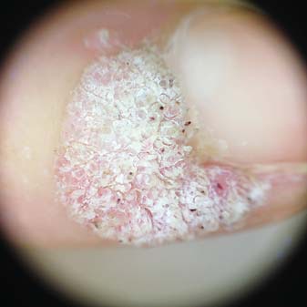
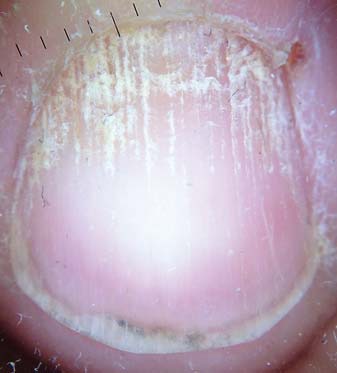
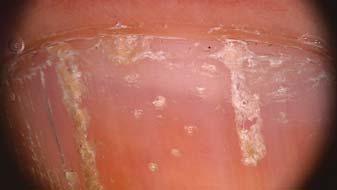
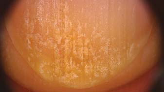
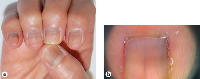
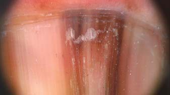
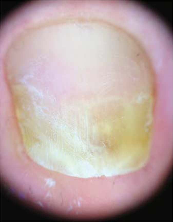
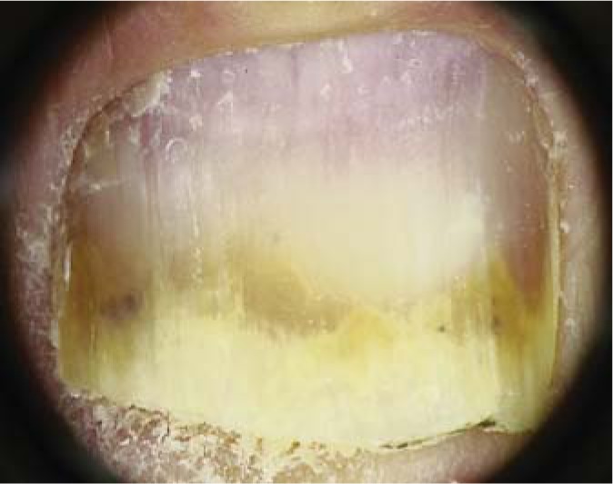
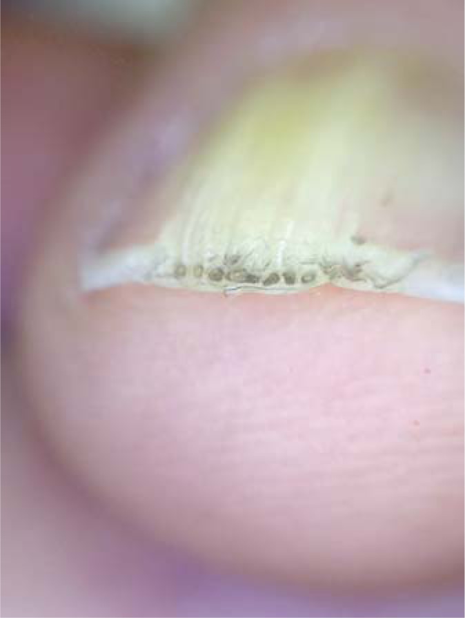
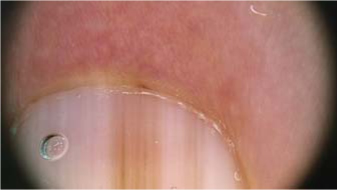
Nail dermoscopy was initially used only in the assessment of nail pigmentation, but now it is widely utilized for the evaluation of many nail disorders. In daily practice, dermoscopy may confirm clinical diagnoses and guides in the management of nail diseases and treatments, permitting a better visualization of symptoms. Dry dermoscopy is required for evaluation of the nail plate surface, while gel as an interface is necessary for assessment of nail pigmentation and onycholysis, as well as for the evaluation of the distal nail margin. In this review, we describe the dermoscopic features of the most important nail disorders, looking at the different areas of the nail. Dermatoscopic changes that usually accompany specific nail diseases are also reviewed.

Keywords: Capillary alterations; Dermoscopy; Hyponychium; Melanonychia; Nail tumor; Onycholysis; Onychoscopy; Splinter hemorrhages; Subungual hyperkeratosis.


Figures

Fig. 1
Fig. 1
Capillaroscopy in dermatomyositis: giant capillaries with arborescent appearance and microhemorrhages. Magnification, ×50.
Fig. 2
Fig. 2
Capillaroscopy in systemic lupus erythematosus: tortuous, oddly shaped loops with serpiginous appearance. Magnification, ×20.
Fig. 3
Fig. 3
Dermoscopy of pyogenic granuloma: reddish mass with irregular vessels and white central area. Magnification, ×10.
Fig. 4
Fig. 4
Dry dermoscopy of periungual warts: hyperkeratotic and rough lesion with small black dots. Magnification, ×10.
Fig. 5
Fig. 5
Dry dermoscopy easily enhances visualization of the longitudinal fissures of the nail plate due to lichen planus with partial anonychia and dorsal pterygium. Magnification, ×10.
Fig. 6
Fig. 6
Dry dermoscopy enhances visualization of the typical surface nail plate alterations with recent trachyonychia in the proximal nail plate. Magnification, ×10.
Fig. 7
Fig. 7
Dry dermoscopy of nail psoriasis with irregular pitting. Magnification, ×20.
Fig. 8
Fig. 8
Lamellar onychoschizia of a fingernail: the distal portion of the nail plate peels off in layers, and horizontal and vertical breakages are easily visible with dry dermoscopy. Magnification, ×20.
Fig. 9
Fig. 9
Dry dermoscopy shows thin fissures on the nail plate and opaqueness of the white spots of nail fragility due to superficial degranulation of the nail plate from chemicals. Magnification, ×20.
Fig. 10
Fig. 10
Dermoscopy with gel shows white bands within the nail plate in transverse leukonychia of the right toenail. Magnification, ×20.
Fig. 11
Fig. 11
The white area in the lunula of a proximal subungual onychomycosis is well visible by dermoscopy. Magnification, ×20.
Fig. 12
Fig. 12
Dermoscopy shows a nail plate with several small white, opaque, and friable patches in white superficial onychomycosis. Magnification, ×20.
Fig. 13
Fig. 13
Dermoscopy shows a bright green color that fades to yellow in case of onycholysis. Note the border of the subungual pigmentation, where the color typically fades into pale green at the margin of detachment. Magnification, ×10.
Fig. 14
Fig. 14
Dermoscopy of onychopapilloma presenting as a longitudinal red band associated with splinter hemorrhages. Magnification, ×10.
Fig. 15
Fig. 15
Dermoscopy of a glomus tumor with a deep red-purple subungual mass. Magnification, ×20.
Fig. 16
Fig. 16
Dry dermoscopy of the third fingernail of a left hand with alternance of multiple and parallel red and white bands in Darier disease. Magnification, ×10.
Fig. 17
Fig. 17
Dermoscopic aspect of distal subungual onycholysis, which is yellow-whitish in color due to the colony's formation and corresponds to a pattern of longitudinal striae. Magnification, ×10.
Fig. 18
Fig. 18
Dermoscopy of dermatophytoma: subungual accumulation of dermatophytes and scales, presenting as a round yellow-orange subungual area, connected by a thin band to the distal edge of the nail plate. Magnification, ×10.
Fig. 19
Fig. 19
Dermoscopy of a recent hematoma (a), presenting as a red-to-black discoloration of the fingernail and proximal fading of the red color, and of a late stage of the same subungual hematoma (b), presenting with a darker color. Magnification, ×10.
Fig. 20
Fig. 20
a Clinical picture of multiple nails affected by melanonychia due to drugs. b Dermoscopy of melanocytic activation, with a gray background of the band and thin grayish regular and parallel lines. b Magnification, ×20.
Fig. 21
Fig. 21
Dermoscopic pattern of a nevus in a 3-year-old child with the presence of a brown background with longitudinal brown-to-black irregular and parallel lines with irregular spacing and thickness. Magnification, ×20.
Fig. 22
Fig. 22
High magnification of the dermoscopic features of nail melanoma, with a brown-to-black background of the band and longitudinal lines irregular in thickness, spacing, color, and parallelism. Magnification, ×20.
Fig. 23
Fig. 23
Dermoscopy of fungal melanonychia with yellow streaks associated with homogeneous pigmentation and brown-black discoloration with irregular accumulation of black pigment and scales under the nail plate. Magnification, ×10.
Fig. 24
Fig. 24
Dermoscopy of the linear and regular margin of an onycholytic area due to traumatic onycholysis. Magnification, ×10.
Fig. 25
Fig. 25
Dermoscopy of nail psoriasis with onycholysis presenting a slightly dented margin of the onycholytic area surrounded by a yellow-orange band and splinter hemorrhages and irregular pitting. Magnification, ×10.
Fig. 26
Fig. 26
Typical dermoscopic aspect of onychomatricoma in the frontal view, with a honeycomb pattern. Magnification, ×10.
Fig. 27
Fig. 27
Dermoscopy of periungual tissue with the micro-Hutchinson sign and longitudinal lines irregular in thickness, spacing, color, and parallelism in a melanoma. Magnification, ×20.

Similar articles

  • Dermoscopic Nail Changes in Psoriasis, Lichen Planus, and Lichen Striatus.
    Sar-Pomian M, Starace MVR, Lencastre A, Piraccini BM, Richert B, Rudnicka L, Trakatelli MG, Iorizzo M. Sar-Pomian M, et al. Skin Appendage Disord. 2024 Aug;10(4):273-292. doi: 10.1159/000538581. Epub 2024 May 8. Skin Appendage Disord. 2024. PMID: 39021761 Free PMC article. Review.
  • Dermoscopy of the Nail Unit.
    Starace M, Alessandrini A, Piraccini BM. Starace M, et al. Dermatol Clin. 2021 Apr;39(2):293-304. doi: 10.1016/j.det.2020.12.008. Epub 2021 Feb 10. Dermatol Clin. 2021. PMID: 33745641 Review.
  • Onychoscopy: Dermoscopy of the Nails.
    Piraccini BM, Alessandrini A, Starace M. Piraccini BM, et al. Dermatol Clin. 2018 Oct;36(4):431-438. doi: 10.1016/j.det.2018.05.010. Dermatol Clin. 2018. PMID: 30201152 Review.
  • Clinical, dermoscopic, and pathologic features of onychopapilloma: A review of 47 cases.
    Tosti A, Schneider SL, Ramirez-Quizon MN, Zaiac M, Miteva M. Tosti A, et al. J Am Acad Dermatol. 2016 Mar;74(3):521-6. doi: 10.1016/j.jaad.2015.08.053. Epub 2015 Oct 27. J Am Acad Dermatol. 2016. PMID: 26518173 Review.
  • Onychoscopy.
    Sathe NC, Saleh HM. Sathe NC, et al. 2024 Oct 6. In: StatPearls [Internet]. Treasure Island (FL): StatPearls Publishing; 2025 Jan–. 2024 Oct 6. In: StatPearls [Internet]. Treasure Island (FL): StatPearls Publishing; 2025 Jan–. PMID: 36256746 Free Books & Documents.

Cited by

References

    1. Piraccini BM, Bruni F, Starace M. Dermoscopy of non-skin cancer nail disorders. Dermatol Ther. 2012;25:594–602. - PubMed
    1. Hasegawa M. Dermoscopy findings of nail fold capillaries in connective tissue disease. J Dermatol. 2011;38:66–70. - PubMed
    1. Cutolo M, Sulli A, Secchi ME, Oliveri M, Pizzorni C. The contribution of capillaroscopy to the differential diagnosis of connective autoimmune diseases. Best Pract Res Clin Rheumatol. 2007;21:1093–1108. - PubMed
    1. Pizzorni C, Sulli A, Smith V, Lladó A, Paolino S, Cutolo M, Ruaro B. Capillaroscopy in 2016: new perspectives in systemic sclerosis. Acta Rheumatol Port. 2016;41:8–14. - PubMed
    1. Shenavandeh S, Zarei Nezhad M. Association of nailfold capillary changes with disease activity, clinical and laboratory findings in patients with dermatomyositis. Med J Islam Repub Iran. 2015;29:233. - PMC - PubMed

LinkOut - more resources