. 2017 May 13:13:69-76.
doi: 10.1016/j.dib.2017.05.026. eCollection 2017 Aug.

Data demonstrating the anti-oxidant role of hemopexin in the heart

Affiliations

Data demonstrating the anti-oxidant role of hemopexin in the heart

Giada Ingoglia et al. Data Brief. .

Abstract

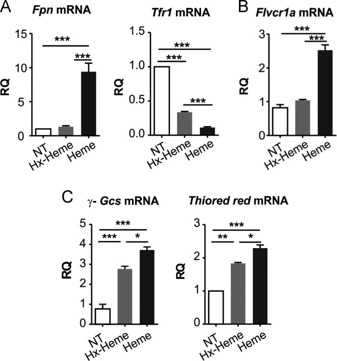
The data presented in this article are related to the research article entitled Hemopexin counteracts systolic dysfunction induced by heme-driven oxidative stress (G. Ingoglia, C. M. Sag, N. Rex, L. De Franceschi, F. Vinchi, J. Cimino, S. Petrillo, S. Wagner, K. Kreitmeier, L. Silengo, F. Altruda, L. S. Maier, E. Hirsch, A. Ghigo and E. Tolosano, 2017) [1]. Data show that heme induces reactive oxygen species (ROS) production in primary cardiomyocytes. H9c2 myoblastic cells treated with heme bound to human Hemopexin (Hx) are protected from heme accumulation and oxidative stress. Similarly, the heme-driven oxidative response is reduced in primary cardiomyocytes treated with Hx-heme compared to heme alone. Our in vivo data show that mouse models of hemolytic disorders, β-thalassemic mice and phenylhydrazine-treated mice, have low serum Hx associated to enhanced expression of heme- and oxidative stress responsive genes in the heart. Hx-/- mice do not show signs of heart fibrosis or overt inflammation. For interpretation and discussion of these data, refer to the research article referenced above.

Keywords: Heart; Heme; Hemopexin; Oxidative stress.


Figures

Fig. 1
Fig. 1
Heme promotes ROS formation in isolated adult rat cardiomyocytes. Data on isolated adult rat cardiomyocytes exposed to heme (5 µM) or vehicle (not-treated, Nt) are shown. ROS were measured by using the fluorescent dye CM-H2DCFDA (Nt, n = 22; heme, n = 17). Two-way ANOVA with Bonferroni post-test analysis was performed. *P < 0.05; #P < 0.05 (#, difference between Nt and heme-treated cells; *, difference between time 0 and time 11 in Nt and heme-treated cells).
Fig. 2
Fig. 2
Hemopexin protects H9c2 cells from heme accumulation and ROS production. Data on H9c2 myoblasts cell line untreated (NT) or treated with either 10 µM Hx-heme complex or 10 µM heme for 8 hours, are shown. (A) Heme content. (B) qRT-PCR analysis of Ho-1 mRNA levels. (C) Western blot analysis of HO-1. (D) ROS content and (E) qRT-PCR analysis of γ-Glutamylcysteine synthetase (γ-Gcs) and Thioredoxin mRNA levels. (F) Western blot analysis of N-Tyr. (G) Immunofluorescence analysis of super-oxide radical formation (super-oxide radical was stained with Mito-sox fluorescent probe. Nuclei were stained with DAPI). Results shown are representative of three independent experiments. One-way analysis of variance with Bonferroni post-test analysis was performed. *P < 0.05; **P < 0.01; ***P < 0.001. Values represent mean ± SEM. AU, arbitrary units; RQ, relative quantity; FIU, fluorescence intensity unit.
Fig. 3
Fig. 3
Hemopexin protects neonatal cardiomyocytes and H9c2 cells from heme accumulation and ROS formation. Data on neonatal cardiomyocytes and H9c2 cells untreated (NT) or treated with either 10 µM Hx-heme complex or 10 µM heme for 8 hours, are shown. (A, C) qRT-PCR analysis of Fpn, Tfr1, γ-Gcs and Thioredoxin reductase mRNA levels of neonatal cardiomyocytes. (B) qRT-PCR analysis of Flvcr1a mRNA levels of H9c2 cells. One-way analysis of variance with Bonferroni post-test analysis was performed. *P < 0.05; **P < 0.01; ***P < 0.001.
Fig. 4
Fig. 4
Hemopexin preserves heme homeostasis in the heart. Data on the heart of wild-type (Wt) and Hx-/- mice are shown. (A) qRT-PCR analysis of Flvcr1a, Fpn, Dmt1 and Tfr1 mRNA levels. (B) Western blot analysis of Tfr1 protein. Results shown are representative of 3 independent experiments. In B, each lane represents an individual animal; E-cadherin (E-Cad) was used as loading control. Unpaired t-test analysis with Welch׳s correction was performed. Values represent mean ± SEM. *P<0.05.
Fig. 5
Fig. 5
Hemopexin loss is not associated with heart fibrosis. Data on Wt and Hx-/- mice are shown. (A) Representative Picrosirius Red staining of heart sections from a Wt and an Hx-/- mouse. ImageJ analysis of Picrosirius Red stained sections is shown on the right. (B) Immunohistochemistry analysis of CD18 expression on heart sections of a Wt and a Hx-/- mouse. (C) qRT-PCR analysis of collagen type I and III, Tnf-α and IL6 mRNA levels in the heart (n = 5). Unpaired t-test analysis with Welch׳s correction was performed. Values represent mean ± SEM. *P<0.05.
Fig. 6
Fig. 6
β-thalassemic mice are hemolytic and accumulate heme in the heart. Data on Wt and β-thalassemic (β-Thal) mice are shown. (A) ELISA quantification of serum Hx. (B) qRT-PCR analysis of Ho-1, Fpn and γ-Gcs mRNA levels in the heart. (C) HO-1 western blot analysis. Unpaired t-test analysis with Welch׳s correction was performed. P < 0.05; ⁎⁎⁎P < 0.001. Values represent mean ± SEM.
Fig. 7
Fig. 7
PHZ-treated mice show an alteration of heme- and oxidative stress-responsive genes in the heart. (A) Western blot of serum Hx of untreated (0) or PHZ-treated Wt mice at 1, 2 or 4 weeks of treatment. (B) qRT-PCR analysis of Ho-1, Flvcr1a, Fpn, Gsr, mRNA levels in the heart of untreated or PHZ-injected mice after 4 weeks of treatment. In A, one-way ANOVA with Bonferroni post-test analysis was performed; in B, unpaired t-test analysis with Welch׳s correction was performed. *P < 0.05; ** P < 0.01; ***P < 0.001.
Fig. 8
Fig. 8
α-tocopherol protects the heart against PHZ-mediated oxidative stress. Data on the heart of PHZ-treated Wt mice administered or not with α-tocopherol are shown. (A) qRT-PCR analysis of γ-Gcs and peroxiredoxin 6 (Prdx6) mRNA levels of PHZ- and PHZ-α-tocopherol-treated mice (n=7) 4 weeks after the treatment. One-way analysis of variance with Bonferroni post-test analysis were performed. P < 0.05; ⁎⁎⁎P < 0.001. Values represent mean ± SEM. RQ, relative quantity.

References

    1. Ingoglia G., Sag C.M., Rex N., De Franceschi L., Vinchi F., Cimino J., Petrillo S., Wagner S., Kreitmeier K., Silengo L., Altruda F., Maier L.S., Hirsch E., Ghigo A., Tolosano E. Hemopexin counteracts systolic dysfunction induced by heme-driven oxidative stress. Free Radic Biol Med. 2017;108:452–464. - PubMed
    1. Tolosano E., Hirsch E., Patrucco E., Camaschella C., Navone R., Silengo L., Altruda F. Defective recovery and severe renal damage after acute hemolysis in hemopexin-deficient mice. Blood. 1999;94(11):3906–3914. - PubMed
    1. Fiorito V., Geninatti Crich S., Silengo L., Aime S., Altruda F., Tolosano E. Lack of Plasma Protein Hemopexin Results in Increased Duodenal Iron Uptake. PLoS One. 2013;8(6):e68146. - PMC - PubMed
    1. Vinchi F., Gastaldi S., Silengo L., Altruda F., Tolosano E. Hemopexin prevents endothelial damage and liver congestion in a mouse model of heme overload. Am J Pathol. 2008;173(1):289–299. - PMC - PubMed
    1. Franco S.S., De Falco L., Ghaffari S., Brugnara C., Sinclair D.A., Matte׳ A., Iolascon A., Mohandas N., Bertoldi M., An X., Siciliano A., Rimmelé P., Cappellini M.D., Michan S., Zoratti E., Anne J., Franceschi L. De. Resveratrol accelerates erythroid maturation by activation of FoxO3 and ameliorates anemia in beta-thalassemic mice. Haematologica. 2014;99(2):267–275. - PMC - PubMed