. 2017 Jul;40(1):182-192.
doi: 10.3892/ijmm.2017.3008. Epub 2017 May 31.

The PI3K/Akt/mTOR pathway is involved in CVB3-induced autophagy of HeLa cells

Affiliations

The PI3K/Akt/mTOR pathway is involved in CVB3-induced autophagy of HeLa cells

Huan Chang et al. Int J Mol Med. 2017 Jul.

Abstract

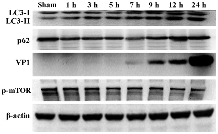
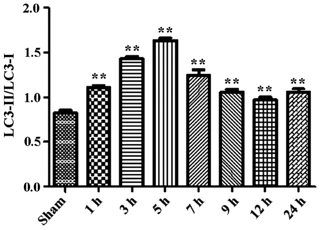
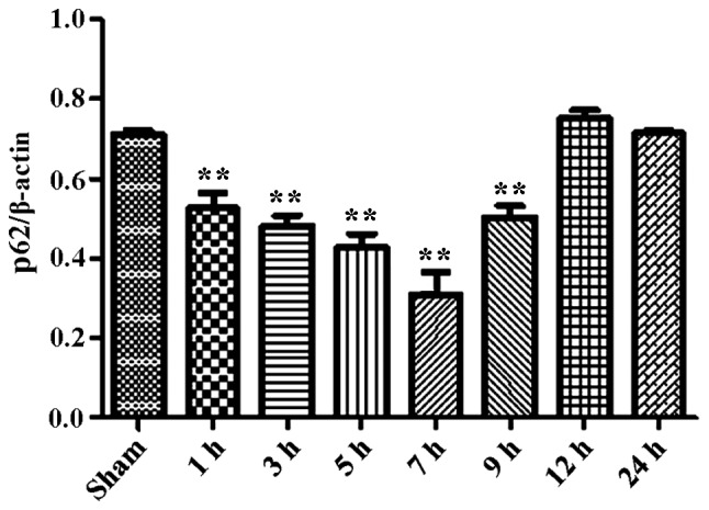
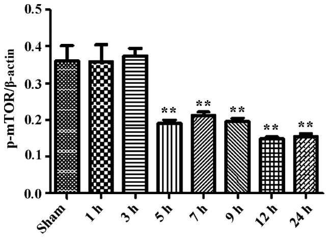
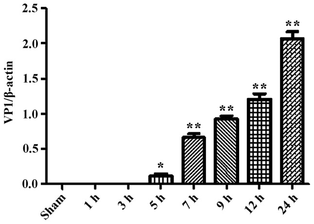
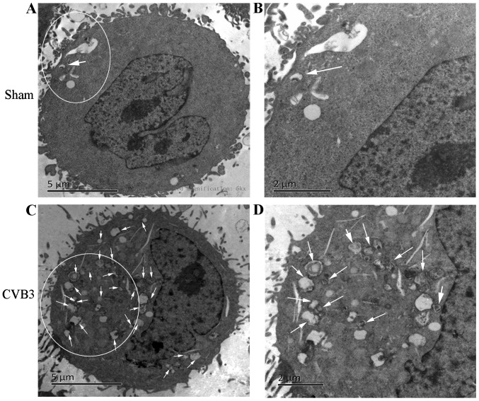
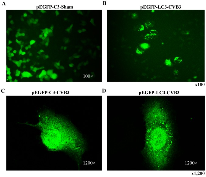
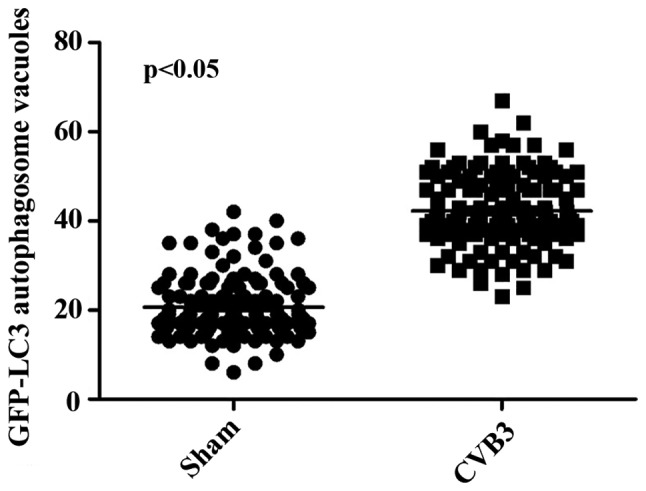
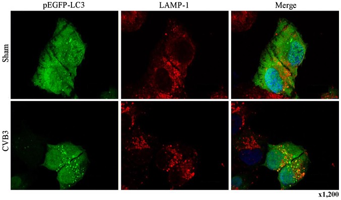
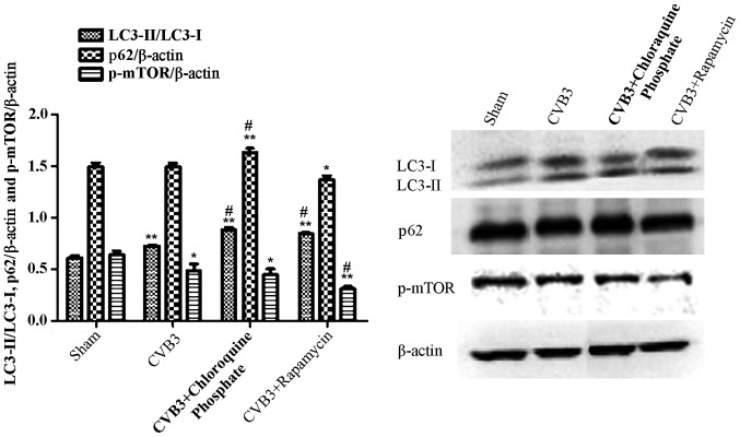
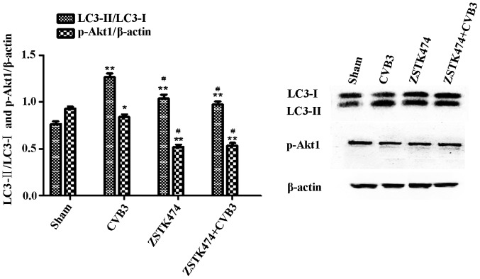
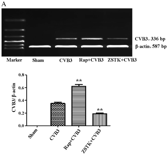
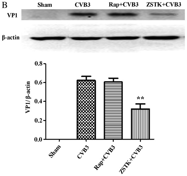
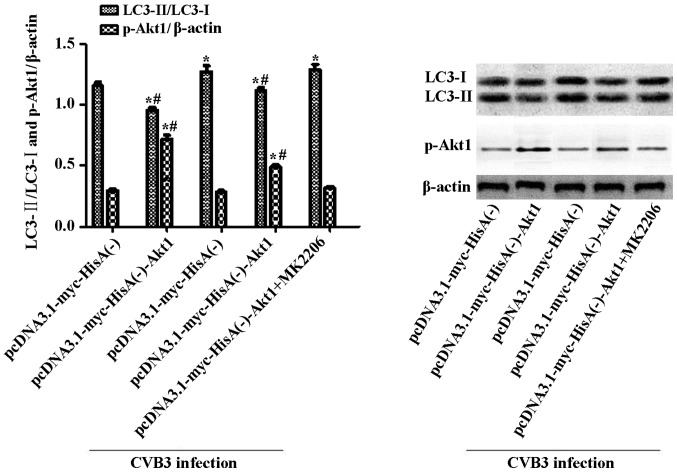
Recent studies have found that viral myocarditis (VMC) associated with coxsackievirus B3 (CVB3) causes autophagy activation after infection, but the specific mechanism is not clear. The present study demonstrated that the phosphatidylinositol 3-kinase (PI3K)/protein kinase B (PKB)/mammalian target of rapamycin (mTOR) signaling pathway participates in CVB3‑induced autophagy. We found that the light chain 3 (LC3)‑Ⅱ/LC3‑I ratio was increased and p62 and p‑mTOR were altered at different times during CVB3 infection. To further assess the effects of this signaling pathway on CVB3 infection and viral replication, we selected 24 h post‑inoculation (h.p.i.) as our research time point to conduct our next study. We inhibited the function of PI3K, Akt1 and mTOR. The outcome showed that inhibition of PI3K with ZSTK474 alleviated autophagy and decreased CVB3 mRNA replication and VP1 expression. Inhibition of mTOR with rapamycin promoted autophagy and viral mRNA replication but did not impact VP1 expression. Inhibition of Akt with MK2206 aggravated autophagy induced by viral infection. In our research, p62 exhibited a decrease at the beginning of infection but then increased as infection time increased. This finding may serve as a clue to elucidate the function of autophagy at different times of infection. However, the details merit further study. In conclusion, our findings suggest that the PI3K/Akt/mTOR signaling pathway participates in the process of autophagy induced by CVB3 infection. This finding may provide a new perspective of CVB3-induced autophagy.


Figures

Figure 1
Figure 1
CVB3 induces an autophagic response at different times during CVB3 infection in HeLa cells. HeLa cells were infected with CVB3, cell lysates were collected at 1, 3, 5, 7, 9, 12 and 24 h.p.i., and mock-infected HeLa cells served as the sham control. LC3, p62, p-mTOR and viral capsid protein VP1 were examined by western blot analysis, and these samples were immunoblotted with an antibody to β-actin to illustrate equal protein loading. Compared with the sham group, the LC3-II/LC3-I ratio increased at 1 h.p.i. then peaked at 5 h.p.i. (p<0.05). p62 exhibited a decrease at 1, 3, 5, 7 and 9 h.p.i. (p<0.01). No changes were noted at 12 and 24 h.p.i. compared with the sham group. p-mTOR showed a decrease after 5 h.p.i. (p<0.05), which was more obvious at 12 and 24 h.p.i. VP1 increased as the time of infection increased. *P<0.05, compared with the sham group; **p<0.01, compared with the sham group. CVB3, coxsackievirus B3; h.p.i., hours post-inoculation; LC3, light chain 3; mTOR, mammalian target of rapamycin.
Figure 1
Figure 1
CVB3 induces an autophagic response at different times during CVB3 infection in HeLa cells. HeLa cells were infected with CVB3, cell lysates were collected at 1, 3, 5, 7, 9, 12 and 24 h.p.i., and mock-infected HeLa cells served as the sham control. LC3, p62, p-mTOR and viral capsid protein VP1 were examined by western blot analysis, and these samples were immunoblotted with an antibody to β-actin to illustrate equal protein loading. Compared with the sham group, the LC3-II/LC3-I ratio increased at 1 h.p.i. then peaked at 5 h.p.i. (p<0.05). p62 exhibited a decrease at 1, 3, 5, 7 and 9 h.p.i. (p<0.01). No changes were noted at 12 and 24 h.p.i. compared with the sham group. p-mTOR showed a decrease after 5 h.p.i. (p<0.05), which was more obvious at 12 and 24 h.p.i. VP1 increased as the time of infection increased. *P<0.05, compared with the sham group; **p<0.01, compared with the sham group. CVB3, coxsackievirus B3; h.p.i., hours post-inoculation; LC3, light chain 3; mTOR, mammalian target of rapamycin.
Figure 2
Figure 2
LC3 and p-mTOR-are altered in HeLa cells at 24 h.p.i. HeLa cells were infected with CVB3. DMEM containing 2% FBS served as the sham control, and rapamycin (Rap) (100 µM) served as a positive control. At 24 h.p.i. cells were collected for western blot analysis. Compared with the sham group, the total amount of LC3 (LC3-I, LC3-II) and the ratio of LC3-II/LC3-I were markedly increased in the infected cells at 24 h.p.i. (p<0.05). The expression of p-mTOR was downregulated (p<0.05). *p<0.05, compared with the sham group; **p<0.01, compared with the sham group. LC3, light chain 3; mTOR, mammalian target of rapamycin; h.p.i., hours post-inoculation; CVB3, coxsackievirus B3; DMEM, Dulbecco's modified Eagle's medium; FBS, fetal bovine serum.
Figure 3
Figure 3
TEM analysis of HeLa cells at 24 h.p.i. HeLa cells were collected at 24 h.p.i. and subjected to electron microscopic observation. Abundant autopha-gosome-like vesicles with double membranes (0.2–0.5 µm in size) clustered in the CVB3-infected group compared with the control group (white arrows). (A and B) The normal structure in the sham group. (C and D) The changes in CVB3-infected HeLa cells. Arrows indicate representative autophagosomes that would be scored positive in (B), with double membranes 0.2–0.5 µm in size. TEM, transmission electron microscopy; h.p.i., hours post-inoculation; CVB3, coxsackievirus B3.
Figure 4
Figure 4
LC3 puncta formation is increased in the HeLa cells at 24 h.p.i. HeLa cells were transfected with plasmid pEGFP-LC3 or the empty vector (pEGFP-C3) using Lipofectamine 2000 (Invitrogen Life Technologies). Twenty-four hours later, fluorescence microscopy demonstrated the successful transduction of cells with pEGFP-LC3 and pEGFP-C3. Then, the cells were seeded in 6-well plates. (B-D) Twenty-four hours later, the cells were infected with CVB3, and (A) mock-infected cells served as the control group. LC3, light chain 3; h.p.i., hours post-inoculation; CVB3, coxsackievirus B3.
Figure 5
Figure 5
Quantification of GFP-LC3 dots in the sham group and infected group. LC3, light chain 3.
Figure 6
Figure 6
Co-localization of GFP-LC3 and LAMP-1 in HeLa cells at 24 h.p.i. HeLa cells were transfected with the pEGFP-LC3 plasmid for 24 h followed by CVB3 infection. Twenty-four hours later, the transfected cells were stained with an antibody specific for the endosomal/lysosomal marker LAMP-1, and the co-localization of GFP-LC3 and LAMP-1 vesicles was examined by confocal microscopy. Single-color fluorescence images of pEGFP-LC3 and LAMP-1 are presented in the 1st and 2nd columns, and a merged view of these two proteins is shown in the 3rd column. A yellow signal signifies co-localization of pEGFP-LC3 and LAMP-1. LC3, light chain 3; LAMP-1, lysosomal-associated membrane protein 1; h.p.i., hours post-inoculation; CVB3, coxsackievirus B3.
Figure 7
Figure 7
Rapamycin aggravates the autophagic reaction induced by CVB3 infection. HeLa cells were treated with 10 nM rapamycin and chloroquine phosphate (CQ group) for 2 h. The cells were washed thrice and infected with CVB3. In the CVB3 group, HeLa cells were incubated with DMEM containing 10% FBS for 2 h and infected with CVB3. The sham group was incubated with DMEM containing 10% FBS. Two hours later, the medium was refreshed with DMEM containing 2% FBS. At 24 h.p.i., cells were collected for western blot analysis. The LC3-II/LC3-I ratio was increased in the CVB3 and rapamycin group (p<0.01), and p-mTOR was decreased in the CVB3 group (p<0.05) and rapamycin group (p<0.01) compared with the sham group. The changes in the LC3-II/LC3-I ratio and p-mTOR were more obvious in the Rap group compared with the CVB3 group (p<0.05). The LC3-II/LC3-I ratio and p62 was increased in the CQ group compared with the CVB3 group (p<0.05). *p<0.05, compared with the sham group; **p<0.01, compared with the sham group; #p<0.05, compared with the CVB3 group. CVB3, coxsackievirus B3; DMEM, Dulbecco's modified Eagle's medium; FBS, fetal bovine serum; h.p.i., hours post-inoculation; LC3, light chain 3; mTOR, mammalian target of rapamycin.
Figure 8
Figure 8
Inhibition of PI3K with ZSTK474 alleviates the autophagic reaction caused by CVB3 infection. HeLa cells were pre-treated with ZSTK474 (50 µM, 2 h) and then infected with CVB3 (ZSTK4747 + CVB3 group) or not (CVB3 group). Mock-infected cells served as the sham control. Cell lysates were collected at 24 h.p.i., and the expression of LC3 and p-Akt1 was determined by western blot analysis. Compared with the sham group, p-Akt1 was decreased in the CVB3 group (p<0.05) and the ZSTK474 group (p<0.01). The LC3-II/LC3-I ratio was increased (p<0.01). The LC3-II/LC3-I ratio was increased in the CVB3 group compared with the ZSTK474 + CVB3 group (p<0.05). *p<0.05, compared with the sham group; **p<0.01, compared with the sham group; #p<0.05, compared with the CVB3 group. PI3K, phosphatidylinositol 3-kinase; CVB3, coxsackievirus B3; h.p.i., hours post-inoculation; LC3, light chain 3; p-Akt1, phosphorylated Akt1.
Figure 9
Figure 9
Rapamycin and ZSTK474 affect viral replication. HeLa cells pre-treated with rapamycin (Rap) (10 nM) and ZSTK474 (ZSTK) (50 µM) were infected with CVB3, and we examined (A) mRNA and (B) viral capsid protein VP1 by semi-quantitative PCR and western blot analysis, respectively. Compared with the CVB3 group, CVB3 mRNA and VP1 expression were decreased in the ZSTK474 group (p<0.01). Rapamycin stimulated viral mRNA synthesis caused by viral infection (p<0.01). **p<0.01, compared with the sham group. CVB3, coxsackievirus B3.
Figure 9
Figure 9
Rapamycin and ZSTK474 affect viral replication. HeLa cells pre-treated with rapamycin (Rap) (10 nM) and ZSTK474 (ZSTK) (50 µM) were infected with CVB3, and we examined (A) mRNA and (B) viral capsid protein VP1 by semi-quantitative PCR and western blot analysis, respectively. Compared with the CVB3 group, CVB3 mRNA and VP1 expression were decreased in the ZSTK474 group (p<0.01). Rapamycin stimulated viral mRNA synthesis caused by viral infection (p<0.01). **p<0.01, compared with the sham group. CVB3, coxsackievirus B3.
Figure 10
Figure 10
Inhibition of Akt1 aggravates the autophagic response caused by CVB3 infection in Akt1-overexpressing cells. Cells overexpressing Akt1 or harboring empty vector alone were infected with CVB3 or not. Cells were collected for western blot analysis at 24 h.p.i. To suppress the overexpression of Akt1, MK2206 (50 µM) was used to pre-treat the pcDNA3.1-myc-HisA(−)-Akt1 HeLa cells. Two hours later, the cells were infected and harvested at 24 h.p.i., and mock-infected cells served as the sham control. In Akt1-overexpressing cell lines, CVB3 infection increased the LC3-II/LC3-I ratio (p<0.05). The ratio was more significantly increased when the infected cells were pre-treated with MK2206 (p<0.05). Under uninfected conditions, the LC3-II/LC3-I ratio was higher in Akt1-overexpressing cells compared with the empty vector cells (p<0.05). *p<0.05, compared with the pcDNA3.1-myc-HisA(−) group; #p<0.05, compared with the pcDNA3.1-myc-HisA(−)-Akt1+CVB3 group. CVB3, coxsackievirus B3; h.p.i., hours post-inoculation; LC3, light chain 3.

References

    1. Yajima T, Knowlton KU. Viral myocarditis: From the perspective of the virus. Circulation. 2009;119:2615–2624. doi: 10.1161/CIRCULATIONAHA.108.766022. - DOI - PubMed
    1. Yajima T. Viral myocarditis: Potential defense mechanisms within the cardiomyocyte against virus infection. Future Microbiol. 2011;6:551–566. doi: 10.2217/fmb.11.40. - DOI - PMC - PubMed
    1. Bowles NE, Ni J, Kearney DL, Pauschinger M, Schultheiss HP, McCarthy R, Hare J, Bricker JT, Bowles KR, Towbin JA. Detection of viruses in myocardial tissues by polymerase chain reaction: Evidence of adenovirus as a common cause of myocarditis in children and adults. J Am Coll Cardiol. 2003;42:466–472. doi: 10.1016/S0735-1097(03)00648-X. - DOI - PubMed
    1. Esfandiarei M, McManus BM. Molecular biology and pathogenesis of viral myocarditis. Annu Rev Pathol. 2008;3:127–155. doi: 10.1146/annurev.pathmechdis.3.121806.151534. - DOI - PubMed
    1. Sin J, Mangale V, Thienphrapa W, Gottlieb RA, Feuer R. Recent progress in understanding coxsackievirus replication, dissemination, and pathogenesis. Virology. 2015;484:288–304. doi: 10.1016/j.virol.2015.06.006. - DOI - PMC - PubMed

MeSH terms

Substances