. 2017 Jul;40(1):164-174.
doi: 10.3892/ijmm.2017.3005. Epub 2017 May 29.

Collagen-derived N-acetylated proline-glycine-proline upregulates the expression of pro-inflammatory cytokines and extracellular matrix proteases in nucleus pulposus cells via the NF-κB and MAPK signaling pathways

Affiliations

Collagen-derived N-acetylated proline-glycine-proline upregulates the expression of pro-inflammatory cytokines and extracellular matrix proteases in nucleus pulposus cells via the NF-κB and MAPK signaling pathways

Chencheng Feng et al. Int J Mol Med. 2017 Jul.

Abstract

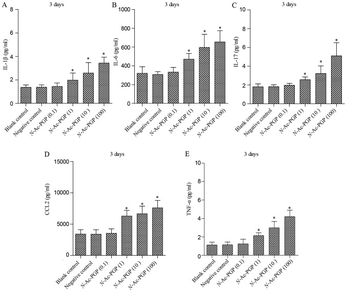
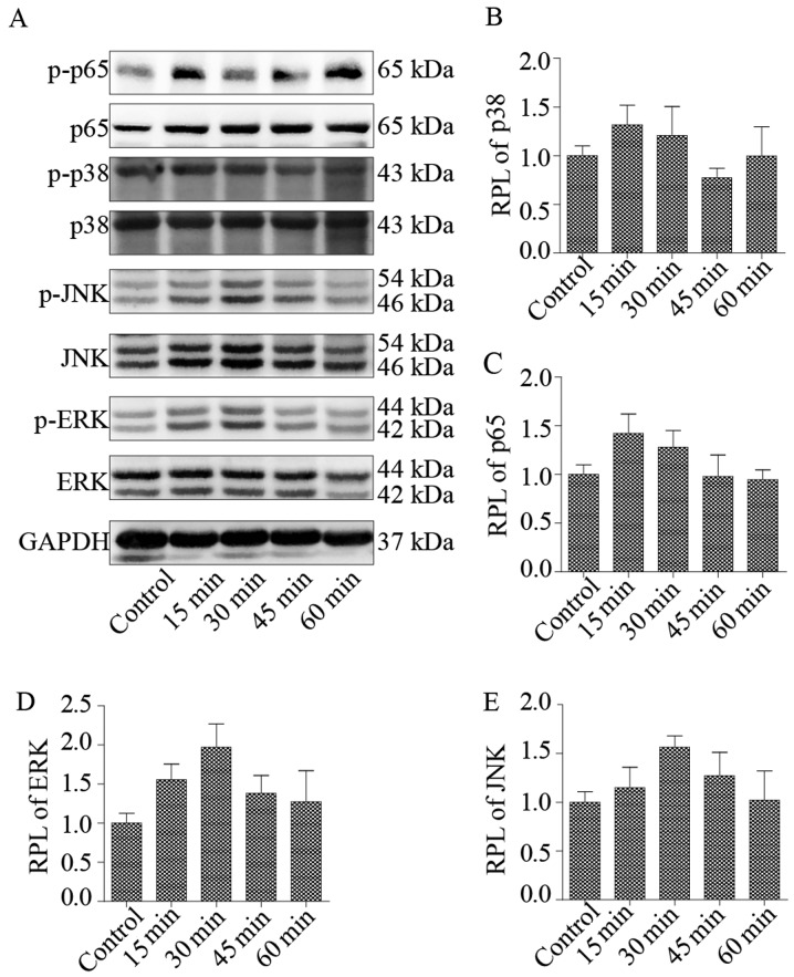
N-acetylated proline-glycine-proline (N-Ac-PGP) is a chemokine involved in inflammatory diseases and is found to accumulate in degenerative discs. N-Ac-PGP has been demonstrated to have a pro-inflammatory effect on human cartilage endplate stem cells. However, the effect of N-Ac-PGP on human intervertebral disc cells, especially nucleus pulposus (NP) cells, remains unknown. The purpose of this study was to investigate the effect of N-Ac-PGP on the expression of pro-inflammatory factors and extracellular matrix (ECM) proteases in NP cells and the molecular mechanism underlying this effect. Therefore, Milliplex assays were used to detect the levels of various inflammatory cytokines in conditioned culture medium of NP cells treated with N-Ac-PGP, including interleukin-1β (IL-1β), IL-6, IL-17, tumor necrosis factor-α (TNF-α) and C-C motif ligand 2 (CCL2). RT-qPCR was also used to determine the expression of pro-inflammatory cytokines and ECM proteases in the NP cells treated with N-Ac-PGP. Moreover, the role of nuclear factor-κB (NF-κB) and mitogen-activated protein kinase (MAPK) signaling pathways in mediating the effect of N-Ac-PGP on the phenotype of NP cells was investigated using specific signaling inhibitors. Milliplex assays showed that NP cells treated with N-Ac-PGP (10 and 100 µg/ml) secreted higher levels of IL-1β, IL-6, IL-17, TNF-α and CCL2 compared with the control. RT-qPCR assays showed that NP cells treated with N-Ac-PGP (100 µg/ml) had markedly upregulated expression of matrix metalloproteinase 3 (MMP3), MMP13, a disintegrin and metalloproteinase with thrombospondin motif 4 (ADAMTS4), ADAMTS5, IL-6, CCL-2, CCL-5 and C-X-C motif chemokine ligand 10 (CXCL10). Moreover, N-Ac-PGP was shown to activate the MAPK and NF-κB signaling pathways in NP cells. MAPK and NF-κB signaling inhibitors suppressed the upregulation of proteases and pro-inflammatory cytokines in NP cells treated with N-Ac-PGP. In conclusion, N-Ac-PGP induces the expression of pro-inflammatory cytokines and matrix catabolic enzymes in NP cells via the NF-κB and MAPK signaling pathways. N-Ac-PGP is a novel therapeutic target for intervertebral disc degeneration.


Figures

Figure 1
Figure 1
Secretion of pro-inflammatory cytokines by nucleus pulposus (NP) cells treated with N-acetylated proline-glycine-proline (N-Ac-PGP) for 1 day. (A–E) The levels of interleukin-1β (IL-1β), IL-6, IL-17, C-C motif ligand 2 (CCL2) and tumor necrosis factor-α (TNF-α) in the conditioned culture media of NP cells treated with N-Ac-PGP. NP cells without any treatment served as the blank control. NP cells treated with phosphate-buffered saline (PBS) served as the negative control. The results are presented as the mean ± SEM. *P<0.05 vs. the blank control. 0.1, 0.1 µg/ml; 1, 1 µg/ml; 10, 10 µg/ml; 100, 100 µg/ml.
Figure 2
Figure 2
Secretion of pro-inflammatory cytokines by nucleus pulposus (NP) cells treated with N-acetylated proline-glycine-proline (N-Ac-PGP) for 3 days. (A–E) The level of interleukin-1β (IL-1β), IL-6, IL-17, C-C motif ligand 2 (CCL2) and tumor necrosis factor-α (TNF-α) in the conditioned culture media of NP cells treated with N-Ac-PGP. NP cells without any treatment served as the blank control. NP cells treated with phosphate-buffered saline (PBS) served as the negative control. The results are presented as the mean ± SEM. *P<0.05 vs. the blank control. 0.1, 0.1 µg/ml; 1, 1 µg/ml; 10, 10 µg/ml; 100, 100 µg/ml.
Figure 3
Figure 3
Secretion of pro-inflammatory cytokines by nucleus pulposus (NP) cells treated with N-acetylated proline-glycine-proline (N-Ac-PGP) for 5 days. (A-E) The level of interleukin-1β (IL-1β), IL-6, IL-17, C-C motif ligand 2 (CCL2) and tumor necrosis factor-α (TNF-α) in the conditioned culture media of NP cells treated with N-Ac-PGP. NP cells without any treatment served as the blank control. NP cells treated with phosphate-buffered saline (PBS) served as the negative control. The results are presented as the mean ± SEM. *P<0.05 vs. the blank control. 0.1, 0.1 µg/ml; 1, 1 µg/ml; 10, 10 µg/ml; 100, 100 µg/ml.
Figure 4
Figure 4
Secretion of pro-inflammatory cytokines by nucleus pulposus (NP) cells treated with N-acetylated proline-glycine-proline (N-Ac-PGP) for 7 days. (A–E) The level of interleukin-1β (IL-1β), IL-6, IL-17, C-C motif ligand 2 (CCL2) and tumor necrosis factor-α (TNF-α) in the conditioned culture media of NP cells treated with N-Ac-PGP. NP cells without any treatment served as the blank control. NP cells treated with phosphate-buffered saline (PBS) served as the negative control. The results are presented as the mean ± SEM. *P<0.05 vs. the blank control. 0.1, 0.1 µg/ml; 1, 1 µg/ml; 10, 10 µg/ml; 100, 100 µg/ml.
Figure 5
Figure 5
Secretion of pro-inflammatory cytokines by nucleus pulposus (NP) cells treated with N-acetylated proline-glycine-proline (N-Ac-PGP, 100 µg/ml) for different durations. (A–E) The level of interleukin-1β (IL-1β), IL-6, IL-17, C-C motif ligand 2 (CCL2) and tumor necrosis factor-α (TNF-α) in the conditioned culture media of NP cells treated with N-Ac-PGP. NP cells without any treatment served as the blank control. The results are presented as the mean ± SEM. *P<0.05 vs. the blank control.
Figure 6
Figure 6
(A–H) Quantitative PCR analysis of matrix degradation enzymes and pro-inflammatory cytokines in NP cells treated with N-acetylated proline-glycine-proline (N-Ac-PGP, 100 µg/ml) for 3 days. NP cells were pretreated with p38 inhibitor (SB202190, SB), JNK inhibitor (SP600125, SP), ERK inhibitor (U0126, U) or nuclear factor-κB (NF-κB) inhibitor (PDTC) for 30 min followed by N-Ac-PGP treatment for signaling inhibition. The results are presented as the mean ± SEM. *P<0.05. NP, nucleus pulposus; ADAMTS, a disintegrin and metalloproteinase with thrombospondin motifs; CCL, C-C motif ligand; CXCL10, C-X-C motif chemokine ligand 10; IL, interleukin; MMP, matrix metalloproteinase.
Figure 7
Figure 7
(A) Western blot analysis of the phosphorylation of p38, ERK, JNK and p65 in nucleus pulposus (NP) cells treated with N-acetylated proline-glycine-proline (N-Ac-PGP, 100 µg/ml) for 15, 30, 45 and 60 min. (B–E) The relative phosphorylation level (RPL) of p38, p65, ERK, JNK in NP cells treated with N-Ac-PGP. NP cells without any treatment served as the control.
Figure 8
Figure 8
(A–D) Western blot analysis of the phosphorylation of ERK, p38, JNK and p65 in nucleus pulposus (NP) cells. NP cells were pre-treated with the ERK (U0126, U, 1 µg/ml), JNK (SP600125, SP, 1 µg/ml), p38 (SB202190, SB, 1 µg/ml) and nuclear factor-κB (NF-κB) (PDTC, 1 µg/ml) signaling inhibitors for 30 min followed by N-acetylated proline-glycine-proline (N-Ac-PGP) treatment (100 µg/ml) for 30, 30, 15 and 15 min respectively. The relative phosphorylation level (RPL) of p38, p65, ERK, JNK in NP cells was calculated. NP cells without any treatment served as the control.
Figure 9
Figure 9
The schema of the signaling pathways investigated in this study. N-acetylated proline-glycine-proline (N-Ac-PGP) upregulates the expression of matrix catabolic and pro-inflammatory genes in nucleus pulposus (NP) cells via the activation of nuclear factor-κB (NF-κB) and mitogen-activated protein kinase (MAPK) signaling pathways.

References

    1. Roberts S, Evans H, Trivedi J, Menage J. Histology and pathology of the human intervertebral disc. J Bone Joint Surg Am. 2006;88(Suppl 2):10–14. - PubMed
    1. Battié MC, Videman T, Kaprio J, Gibbons LE, Gill K, Manninen H, Saarela J, Peltonen L. The twin spine study: contributions to a changing view of disc degeneration. Spine J. 2009;9:47–59. doi: 10.1016/j.spinee.2008.11.011. - DOI - PubMed
    1. Xing QJ, Liang QQ, Bian Q, Ding DF, Cui XJ, Shi Q, Wang YJ. Leg amputation accelerates senescence of rat lumbar intervertebral discs. Spine. 2010;35:E1253–E1261. doi: 10.1097/BRS.0b013e3181e7d087. - DOI - PubMed
    1. Wang D, Nasto LA, Roughley P, Leme AS, Houghton AM, Usas A, Sowa G, Lee J, Niedernhofer L, Shapiro S, et al. Spine degeneration in a murine model of chronic human tobacco smokers. Osteoarthritis Cartilage. 2012;20:896–905. doi: 10.1016/j.joca.2012.04.010. - DOI - PMC - PubMed
    1. Stirling A, Worthington T, Rafiq M, Lambert PA, Elliott TS. Association between sciatica and Propionibacterium acnes. Lancet. 2001;357:2024–2025. doi: 10.1016/S0140-6736(00)05109-6. - DOI - PubMed