Review
. 2017:994:275-284.
doi: 10.1007/978-3-319-55947-6_15.

Glycan Markers as Potential Immunological Targets in Circulating Tumor Cells

Affiliations
Review

Glycan Markers as Potential Immunological Targets in Circulating Tumor Cells

Denong Wang et al. Adv Exp Med Biol. 2017.

Abstract

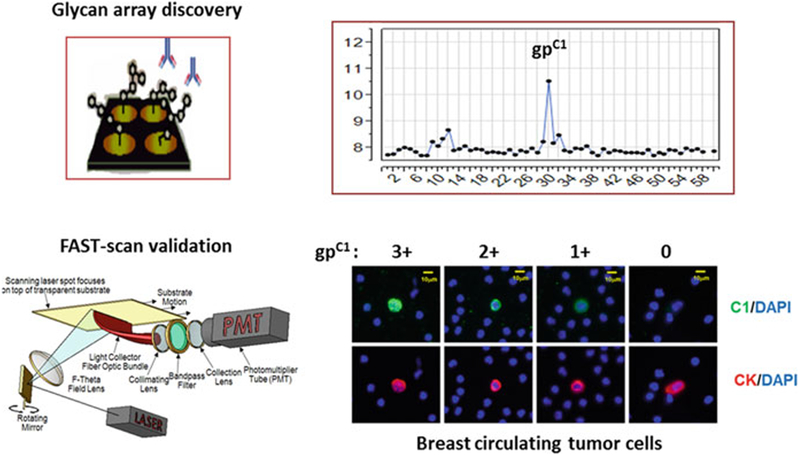
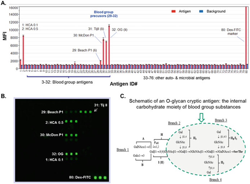
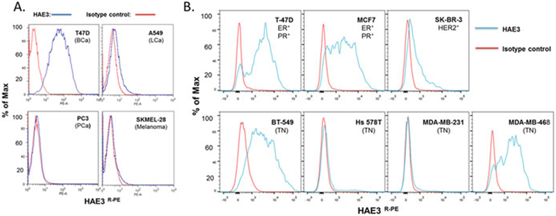
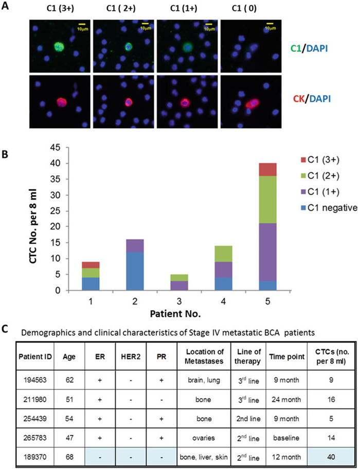
We present here an experimental approach for exploring a new class of tumor biomarkers that are overexpressed by circulating tumor cells (CTCs) and are likely targetable in immunotherapy against tumor metastasis. Using carbohydrate microarrays, anti-tumor monoclonal antibodies (mAbs) were scanned against a large panel of carbohydrate antigens to identify potential tumor glycan markers. Subsequently, flow cytometry and fiber-optic array scanning technology (FAST) were applied to determine whether the identified targets are tumor-specific cell-surface markers and are, therefore, likely suitable for targeted immunotherapy. Finally, the tumor glycan-specific antibodies identified were validated using cancer patients' blood samples for their performance in CTC-detection and immunotyping analysis. In this article, identifying breast CTC-specific glycan markers and targeting mAbs serve as examples to illustrate this tumor biomarker discovery strategy.

Keywords: Breast cancer; Breast circulating tumor cells; Carbohydrate microarray; Glycan markers.


Conflict of interest statement

Conflict of Interest There are no conflicts of interest to declare for any of the authors involved in this work.

Figures

Fig. 15.1
Fig. 15.1
Glycan array discovery and FAST-scan validation of a novel glycan marker gpC1 of bCTCs (Adapted from Wang et al. 2015a)
Fig. 15.2
Fig. 15.2
Carbohydrate microarray analysis of anti-epiglycanin mAb HAE3. Seventy-six glyco-proteins, glycoconjugates, and polysaccharides were spotted in triplicates in 1 to 2 dilutions to yield the customized microarrays for antibody screening. (a) Microarray detections were shown as the mean fluorescent intensities (MFIs) of each microspot with antigen-binding signal in red and background reading in blue. Each error bar is constructed using one standard deviation from the mean of triplicate detections. The labeled antigens include HCA (ID# 1 and 2), a number of blood group precursors (29#–32#), and a microarray spotting marker (80#). (b) Images of a microarray stained with HAE3 (5 μg/ml). (c) Schematic of a blood group substance structure with the conserved O-glycan core highlighted (Adapted from Wang et al. 2015b)
Fig. 15.3
Fig. 15.3
HAE3 cell surface staining detected selective expression of the HAE3-cryptic glycan markers in human cancer cell lines. (a) Four tumor cell lines, T-47D, A549, PC3, and SKMEL-28, were stained with the C1 preparation of HAE3 (IgM) at 1:6 dilution or with an isotype control IgM,9.14.7 (5.0 μg/ml). (b) Seven breast cancer cell lines were stained with purified mAb HAE3(5.0 μg/ml) or 9.14.7 (5.0 μg/ml). These cell lines are T-47D, MCF-7, SK-BR-3, BT-549, Hs578T, MDA-MB-231, and MDA-MB-468. An R-PE-conjugated goat anti-mouse IgM antibody was applied to quantify the cell surface-captured IgM antibodies. Blue line: HAE3 stain; Red line:9.14.7 IgM isotype control (Adapted from Wang et al. 2015b)
Fig. 15.4
Fig. 15.4
Glycan marker gpC1 is expressed in significant numbers of CTCs in Stage IV breast cancer patients. (a) FAST-scan images of bCTCs. Upper panels: Co-staining of C1 (green) and DAPI (blue); Bottom panels: co-staining of anti-CK (red) and DAPI (blue). (b) Distribution of gpC1–positive and -negative bCTCs in five subjects. C1-staining of bCTCs was semi-quantitatively measured by the FAST scan as antibody negative (blue), 1+ (purple), 2+ (green), and 3+ (red) as described. A patient with triple-negative BCa (ID# 189370) was measured gpC1–positive in 37 of 40 CTCs with 50% scored as strong positive (2+ and 3+). (ER, estrogen receptor; PR, progesterone receptor; HER2, human epidermal growth factor receptor 2). (c) A summary of patients’ demographics and clinical characteristics (Adapted from Wang et al. 2015a)

References

    1. Abd Hamid UM, Royle L, Saldova R et al. (2008) A strategy to reveal potential glycan markers from serum glycoproteins associated with breast cancer progression. Glycobiology 18(12):1105–1118 - PubMed
    1. Anders CK, Carey LA (2009) Biology, metastatic patterns, and treatment of patients with triple-negative breast cancer. Clin Breast Cancer 9(Suppl 2):S73–S81 - PMC - PubMed
    1. Apostolopoulos V, Pietersz GA, Tsibanis A et al. (2006) Pilot phase III immunotherapy study in early-stage breast cancer patients using oxidized mannan-MUC1 [ISRCTN71711835]. Breast Cancer Ress: BCR 8(3):R27 - PMC - PubMed
    1. Brenton JD, Carey LA, Ahmed AA et al. (2005) Molecular classification and molecular forecasting of breast cancer: ready for clinical application? J Clin Oncol Off J Am Soc Clin Oncol 23(29):7350–7360 - PubMed
    1. Chatterjee SK, Zetter BR (2005) Cancer biomarkers: knowing the present and predicting the future. Future Oncol 1(1):37–50 - PubMed

Publication types