. 2017 Aug;65(8):1350-1360.
doi: 10.1002/glia.23167. Epub 2017 May 31.

Acutely damaged axons are remyelinated in multiple sclerosis and experimental models of demyelination

Affiliations

Acutely damaged axons are remyelinated in multiple sclerosis and experimental models of demyelination

Verena Schultz et al. Glia. 2017 Aug.

Abstract

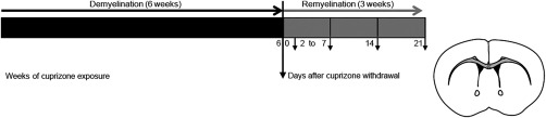
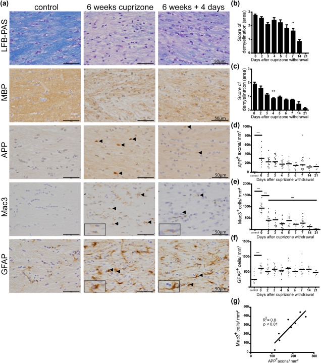
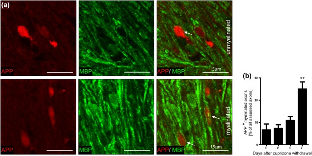
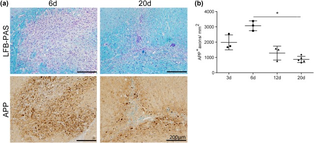
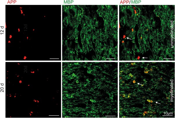
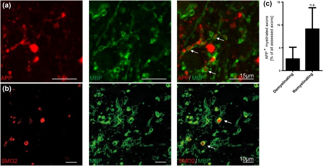
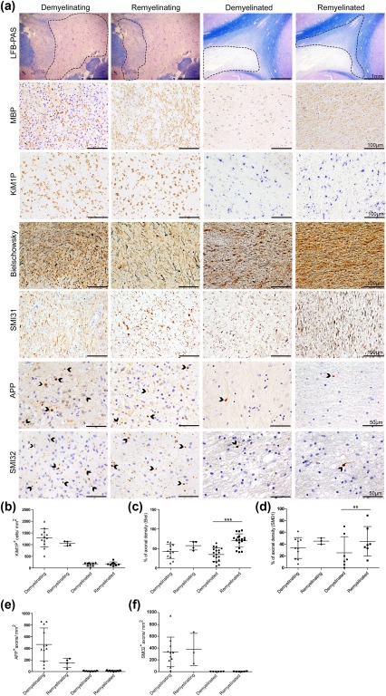
Remyelination is in the center of new therapies for the treatment of multiple sclerosis to resolve and improve disease symptoms and protect axons from further damage. Although remyelination is considered beneficial in the long term, it is not known, whether this is also the case early in lesion formation. Additionally, the precise timing of acute axonal damage and remyelination has not been assessed so far. To shed light onto the interrelation between axons and the myelin sheath during de- and remyelination, we employed cuprizone- and focal lysolecithin-induced demyelination and performed time course experiments assessing the evolution of early and late stage remyelination and axonal damage. We observed damaged axons with signs of remyelination after cuprizone diet cessation and lysolecithin injection. Similar observations were made in early multiple sclerosis lesions. To assess the correlation of remyelination and axonal damage in multiple sclerosis lesions, we took advantage of a cohort of patients with early and late stage remyelinated lesions and assessed the number of APP- and SMI32- positive damaged axons and the density of SMI31-positive and silver impregnated preserved axons. Early de- and remyelinating lesions did not differ with respect to axonal density and axonal damage, but we observed a lower axonal density in late stage demyelinated multiple sclerosis lesions than in remyelinated multiple sclerosis lesions. Our findings suggest that remyelination may not only be protective over a long period of time, but may play an important role in the immediate axonal recuperation after a demyelinating insult.

Keywords: axonal damage; multiple sclerosis; remyelination.


Figures

Figure 1
Figure 1
Design of the cuprizone experiments to investigate the timing and relation of acute axonal damage and remyelination. 7 to 8‐week‐old male C57BL/6J mice were fed for 6 weeks with a 0.25% cuprizone diet to induce demyelination in the corpus callosum. Demyelinated lesions remyelinate after cuprizone withdrawal. To study early and late remyelination, CNS tissue was harvested from days 2 to 7, and on days 14 and 21 after cuprizone diet cessation
Figure 2
Figure 2
Comprehensive assessment of white matter pathology early and late after cuprizone‐induced demyelination. (a) Representative images of the medial corpus callosum of control mice and mice after cuprizone challenge for 6 weeks and after 4 days of recovery. Myelin assessment by LFB‐PAS histochemistry and MBP immunohistochemistry (IHC) indicated an almost complete demyelination of the corpus callosum after 6 weeks of cuprizone feeding. Significant remyelination was observed 4 days after diet cessation. Acute axonal damage (APP+ axons) decreased during remyelination in the cuprizone model. Microglial cells were activated during demyelination. The number of Mac3+ activated microglia declined during remyelination in comparison to 6 weeks of demyelination, while the density of reactive astrocytes (GFAP+ cells) did not change during the period of remyelination observed here. (b, c) The demyelinated area in the corpus callosum was reduced during remyelination as judged by semi‐quantitative evaluation of LFB‐PAS and MBP IHC. The demyelinated area was significantly reduced after 4 and 7 days (*p < 0.05; **p < 0.01; n = 5‐17; mean ± SEM). (d) Acute axonal damage was observed after cuprizone‐induced demyelination in the corpus callosum and was decreased already after 2 days of recovery. The density of APP+ axons decreased continuously during remyelination (*p < 0.05; ***p < 0.001; n = 5‐17; median). (e) Three days after cuprizone withdrawal the number of activated microglia was significantly diminished and almost absent after 3 weeks of remyelination (***p < 0.001; n = 5‐17; mean ± SEM). (f) The evaluation of immunoreactivity for GFAP revealed a persistent activation of astrocytes. (g) The activation of microglia correlated with the amount of damaged axons during recovery after cuprizone diet ingestion (p= 0.0028; R2= 0.7981; n = 8; mean)
Figure 3
Figure 3
The percentage of myelinated damaged axons increased during remyelination. (a) Unmyelinated and myelinated APP+ axons were observed in the corpus callosum of mice after demyelination and during remyelination. A representative image of the corpus callosum 3 and 4 days after withdrawal of cuprizone is shown. (b) APP+ axons were analyzed in 30 randomly selected areas with at least one APP+ axon (approx. 50 axons) in the corpus callosum of mice by confocal microscopy. After 1 week of recovery the number of myelinated APP+ axons increased significantly indicating remyelination of damaged axons (**p < 0.01; n = 3‐7; mean ± SEM)
Figure 4
Figure 4
Significant reduction of acute axonal damage during lesion repair in lysolecithin‐induced demyelination. (a) Representative images of lysolecithin‐induced lesions in the rat corpus callosum on days 6 and 20 postinjection. LFB‐PAS histochemistry revealed almost complete demyelination after 6 days and almost complete remyelination after 20 days. APP+ axons were observed in lysolecithin‐induced lesions. (b) The number of APP+ axons in the lesions increased until day 6 and showed a significant decrease on day 20 after lysolecithin injection (*p<0.05; n = 3‐5; mean ± SD)
Figure 5
Figure 5
APP+ axons are remyelinated in lysolecithin‐induced lesions. Unmyelinated and myelinated APP+ axons were detected in lysolecithin‐induced lesions during lesion repair. A representative image 12 and 20 days after lysolecithin injection is shown
Figure 6
Figure 6
Myelinated APP+ and SMI32+ axons were found in early remyelinating multiple sclerosis lesions. (a, b) Representative confocal image of myelinated APP+ and SMI32+ axons (arrows) in an early remyelinating multiple sclerosis lesion. (c) A higher number of myelinated axons with axonal transport disturbance were observed in remyelinating compared with demyelinating multiple sclerosis lesions. This finding suggests that damaged axons in multiple sclerosis lesions are indeed capable of gaining a new myelin sheath (n = 3, mean ± SEM)
Figure 7
Figure 7
Less axonal loss in remyelinated multiple sclerosis lesions. (a) LFB‐PAS histochemistry shows examples of early (demyelinating and remyelinating) and late stage (demyelinated and remyelinated) multiple sclerosis lesions. (Re)myelination in the lesions was confirmed by immunohistochemistry for myelin basic protein (MBP). (b) Early stage lesions exhibited a higher density of KiM1P+ macrophages/activated microglia compared with late stage lesions. (a, c, d) Axonal loss was observed in all lesion stages with a higher axonal density in remyelinated multiple sclerosis lesions. (a) APP+ and SMI32+ axons reflecting axonal injury were detected in all types of lesions investigated. (e, f) Axonal injury and KiM1P+ macrophages/activated microglia occurred more frequently in early than in late stage multiple sclerosis lesions with no significant difference between lesions displaying demyelination and lesions showing remyelination. (c, d) Remyelinated multiple sclerosis lesions revealed a higher axonal density than early stage lesions and demyelinated lesions (** p < 0.01; *** p < 0.001; n = 3‐18; mean ± SD)

Similar articles

Cited by

References

    1. Bramlett, HM. , Kraydieh, S. , Green, E. J. , & Dietrich, W. D. (1997). Temporal and regional patterns of axonal damage following traumatic brain injury: a beta‐amyloid precursor protein immunocytochemical study in rats. Journal of Neuropathology & Experimental Neurology 56:1132–1141. - PubMed
    1. Brück, W. , Porada, P. , Poser, S. , Rieckmann, P. , Hanefeld, F. , Kretzschmar, HA. , & Lassmann, H . (1995). Monocyte/macrophage differentiation in early multiple sclerosis lesions. Annals of Neurology 38:788–796. - PubMed
    1. Brück, W. , Schmied, M. , Suchanek, G. , Brück, Y. , Breitschopf, H , Poser, S. , … Lassmann, H. (1994). Oligodendrocytes in the early course of multiple sclerosis. Annals of Neurology 35:65–73. - PubMed
    1. Clark, K. C. , Josephson, A. , Benusa, S. D. , Hartley, R. K. , Baer, M. , Thummala, S. , … Dupree, J. L. (2016). Compromised axon initial segment integrity in EAE is preceded by microglial reactivity and contact. Glia 64:1190–1209. - PubMed
    1. Cunningham, C. (2013). Microglia and neurodegeneration: the role of systemic inflammation. Glia 61:71–90. - PubMed

Publication types

MeSH terms