

c-Jun N-Terminal Kinases (JNKs) Are Critical Mediators of Osteoblast Activity In Vivo
- PMID: 28561373
- PMCID: PMC5599178
- DOI: 10.1002/jbmr.3184
c-Jun N-Terminal Kinases (JNKs) Are Critical Mediators of Osteoblast Activity In Vivo
Abstract
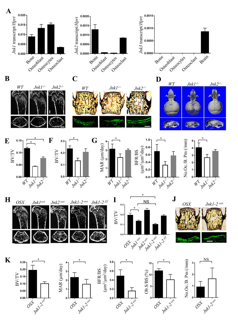
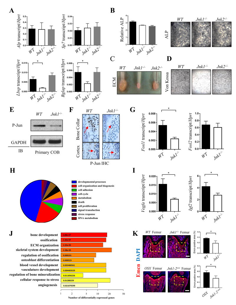
The c-Jun N-terminal kinases (JNKs) are ancient and evolutionarily conserved regulators of proliferation, differentiation, and cell death responses. Currently, in vitro studies offer conflicting data about whether the JNK pathway augments or represses osteoblast differentiation, and the contribution of the JNK pathway to regulation of bone mass in vivo remains unclear. Here we show that Jnk1-/- mice display severe osteopenia due to impaired bone formation, whereas Jnk2-/- mice display a mild osteopenia only evident in long bones. In order to both confirm that these effects were osteoblast intrinsic and assess whether redundancy with JNK1 masks a potential contribution of JNK2, mice with a conditional deletion of both JNK1 and JNK2 floxed conditional alleles in osteoblasts (Jnk1-2osx ) were bred. These mice displayed a similar degree of osteopenia to Jnk1-/- mice due to decreased bone formation. In vitro, Jnk1-/- osteoblasts display a selective defect in the late stages of osteoblast differentiation with impaired mineralization activity. Downstream of JNK1, phosphorylation of JUN is impaired in Jnk1-/- osteoblasts. Transcriptome analysis showed that JNK1 is required for upregulation of several osteoblast-derived proangiogenic factors such as IGF2 and VEGFa. Accordingly, JNK1 deletion results in a significant reduction skeletal vasculature in mice. Taken together, this study establishes that JNK1 is a key mediator of osteoblast function in vivo and in vitro. © 2017 American Society for Bone and Mineral Research.
Keywords: ANGIOGENESIS; BONE FORMATION; JNK; JUN; MAPK; OSTEOBLASTS.
© 2017 American Society for Bone and Mineral Research.
Conflict of interest statement
Conflict of Interest: All authors state that they have no conflicts of interest.
Figures
References
-
- Kyriakis JM, Banerjee P, Nikolakaki E, et al. The stress-activated protein kinase subfamily of c-Jun kinases. Nature. 1994;369:156–60. - PubMed
-
- Hibi M, Lin A, Smeal T, Minden A, Karin M. Identification of an oncoprotein- and UV-responsive protein kinase that binds and potentiates the c-Jun activation domain. Genes & development. 1993;7:2135–48. - PubMed
-
- Derijard B, Hibi M, Wu IH, et al. JNK1: a protein kinase stimulated by UV light and Ha-Ras that binds and phosphorylates the c-Jun activation domain. Cell. 1994;76:1025–37. - PubMed
-
- Weston CR, Davis RJ. The JNK signal transduction pathway. Current opinion in genetics & development. 2002;12:14–21. - PubMed
MeSH terms
Substances
Grants and funding
LinkOut - more resources
Full Text Sources
Other Literature Sources
Research Materials
Miscellaneous
