

Friedreich's ataxia induced pluripotent stem cell-derived cardiomyocytes display electrophysiological abnormalities and calcium handling deficiency
- PMID: 28562313
- PMCID: PMC5472743
- DOI: 10.18632/aging.101247
Friedreich's ataxia induced pluripotent stem cell-derived cardiomyocytes display electrophysiological abnormalities and calcium handling deficiency
Abstract
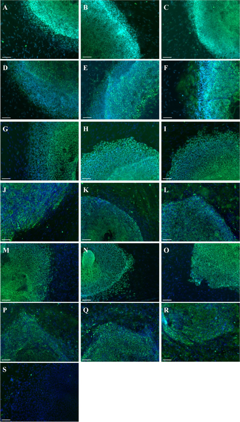
We sought to identify the impacts of Friedreich's ataxia (FRDA) on cardiomyocytes. FRDA is an autosomal recessive degenerative condition with neuronal and non-neuronal manifestations, the latter including progressive cardiomyopathy of the left ventricle, the leading cause of death in FRDA. Little is known about the cellular pathogenesis of FRDA in cardiomyocytes. Induced pluripotent stem cells (iPSCs) were derived from three FRDA individuals with characterized GAA repeats. The cells were differentiated into cardiomyocytes to assess phenotypes. FRDA iPSC- cardiomyocytes retained low levels of FRATAXIN (FXN) mRNA and protein. Electrophysiology revealed an increased variation of FRDA- cardiomyocyte beating rates which was prevented by addition of nifedipine, suggestive of a calcium handling deficiency. Finally, calcium imaging was performed and we identified small amplitude, diastolic and systolic calcium transients confirming a deficiency in calcium handling. We defined a robust FRDA cardiac-specific electrophysiological profile in patient-derived iPSCs which could be used for high throughput compound screening. This cell-specific signature will contribute to the identification and screening of novel treatments for this life-threatening disease.
Keywords: Friedreich’s ataxia; cardiomyopathy; induced pluripotent stem cells; modelling.
Conflict of interest statement
The authors have no conflicts of interest to declare.
Figures


Similar articles
-
Correlation between frataxin expression and contractility revealed by in vitro Friedreich's ataxia cardiac tissue models engineered from human pluripotent stem cells.Stem Cell Res Ther. 2019 Jul 8;10(1):203. doi: 10.1186/s13287-019-1305-y. Stem Cell Res Ther. 2019. PMID: 31286988 Free PMC article.
-
Excision of the expanded GAA repeats corrects cardiomyopathy phenotypes of iPSC-derived Friedreich's ataxia cardiomyocytes.Stem Cell Res. 2019 Oct;40:101529. doi: 10.1016/j.scr.2019.101529. Epub 2019 Aug 7. Stem Cell Res. 2019. PMID: 31446150 Free PMC article.
-
Efficient attenuation of Friedreich's ataxia (FRDA) cardiomyopathy by modulation of iron homeostasis-human induced pluripotent stem cell (hiPSC) as a drug screening platform for FRDA.Int J Cardiol. 2016 Jan 15;203:964-71. doi: 10.1016/j.ijcard.2015.11.101. Epub 2015 Nov 17. Int J Cardiol. 2016. PMID: 26625322
-
Cellular pathophysiology of Friedreich's ataxia cardiomyopathy.Int J Cardiol. 2022 Jan 1;346:71-78. doi: 10.1016/j.ijcard.2021.11.033. Epub 2021 Nov 16. Int J Cardiol. 2022. PMID: 34798207 Review.
-
Neurodegeneration in Friedreich's ataxia: from defective frataxin to oxidative stress.Oxid Med Cell Longev. 2013;2013:487534. doi: 10.1155/2013/487534. Epub 2013 Jul 9. Oxid Med Cell Longev. 2013. PMID: 23936609 Free PMC article. Review.
Cited by
-
Antioxidant Therapies and Oxidative Stress in Friedreich´s Ataxia: The Right Path or Just a Diversion?Antioxidants (Basel). 2020 Jul 24;9(8):664. doi: 10.3390/antiox9080664. Antioxidants (Basel). 2020. PMID: 32722309 Free PMC article. Review.
-
Patient-derived iPSC models of Friedreich ataxia: a new frontier for understanding disease mechanisms and therapeutic application.Transl Neurodegener. 2023 Sep 20;12(1):45. doi: 10.1186/s40035-023-00376-8. Transl Neurodegener. 2023. PMID: 37726850 Free PMC article. Review.
-
Correlation between frataxin expression and contractility revealed by in vitro Friedreich's ataxia cardiac tissue models engineered from human pluripotent stem cells.Stem Cell Res Ther. 2019 Jul 8;10(1):203. doi: 10.1186/s13287-019-1305-y. Stem Cell Res Ther. 2019. PMID: 31286988 Free PMC article.
-
Cardiac calcium regulation in human induced pluripotent stem cell cardiomyocytes: Implications for disease modeling and maturation.Front Cell Dev Biol. 2023 Jan 18;10:986107. doi: 10.3389/fcell.2022.986107. eCollection 2022. Front Cell Dev Biol. 2023. PMID: 36742199 Free PMC article. Review.
-
Cellular models for human cardiomyopathy: What is the best option?World J Cardiol. 2019 Oct 26;11(10):221-235. doi: 10.4330/wjc.v11.i10.221. World J Cardiol. 2019. PMID: 31754410 Free PMC article. Review.
References
-
- Delatycki MB, Paris DB, Gardner RJ, Nicholson GA, Nassif N, Storey E, MacMillan JC, Collins V, Williamson R, Forrest SM. Clinical and genetic study of Friedreich ataxia in an Australian population. Am J Med Genet. 1999;87:168–74. doi: 10.1002/(SICI)1096-8628(19991119)87:2<168::AID-AJMG8>3.0.CO;2-2. - DOI - PubMed
Publication types
MeSH terms
Substances
LinkOut - more resources
Full Text Sources
Other Literature Sources
Medical
Miscellaneous
