

From medical imaging data to 3D printed anatomical models
- PMID: 28562693
- PMCID: PMC5451060
- DOI: 10.1371/journal.pone.0178540
From medical imaging data to 3D printed anatomical models
Abstract
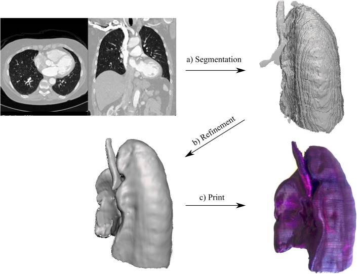
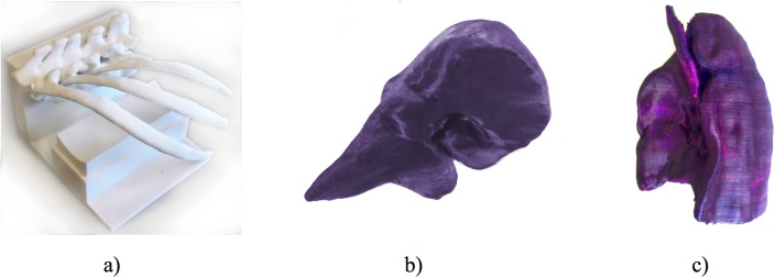
Anatomical models are important training and teaching tools in the clinical environment and are routinely used in medical imaging research. Advances in segmentation algorithms and increased availability of three-dimensional (3D) printers have made it possible to create cost-efficient patient-specific models without expert knowledge. We introduce a general workflow that can be used to convert volumetric medical imaging data (as generated by Computer Tomography (CT)) to 3D printed physical models. This process is broken up into three steps: image segmentation, mesh refinement and 3D printing. To lower the barrier to entry and provide the best options when aiming to 3D print an anatomical model from medical images, we provide an overview of relevant free and open-source image segmentation tools as well as 3D printing technologies. We demonstrate the utility of this streamlined workflow by creating models of ribs, liver, and lung using a Fused Deposition Modelling 3D printer.
Conflict of interest statement
Figures
References
-
- McGaghie WC, Issenberg SB, Petrusa ER, Scalese RJ. A critical review of simulation-based medical education research: 2003–2009. Med Educ. Wiley Online Library; 2010;44: 50–63. doi: 10.1111/j.1365-2923.2009.03547.x - DOI - PubMed
-
- Evans L V, Dodge KL, Shah TD, Kaplan LJ, Siegel MD, Moore CL, et al. Simulation training in central venous catheter insertion: Improved performance in clinical practice. Acad Med. 2010;85: 1462–1469. doi: 10.1097/ACM.0b013e3181eac9a3 - DOI - PubMed
-
- Müller A, Krishnan KG, Uhl E, Mast G. The application of rapid prototyping techniques in cranial reconstruction and preoperative planning in neurosurgery. J Craniofac Surg. 2003;14: 899–914. - PubMed
-
- Rengier F, Mehndiratta A, Von Tengg-Kobligk H, Zechmann CM, Unterhinninghofen R, Kauczor HU, et al. 3D printing based on imaging data: Review of medical applications. Int J Comput Assist Radiol Surg. 2010;5: 335–341. doi: 10.1007/s11548-010-0476-x - DOI - PubMed
-
- Kusaka M, Sugimoto M, Fukami N, Sasaki H, Takenaka M, Anraku T, et al. Initial experience with a tailor-made simulation and navigation program using a 3-D printer model of kidney transplantation surgery. Transplant Proc. Elsevier Inc.; 2015;47: 596–599. - PubMed
MeSH terms
LinkOut - more resources
Full Text Sources
Other Literature Sources
