

Hepatitis B virus DNA detection by in situ hybridization in human hepatocellular carcinoma
- PMID: 2856435
- PMCID: PMC4532137
- DOI: 10.3904/kjim.1988.3.1.24
Hepatitis B virus DNA detection by in situ hybridization in human hepatocellular carcinoma
Abstract
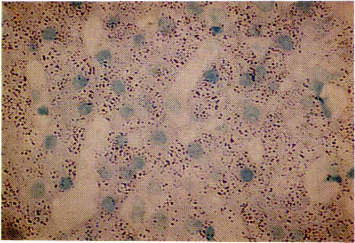
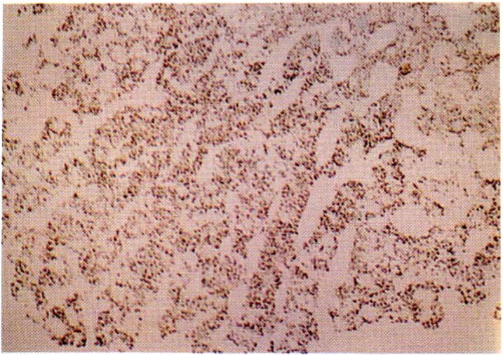
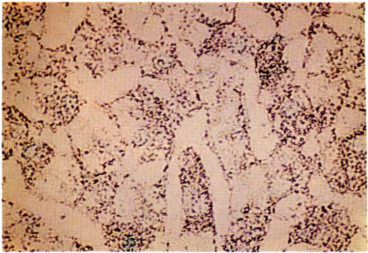
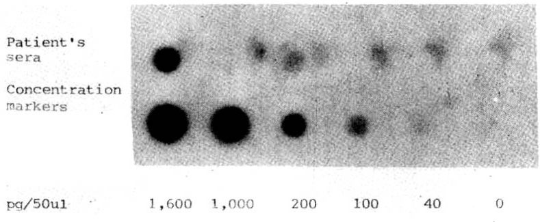
The distribution of hepatitis B virus (HBV) DNA in tumor tissue sections from six Korean patients with HBsAg positive hepatocellular carcinoma (HCC) was examined by in situ hybridization using a biotin-labeled recombinant, cloned HBV DNA probe. All patients tested were positive for both HBeAg and anti-HBc in their sera. HBV DNA was distributed abundantly in the cytoplasm and rarely in the nuclei of tumor cells. The validity of the in situ hybridization assay was confirmed by the dot blotting technique using a 32P-labeled HBV DNA probe obtained by nick translation. In conclusion, it is speculated that integration of HBV DNA into host DNA as well as persistant amplified replication of the HBV DNA within the hepatocytes is linked etiologically to the development of human hepatocellular carcinoma.
Figures
References
-
- Shafritz DA, Kew MC. Identification of integrated hepatitis B virus DNA sequences in human hepatocellular carcinoma. Hepatology. 1981;1:1. - PubMed
-
- Lieberman HM, LaBrecque DR, Kew MC, Hadziyannis SJ, Shafritz DA. Detection of hepatitis B virus DNA directly in human serum by a simplified molecular hybridization test: Comparison to HBeAg/anti-HBe status in HBsAg carriers. Hepatology. 1983;3:285. - PubMed
-
- Brechot C, Hadchovel M, Scotto J. Detection of hepatitis B virus DNA in liver and serum: A direct appraisal of the chronic carrier state. Lancet. 1981;2:765. - PubMed
-
- Sninsky JJ, Cohen SN. Specialized cloning vectors for hepatitis B virus gene expression in Escherichia coli. Hepatology. 1982;2:72S.
MeSH terms
Substances
LinkOut - more resources
Full Text Sources
Medical