

Sequential Steps of CRAC Channel Activation
- PMID: 28564609
- PMCID: PMC5519287
- DOI: 10.1016/j.celrep.2017.05.025
Sequential Steps of CRAC Channel Activation
Abstract
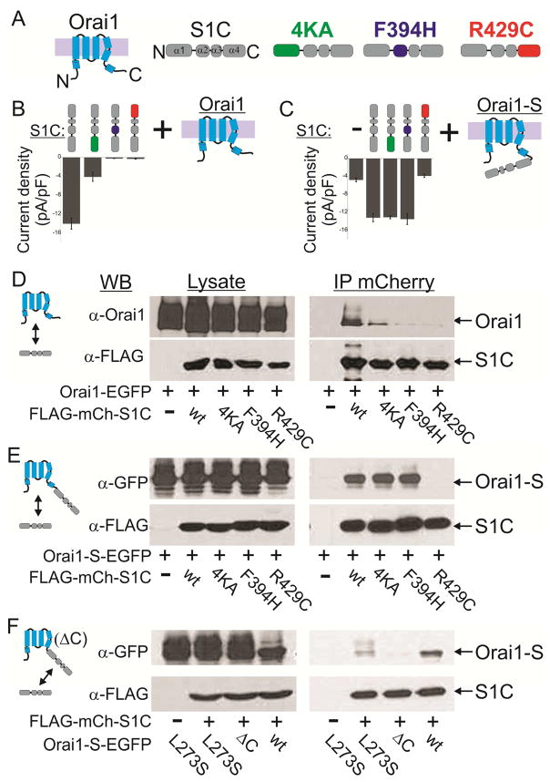
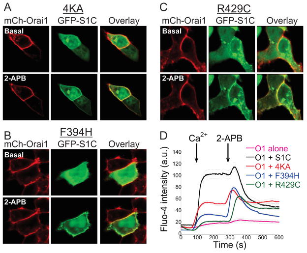
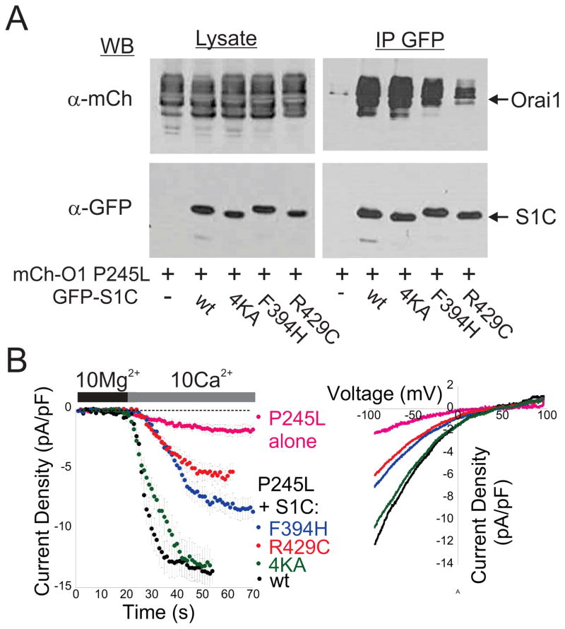
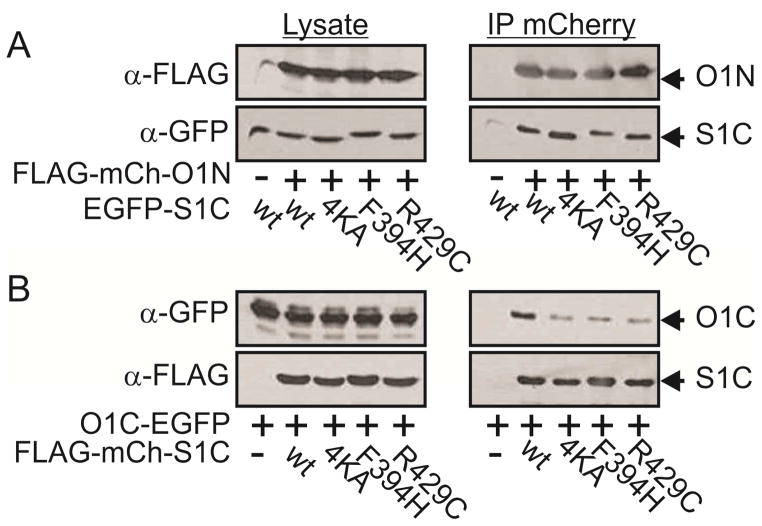
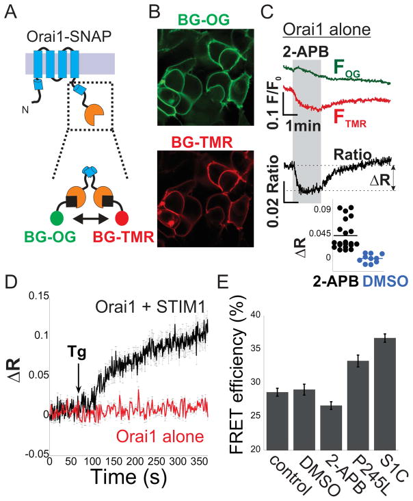
Interaction between the endoplasmic reticulum protein STIM1 and the plasma membrane channel ORAI1 generates calcium signals that are central for diverse cellular functions. How STIM1 binds and activates ORAI1 remains poorly understood. Using electrophysiological, optical, and biochemical techniques, we examined the effects of mutations in the STIM1-ORAI1 activating region (SOAR) of STIM1. We find that SOAR mutants that are deficient in binding to resting ORAI1 channels are able to bind to and boost activation of partially activated ORAI1 channels. We further show that the STIM1 binding regions on ORAI1 undergo structural rearrangement during channel activation. The results suggest that activation of ORAI1 by SOAR occurs in multiple steps. In the first step, SOAR binds to ORAI1, partially activates the channel, and induces a rearrangement in the SOAR-binding site of ORAI1. That rearrangement of ORAI1 then permits sequential steps of SOAR binding, via distinct molecular interactions, to fully activate the channel.
Keywords: CRAC channel; ORAI1; STIM1; calcium; gating.
Copyright © 2017 The Authors. Published by Elsevier Inc. All rights reserved.
Figures

Similar articles
-
Defects in the STIM1 SOARα2 domain affect multiple steps in the CRAC channel activation cascade.Cell Mol Life Sci. 2021 Oct;78(19-20):6645-6667. doi: 10.1007/s00018-021-03933-4. Epub 2021 Sep 8. Cell Mol Life Sci. 2021. PMID: 34498097 Free PMC article.
-
Ca2+/Calmodulin Binding to STIM1 Hydrophobic Residues Facilitates Slow Ca2+-Dependent Inactivation of the Orai1 Channel.Cell Physiol Biochem. 2020 Mar 17;54(2):252-270. doi: 10.33594/000000218. Cell Physiol Biochem. 2020. PMID: 32176842
-
STIM1 dimers undergo unimolecular coupling to activate Orai1 channels.Nat Commun. 2015 Sep 24;6:8395. doi: 10.1038/ncomms9395. Nat Commun. 2015. PMID: 26399906 Free PMC article.
-
The STIM-Orai coupling interface and gating of the Orai1 channel.Cell Calcium. 2017 May;63:8-13. doi: 10.1016/j.ceca.2017.01.001. Epub 2017 Jan 8. Cell Calcium. 2017. PMID: 28087079 Free PMC article. Review.
-
The STIM-Orai Pathway: Conformational Coupling Between STIM and Orai in the Activation of Store-Operated Ca2+ Entry.Adv Exp Med Biol. 2017;993:83-98. doi: 10.1007/978-3-319-57732-6_5. Adv Exp Med Biol. 2017. PMID: 28900910 Free PMC article. Review.
Cited by
-
RGS10 physically and functionally interacts with STIM2 and requires store-operated calcium entry to regulate pro-inflammatory gene expression in microglia.Cell Signal. 2021 Jul;83:109974. doi: 10.1016/j.cellsig.2021.109974. Epub 2021 Mar 9. Cell Signal. 2021. PMID: 33705894 Free PMC article.
-
Numbers count: How STIM and Orai stoichiometry affect store-operated calcium entry.Cell Calcium. 2019 May;79:35-43. doi: 10.1016/j.ceca.2019.02.002. Epub 2019 Feb 12. Cell Calcium. 2019. PMID: 30807904 Free PMC article. Review.
-
Ca2+ and lipid signals hold hands at endoplasmic reticulum-plasma membrane contact sites.J Physiol. 2018 Jul;596(14):2709-2716. doi: 10.1113/JP274957. Epub 2018 Jan 4. J Physiol. 2018. PMID: 29210464 Free PMC article. Review.
-
Molecular basis of allosteric Orai1 channel activation by STIM1.J Physiol. 2020 May;598(9):1707-1723. doi: 10.1113/JP276550. Epub 2019 May 1. J Physiol. 2020. PMID: 30950063 Free PMC article. Review.
-
Structural features of STIM and Orai underlying store-operated calcium entry.Curr Opin Cell Biol. 2019 Apr;57:90-98. doi: 10.1016/j.ceb.2018.12.012. Epub 2019 Feb 1. Curr Opin Cell Biol. 2019. PMID: 30716649 Free PMC article. Review.
References
-
- Feske S, Gwack Y, Prakriya M, Srikanth S, Puppel SH, Tanasa B, Hogan PG, Lewis RS, Daly M, Rao A. A mutation in Orai1 causes immune deficiency by abrogating CRAC channel function. Nature. 2006;441:179–185. - PubMed
Publication types
MeSH terms
Substances
Grants and funding
LinkOut - more resources
Full Text Sources
Other Literature Sources
