

Genomic instability induced in distant progeny of bystander cells depends on the connexins expressed in the irradiated cells
- PMID: 28565963
- PMCID: PMC9233304
- DOI: 10.1080/09553002.2017.1334980
Genomic instability induced in distant progeny of bystander cells depends on the connexins expressed in the irradiated cells
Abstract
Purpose: To examine the time window during which intercellular signaling though gap junctions mediates non-targeted (bystander) effects induced by moderate doses of ionizing radiation; and to investigate the impact of gap junction communication on genomic instability in distant progeny of bystander cells.
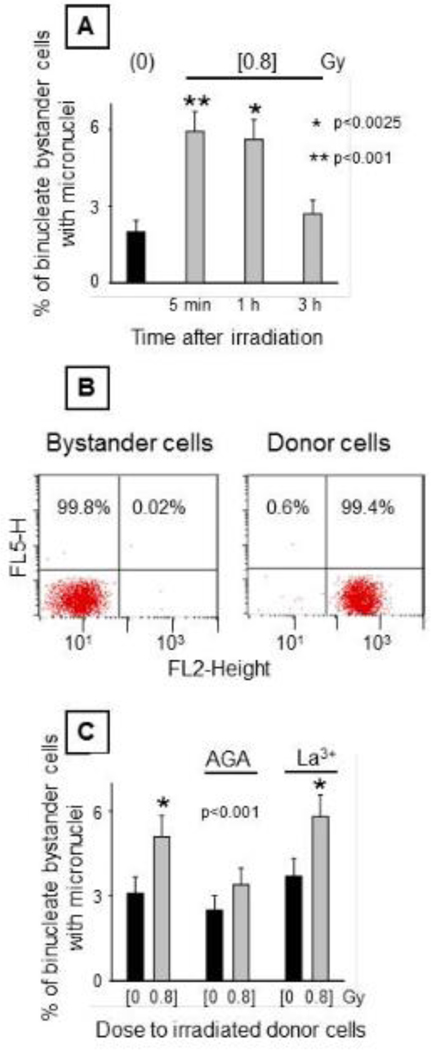
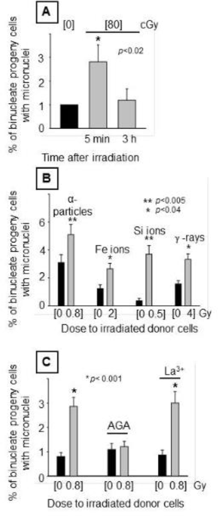
Materials and methods: A layered cell culture system was developed to investigate the propagation of harmful effects from irradiated normal or tumor cells that express specific connexins to contiguous bystander normal human fibroblasts. Irradiated cells were exposed to moderate mean absorbed doses from 3.7 MeV α particle, 1000 MeV/u iron ions, 600 MeV/u silicon ions, or 137Cs γ rays. Following 5 h of co-culture, pure populations of bystander cells, unexposed to secondary radiation, were isolated and DNA damage and oxidative stress was assessed in them and in their distant progeny (20-25 population doublings).
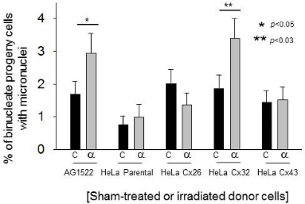
Results: Increased frequency of micronucleus formation and enhanced oxidative changes were observed in bystander cells co-cultured with confluent cells exposed to either sparsely ionizing (137Cs γ rays) or densely ionizing (α particles, energetic iron or silicon ions) radiations. The irradiated cells propagated signals leading to biological changes in bystander cells within 1 h of irradiation, and the effect required cellular coupling by gap junctions. Notably, the distant progeny of isolated bystander cells also exhibited increased levels of spontaneous micronuclei. This effect was dependent on the type of junctional channels that coupled the irradiated donor cells with the bystander cells. Previous work showed that gap junctions composed of connexin26 (Cx26) or connexin43 (Cx43) mediate toxic bystander effects within 5 h of co-culture, whereas gap junctions composed of connexin32 (Cx32) mediate protective effects. In contrast, the long-term progeny of bystander cells expressing Cx26 or Cx43 did not display elevated DNA damage, whereas those coupled by Cx32 had enhanced DNA damage.
Conclusions: In response to moderate doses from either sparsely or densely ionizing radiations, toxic and protective effects are rapidly communicated to bystander cells through gap junctions. We infer that bystander cells damaged by the initial co-culture (expressing Cx26 or Cx43) die or undergo proliferative arrest, but that the bystander cells that were initially protected (expressing Cx32) express DNA damage upon sequential passaging. Together, the results inform the roles that intercellular communication play under stress conditions, and aid assessment of the health risks of exposure to ionizing radiation. Identification of the communicated molecules may enhance the efficacy of radiotherapy and help attenuate its debilitating side-effects.
Keywords: Bystander effects; channel permeability; gap junctions; genomic instability; non-targeted effects; radiation quality.
Conflict of interest statement
DISCLOSURES
The authors declare that they have no competing or conflicting interests.
Figures


Similar articles
-
Connexins and cyclooxygenase-2 crosstalk in the expression of radiation-induced bystander effects.Br J Cancer. 2014 Jul 8;111(1):125-31. doi: 10.1038/bjc.2014.276. Epub 2014 May 27. Br J Cancer. 2014. PMID: 24867691 Free PMC article.
-
Genetic changes in progeny of bystander human fibroblasts after microbeam irradiation with X-rays, protons or carbon ions: the relevance to cancer risk.Int J Radiat Biol. 2015 Jan;91(1):62-70. doi: 10.3109/09553002.2014.950715. Int J Radiat Biol. 2015. PMID: 25084840
-
Human cell responses to ionizing radiation are differentially affected by the expressed connexins.J Radiat Res. 2013 Mar 1;54(2):251-9. doi: 10.1093/jrr/rrs099. Epub 2012 Nov 8. J Radiat Res. 2013. PMID: 23139176 Free PMC article.
-
Oxidative metabolism, gap junctions and the ionizing radiation-induced bystander effect.Oncogene. 2003 Oct 13;22(45):7050-7. doi: 10.1038/sj.onc.1206961. Oncogene. 2003. PMID: 14557810 Review.
-
[Radiation-induced bystander effect: the important part of ionizing radiation response. Potential clinical implications].Postepy Hig Med Dosw (Online). 2009 Aug 18;63:377-88. Postepy Hig Med Dosw (Online). 2009. PMID: 19724078 Review. Polish.
Cited by
-
Intercellular communications-redox interactions in radiation toxicity; potential targets for radiation mitigation.J Cell Commun Signal. 2019 Mar;13(1):3-16. doi: 10.1007/s12079-018-0473-3. Epub 2018 Jun 17. J Cell Commun Signal. 2019. PMID: 29911259 Free PMC article. Review.
-
Evaluation of the role of mitochondria in the non-targeted effects of ionizing radiation using cybrid cellular models.Sci Rep. 2020 Apr 9;10(1):6131. doi: 10.1038/s41598-020-63011-w. Sci Rep. 2020. PMID: 32273537 Free PMC article.
-
Molecular Pathogenesis of Radiation-Induced Cell Toxicity in Stem Cells.Int J Mol Sci. 2017 Dec 18;18(12):2749. doi: 10.3390/ijms18122749. Int J Mol Sci. 2017. PMID: 29258244 Free PMC article. Review.
-
Low-Dose Non-Targeted Effects and Mitochondrial Control.Int J Mol Sci. 2023 Jul 14;24(14):11460. doi: 10.3390/ijms241411460. Int J Mol Sci. 2023. PMID: 37511215 Free PMC article. Review.
-
Assessment of Genomic Instability in Medical Workers Exposed to Chronic Low-Dose X-Rays in Northern China.Dose Response. 2019 Nov 28;17(4):1559325819891378. doi: 10.1177/1559325819891378. eCollection 2019 Oct-Dec. Dose Response. 2019. PMID: 31819742 Free PMC article.
References
-
- Autsavapromporn N, Suzuki M, Funayama T, Usami N, Plante I, Yokota Y, Mutou Y, Ikeda H, Kobayashi K, Kobayashi Y, et al. 2013. Gap junction communication and the propagation of bystander effects induced by microbeam irradiation in human fibroblast cultures: the impact of radiation quality. Radiat Res. Oct;180:367–375. - PMC - PubMed
-
- Ayad WA, Locke D, Koreen IV, Harris AL. 2006. Heteromeric, but not homomeric, connexin channels are selectively permeable to inositol phosphates. J Biol Chem.281:16727–16739. - PubMed
Publication types
MeSH terms
Substances
Grants and funding
LinkOut - more resources
Full Text Sources
Other Literature Sources
Miscellaneous