

The anteroposterior and primary-to-posterior limbic ratios as MRI-derived volumetric markers of Alzheimer's disease
- PMID: 28566144
- PMCID: PMC5535801
- DOI: 10.1016/j.jns.2017.04.046
The anteroposterior and primary-to-posterior limbic ratios as MRI-derived volumetric markers of Alzheimer's disease
Abstract
Background/aims: Alzheimer's disease (AD) shows a characteristic pattern of brain atrophy, with predominant involvement of posterior limbic structures, and relative preservation of rostral limbic and primary cortical regions. We aimed to investigate the diagnostic utility of two gray matter volume ratios based on this pattern, and to develop a fully automated method to calculate them from unprocessed MRI files.
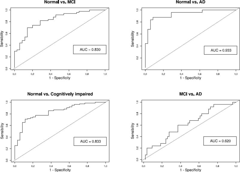
Patients and methods: Cross-sectional study of 118 subjects from the ADNI database, including normal controls and patients with mild cognitive impairment (MCI) and AD. Clinical variables and 3T T1-weighted MRI files were analyzed. Regional gray matter and total intracranial volumes were calculated with a shell script (gm_extractor) based on FSL. Anteroposterior and primary-to-posterior limbic ratios (APL and PPL) were calculated from these values. Diagnostic utility of variables was tested in logistic regression models using Bayesian model averaging for variable selection. External validity was evaluated with bootstrap sampling and a test set of 60 subjects.
Results: gm_extractor showed high test-retest reliability and high concurrent validity with FSL's FIRST. Volumetric measurements agreed with the expected anatomical pattern associated with AD. APL and PPL ratios were significantly different between groups, and were selected instead of hippocampal and entorhinal volumes to differentiate normal from MCI or cognitively impaired (MCI plus AD) subjects.
Conclusion: APL and PPL ratios may be useful components of models aimed to differentiate normal subjects from patients with MCI or AD. These values, and other gray matter volumes, may be reliably calculated with gm_extractor.
Keywords: Alzheimer disease (MeSH); Magnetic resonance imaging (MeSH); Mild cognitive impairment; Volumetry.
Copyright © 2017 Elsevier B.V. All rights reserved.
Conflict of interest statement
AJH and SES have no conflict of interest to report.
Figures


Similar articles
-
Comparative analysis of methods of volume adjustment in hippocampal volumetry for the diagnosis of Alzheimer disease.J Neuroradiol. 2020 Mar;47(2):161-165. doi: 10.1016/j.neurad.2019.02.004. Epub 2019 Mar 8. J Neuroradiol. 2020. PMID: 30857897
-
Classification of Alzheimer's disease and prediction of mild cognitive impairment-to-Alzheimer's conversion from structural magnetic resource imaging using feature ranking and a genetic algorithm.Comput Biol Med. 2017 Apr 1;83:109-119. doi: 10.1016/j.compbiomed.2017.02.011. Epub 2017 Feb 27. Comput Biol Med. 2017. PMID: 28260614
-
Structural correlates of mild cognitive impairment: A clinicovolumetric study.Neurol India. 2018 Mar-Apr;66(2):370-376. doi: 10.4103/0028-3886.227298. Neurol India. 2018. PMID: 29547157
-
Potential Role of Neuroimaging Markers for Early Diagnosis of Dementia in Primary Care.Curr Alzheimer Res. 2018;15(1):18-27. doi: 10.2174/1567205014666170908093846. Curr Alzheimer Res. 2018. PMID: 28891447 Review.
-
Fully-automated volumetric MRI with normative ranges: translation to clinical practice.Behav Neurol. 2009;21(1):21-8. doi: 10.3233/BEN-2009-0226. Behav Neurol. 2009. PMID: 19847042 Free PMC article. Review.
Cited by
-
Adapting UK Biobank imaging for use in a routine memory clinic setting: The Oxford Brain Health Clinic.Neuroimage Clin. 2022;36:103273. doi: 10.1016/j.nicl.2022.103273. Epub 2022 Nov 21. Neuroimage Clin. 2022. PMID: 36451375 Free PMC article.
References
-
- Scheltens P, Blennow K, Breteler MM, de Strooper B, Frisoni GB, Salloway S, Van der Flier WM. Alzheimer's disease. Lancet. 2016;388(10043):505–17. - PubMed
-
- Winblad B, Amouyel P, Andrieu S, Ballard C, Brayne C, Brodaty H, Cedazo-Minguez A, Dubois B, Edvardsson D, Feldman H, Fratiglioni L, Frisoni GB, Gauthier S, Georges J, Graff C, Iqbal K, Jessen F, Johansson G, Jönsson L, Kivipelto M, Knapp M, Mangialasche F, Melis R, Nordberg A, Rikkert MO, Qiu C, Sakmar TP, Scheltens P, Schneider LS, Sperling R, Tjernberg LO, Waldemar G, Wimo A, Zetterberg H. Defeating Alzheimer's disease and other dementias: a priority for European science and society. Lancet Neurol. 2016;15(5):455–532. - PubMed
-
- Vogt BA, Finch DM, Olson CR. Functional heterogeneity in cingulate cortex: the anterior executive and posterior evaluative regions. Cereb Cortex. 1992;2(6):435–43. - PubMed
-
- Mosconi L, Pupi A, De Cristofaro MT, Fayyaz M, Sorbi S, Herholz K. Functional interactions of the entorhinal cortex: an 18F-FDG PET study on normal aging and Alzheimer's disease. J Nucl Med. 2004;45(3):382–92. - PubMed
Publication types
MeSH terms
Grants and funding
LinkOut - more resources
Full Text Sources
Other Literature Sources
Medical
