. 2017 Jun:20:120-126.
doi: 10.1016/j.ebiom.2017.05.012. Epub 2017 May 17.

Local Somatodendritic Translation and Hyperphosphorylation of Tau Protein Triggered by AMPA and NMDA Receptor Stimulation

Affiliations

Local Somatodendritic Translation and Hyperphosphorylation of Tau Protein Triggered by AMPA and NMDA Receptor Stimulation

Shunsuke Kobayashi et al. EBioMedicine. 2017 Jun.

Abstract

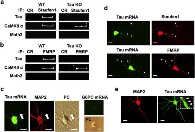
Tau is a major component of the neurofibrillary tangles (NFT) that represent a pathological hallmark of Alzheimer's disease (AD). Although generally considered an axonal protein, Tau is found in the somato-dendritic compartment of degenerating neurons and this redistribution is thought to be a trigger of neurodegeneration in AD. Here, we show the presence of tau mRNA in a dendritic ribonucleoprotein (RNP) complex that includes Ca2+-calmodulin dependent protein kinase (CaMK)IIα mRNA and that is translated locally in response to glutamate stimulation. Further, we show that Tau mRNA is a component of mRNP granules that contain RNA-binding proteins, and that it interacts with Myosin Va, a postsynaptic motor protein; these findings suggest that tau mRNA is transported into dendritic spines. We also report that tau mRNA localized in the somato-dendritic component of primary hippocampal cells and that a sub-toxic concentration of glutamate enhances local translation and hyperphosphorylation of tau, effects that are blocked by the gluatamatergic antagonists MK801 and NBQX. These data thus demonstrate that alpha-amino-3-hydroxy-5-methyl-4-isoxazole propionic acid (AMPA) and N-methyl-d-aspartate (NMDA) stimulation redistributes tau to the somato-dendritic region of neurons where it may trigger neurodegeneration.

Keywords: AMPA and NMDA receptors; Local translation; RNP particle; Somatodendritic localization of tau; Tau mRNA.


Figures

Image 2
Graphical abstract
Fig. 1
Fig. 1
Tau mRNA is a constituent of FMRP- and Staufen1-positive dendritic RNP granules. Tau and CaMKIIα mRNAs detectable in (a) Staufen1- and (b) FMRP-positive immunoprecipitates from RNP granules (fraction 13; Supplemental Fig. 1) from hippocampi of wild type (WT), but not tau knockout (KO), mice. The two images on the left of panel c show tau mRNA in a neuron (fluorescence in situ hybridization: Alexa Fluor 488) whose dendrites are MAP2-immunostained (Alexa Fluor 555); a phase contrast (PC) image (third-from-left) and a negative control (G6PC mRNA, fourth-from-left) are also shown. Liver- and kidney-specific G6PC mRNA was not detected. Arrows indicate axons. Panel (d) shows Staufen1- and FMRP-immunopositive neurons that co-express tau mRNA. Arrowheads indicate granule structures in dendrites expressing tau mRNA and Satufen1 or FMRP. Panel (e) shows the neuronal distribution of tau mRNA as revealed by confocal microscopy. Arrowheads (inset) indicate dendritic spines. Scale bar: 10 μm.
Fig. 2
Fig. 2
Glutamate stimulates local translation of tau protein from RNP granules in dendrites. Tau protein immunoreactivity was observed 25 min after brief (5 min) application of glutamate (0–0.5 mM) to hippocampal cultures (a-e). Quantitative analysis of tau (Alexa Fluor 555) immunoreactivity is shown in panels (b-e) as relative tau protein levels (fluorescence intensity) in dendrites (c), axons (d) and soma (e) in glutamate-treated and untreated control (CR) hippocampal neurons; for this, approximately 30 neurons were evaluated. Data, expressed as mean ± SEM, were normalized to intensity of nuclear DAPI staining. **P < 0.01 (Student's t-test). Scale bar: 10 μm. To demonstrate that glutamate triggers translational elongation in dendrites, primary hippocampal neurons were treated with glutamate (0.5 mM, 5 min) with or without cycloheximide (CHX, 20 μg/ml). Tau protein expression was rapidly upregulated (within 25 min of glutamate application) in MAP2-immunopositive dendrites of cells, and effect blocked by CHX (f). To quantify levels of tau protein, signal intensities obtained for approximately 30 dendrites (glutamate-treated or glutamate/CHX-treated neurons) using the NIH ImageJ software package were normalized to fluorescence intensity of each corresponding DAPI-stained neuron. Data represent means and standard errors. **P < 0.01 versus control or in presence of CHX (Student's t-test). Scale bar: 10 μm.
Fig. 3
Fig. 3
AMPA and NMDA receptor activation stimulates local translation of tau protein in dendrites. Glutamate (0.5 mM) effects were blocked by NBQX (50 μM) and MK801 (10 μM), the respective pharmacological antagonists of AMPA and NMDA receptors (a). Quantitative analysis of tau (Alexa Fluor 555) immunoreactivity is shown in panel (b) after normalization to tau protein levels (fluorescence intensity) in dendrites in glutamate-treated and untreated control hippocampal neurons; for this, approximately 80 neurons were evaluated. Data, expressed as mean ± SEM **P < 0.01 (Student's t-test). Scale bar: 10 μm.
Fig. 4
Fig. 4
Glutamate induces mis-localization of phosphorylated tau to somatodendrites. Differentiated hippocampal neurons were exposed to glutamate (0.5 mM, 5 min) and, after 25 min, immunostained using antibodies against MAP2 (visualized with Alexa Fluor 488) and AT8 (Alexa Fluor 555) (a); semi-quantitative levels of phospho-tau (AT8) were derived by normalizing signal intensities to those obtained in untreated control (CR) cells (b). PC, phase-contrast images of corresponding cells; arrows, axon; scale bar, 10 μm. Glutamate effects were lost in neurons exposed to the AMPA and NMDA receptor antagonists NBQX (50 μM) and MK801(10 μM) or to LiCl (inhibitor of GSK-3β, 5 mM) (c). Inset shows magnified image of AT8-positive dendritic spines (white arrowheads).

Comment in

  • Dendritic TAU-telidge.
    Wolozin B, Sotiropoulos I. Wolozin B, et al. EBioMedicine. 2017 Jun;20:3-4. doi: 10.1016/j.ebiom.2017.05.013. Epub 2017 May 17. EBioMedicine. 2017. PMID: 28529034 Free PMC article. No abstract available.

References

    1. Amadoro G., Ciotti M.T., Costanzi M., Cestari V., Calissano P., Canu N. NMDA receptor mediates tau-induced neurotoxicity by calpain and ERK/MAPK activation. Proc. Natl. Acad. Sci. U. S. A. 2006;103:2892–2897. - PMC - PubMed
    1. Balasanyan V., Arnold D.B. Actin and myosin-dependent localization of mRNA to dendrites. PLoS One. 2014;9:e92349. - PMC - PubMed
    1. Biegon A., Fry P.A., Paden C.M., Alexandrovich A., Tsenter J., Shohami E. Dynamic changes in N-methyl-D-aspartate receptors after closed head injury in mice: implications for treatment of neurological and cognitive deficits. Proc. Natl. Acad. Sci. U. S. A. 2004;101:5117–5122. - PMC - PubMed
    1. Braak E., Braak H., Mandelkow E.-M. A sequence of cytoskeleton changes related to the formation of neurofibrillary tangles and neuropil threads. Acta Neuropathol. 1994;87:554–567. - PubMed
    1. Bramham C.R., Wells D.G. Dendritic mRNA: transport, translation and function. Nat. Rev. Neurosci. 2007;8:776–789. - PubMed

MeSH terms