. 2017 Sep 8;292(36):14747-14763.
doi: 10.1074/jbc.M117.786491. Epub 2017 May 31.

Microtubule-assisted altered trafficking of astrocytic gap junction protein connexin 43 is associated with depletion of connexin 47 during mouse hepatitis virus infection

Affiliations

Microtubule-assisted altered trafficking of astrocytic gap junction protein connexin 43 is associated with depletion of connexin 47 during mouse hepatitis virus infection

Rahul Basu et al. J Biol Chem. .

Abstract

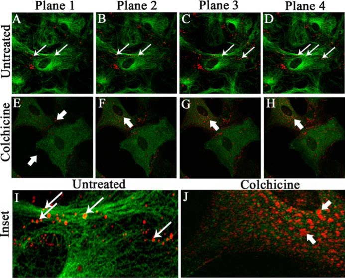
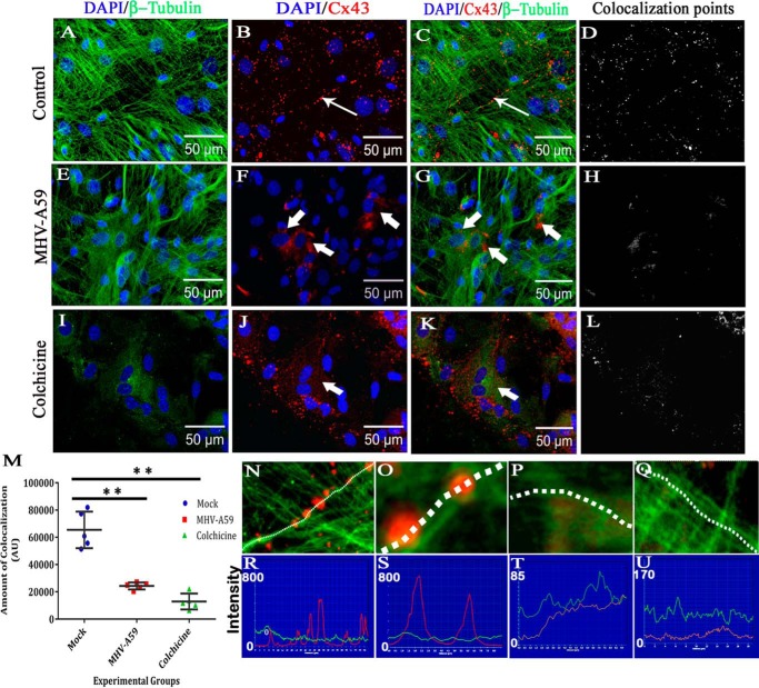
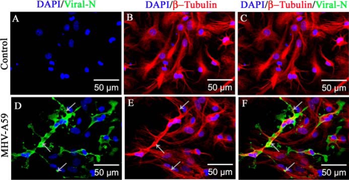
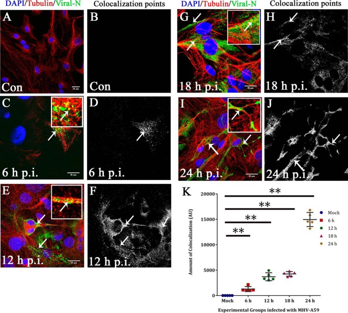
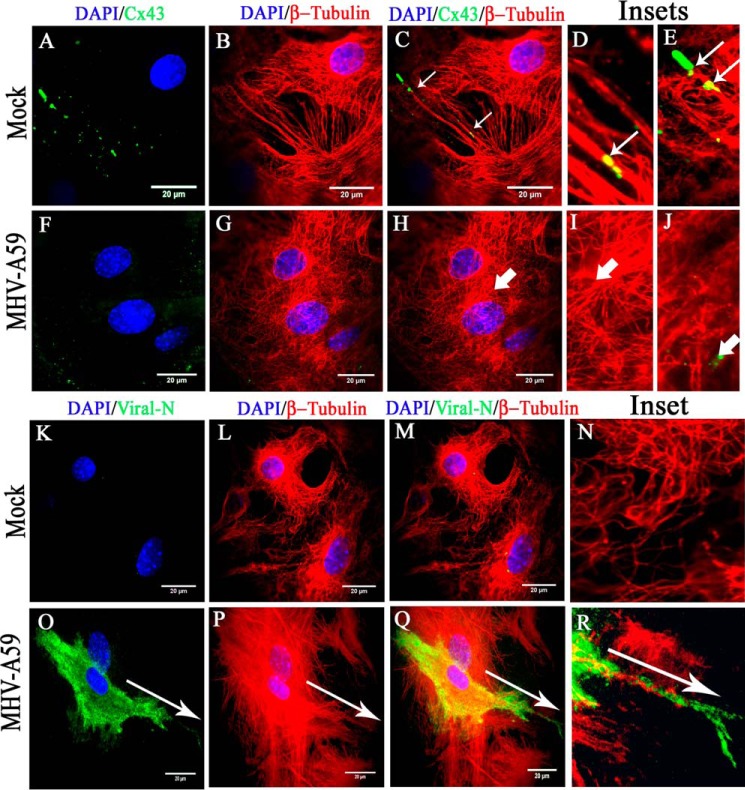
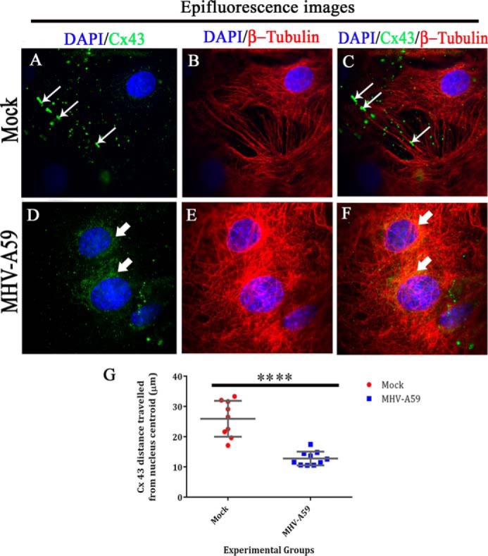
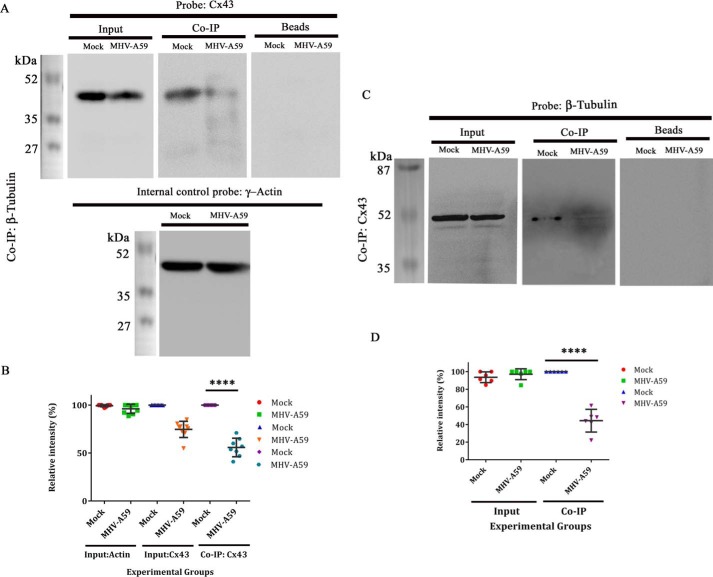
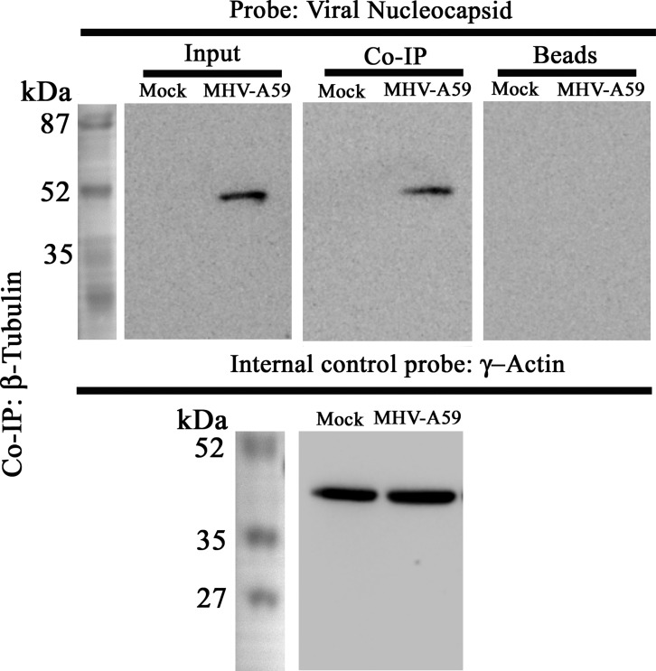
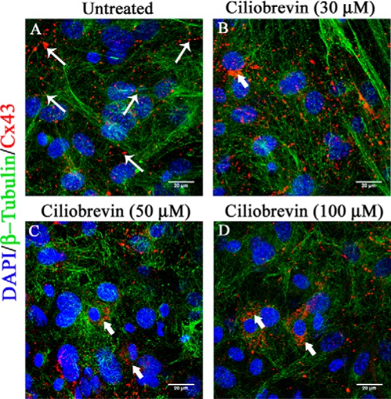
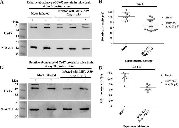
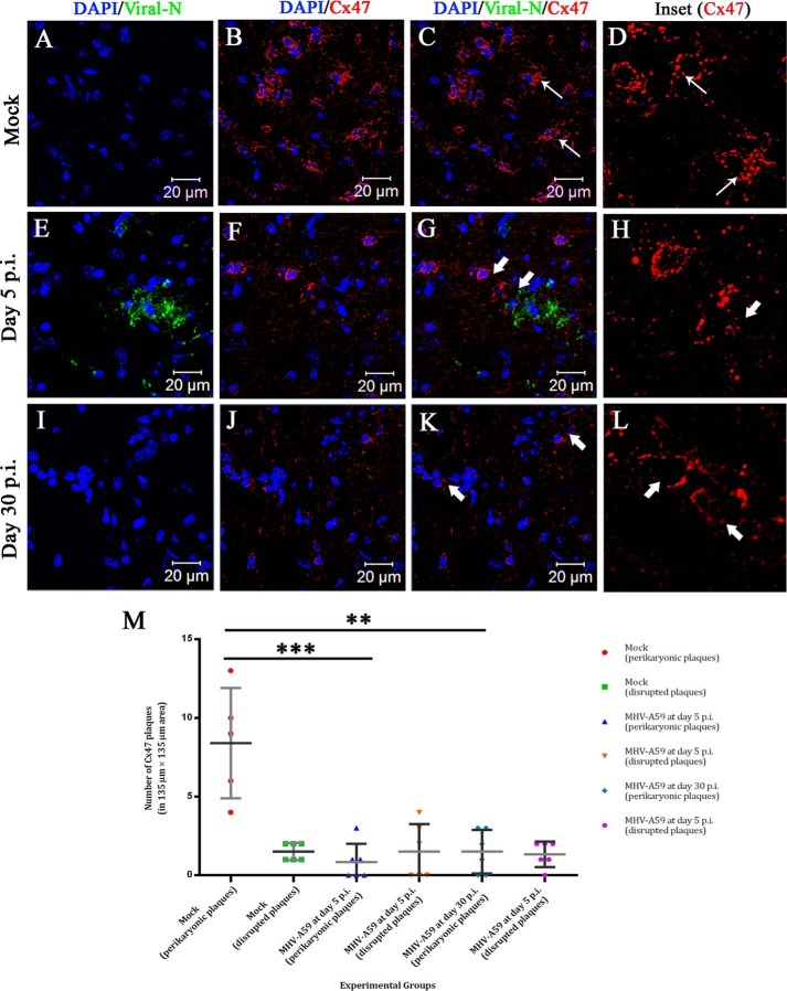
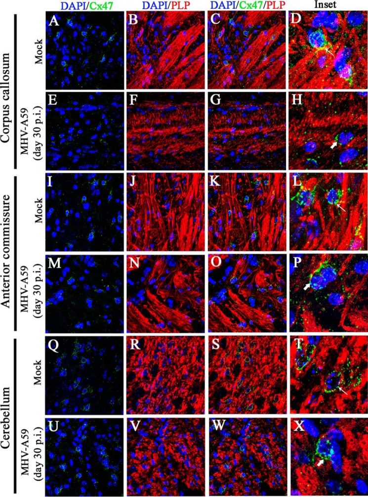
Gap junctions (GJs) are important for maintenance of CNS homeostasis. GJ proteins, connexin 43 (Cx43) and connexin 47 (Cx47), play a crucial role in production and maintenance of CNS myelin. Cx43 is mainly expressed by astrocytes in the CNS and forms gap junction intercellular communications between astrocytes-astrocytes (Cx43-Cx43) and between astrocytes-oligodendrocytes (Cx43-Cx47). Mutations of these connexin (Cx) proteins cause dysmyelinating diseases in humans. Previously, it has been shown that Cx43 localization and expression is altered due to mouse hepatitis virus (MHV)-A59 infection both in vivo and in vitro; however, its mechanism and association with loss of myelin protein was not elaborated. Thus, we explored potential mechanisms by which MHV-A59 infection alters Cx43 localization and examined the effects of viral infection on Cx47 expression and its association with loss of the myelin marker proteolipid protein. Immunofluorescence and total internal reflection fluorescence microscopy confirmed that MHV-A59 used microtubules (MTs) as a conduit to reach the cell surface and restricted MT-mediated Cx43 delivery to the cell membrane. Co-immunoprecipitation experiments demonstrated that Cx43-β-tubulin molecular interaction was depleted due to protein-protein interaction between viral particles and MTs. During acute MHV-A59 infection, oligodendrocytic Cx47, which is mainly stabilized by Cx43 in vivo, was down-regulated, and its characteristic staining remained disrupted even at chronic phase. The loss of Cx47 was associated with loss of proteolipid protein at the chronic stage of MHV-A59 infection.

Keywords: astrocyte; connexin; gap junction; hepatitis virus; microtubule; myelin; oligodendrocyte; tubulin.


Conflict of interest statement

The authors declare that they have no conflicts of interest with the contents of this article

Figures

Figure 1.
Figure 1.
Localization of Cx43 in primary astrocytes upon colchicine treatment. Primary astrocytes were treated with 100 μm colchicine for 24 h. Control cells were maintained in parallel. Cells were subjected to double-label immunofluorescence with rabbit anti-Cx43 antiserum (red) and mouse anti-β-tubulin antiserum (green). Z-stacking was obtained by a confocal microscope from the base of the cells, at the coverslip (Plane 1), to the medial part of the cells (Plane 4), to observe the distribution of Cx43 with microtubule morphology. Untreated cells showed the presence of Cx43 at both the basal (A, thin arrow) and medial parts of cells (B–D). In contrast, at the basal stack of colchicine-treated cells (E, thick arrow), the presence of Cx43 was minimal. The medial stacks showed the presence of Cx43 mostly inside the cells, showing that Cx43 delivery was restricted upon MT disruption, which was confirmed by disrupted β-tubulin staining (F–H, thick arrow). Digitally magnified insets showed that Cx43 was present on MT threads in a single focal plane, and colocalization was evident, specifically where intensities of Cx43 and β-tubulin were similar (I, ringlike yellow spots, thin arrow). Colchicine-treated cells showed smearlike disrupted β-tubulin signal, whereas Cx43 surface localization was restricted (J, thick arrow).
Figure 2.
Figure 2.
Altered association of Cx43 with microtubule in primary astrocytes upon MHV-A59 infection and colchicine treatment. Primary astrocytes were infected with MHV-A59 at an MOI of 2 or treated with 100 μm colchicine for 24 h. Control cells were maintained in parallel. Cells were subjected to double-label immunofluorescence with rabbit anti-Cx43 antiserum (red) and mouse anti-β-tubulin antiserum (green). Cells were counterstained with DAPI (blue). In control astrocytes, punctate Cx43 (B, thin arrow) was observed to be closely associated with β-tubulin (A), and Cx43 staining was aligned along the typical radial structure of MTs (C, thin arrow). Spots of colocalized signal are shown in D. Upon infection of the cells with MHV-A59, Cx43 was retained in the intracellular compartment (F, thick arrow). Although the MT morphology appeared to be normal (E), intracellular compartment–retained Cx43 had a minimal association with the MT network (G, thick arrow). The intensity and number of colocalization spots were reduced (H). Upon colchicine treatment, the MT network was depolymerized in the primary astrocytes, and diffuse tubulin staining was observed in the cytosol (I). Colchicine treatment showed retention of Cx43 in the intracellular compartment, predominantly in the perinuclear area (J and K, thick arrow). The number of colocalization spots was reduced significantly (L). The number of colocalization points compared between experimental groups showed there was an ∼62.8% reduction in virus-infected cells compared with mock-infected cells (**, p < 0.01; Mann–Whitney U test) and ∼80.3% reduction in colchicine-treated cells compared with mock-infected cells (**, p < 0.01; Mann-Whiney U test). Comparison of all three groups by Kruskal–Wallis ANOVA showed that differences were significant (****, p < 0.0001; M). Five fields were analyzed for each group from n = 3 experiments. Digitally magnified images show that Cx43 molecules were aligned along a MT thread (N and O), whereas intracellular compartment–retained Cx43 did not show such alignment (P and Q). Association of Cx43 molecules on a single MT thread is shown (red line, Cx43; green line, tubulin). Cx43 molecules showed high-intensity peaks on MT threads in mock-infected cells (R and S), but not in virus-infected cells (T and U). Error bars, S.D. AU, arbitrary units.
Figure 3.
Figure 3.
Association of MHV-A59 virus particles with microtubules in primary astrocytes. Primary astrocytes were infected with MHV-A59 at an MOI of 2, and mock-infected cells were maintained in parallel. Cells were subjected to double-label immunofluorescence with rabbit anti-β-tubulin antiserum (red) and mouse anti-N antiserum (green). Cells were counterstained with DAPI (blue), and merged image projection (mip) signals were imaged with an apotome microscope. No viral N–specific signal was observed in mock-infected cells (A), but viral N staining was observed to be dispersed from the perinuclear space to the surface of MHV-A59–infected cells (D, arrow). MT morphology is shown in mock-infected (B) and virus-infected (E, arrow) cells. MHV-A59–infected cells showed the virus-specific signal colocalizing with the MTs. Specifically, near the cell periphery, viral N signal was present on MT threads (F, arrow). As expected, no viral N signal was seen in mock-infected cells (C).
Figure 4.
Figure 4.
Kinetics of viral particle spread along microtubule threads. To observe viral spread along MT threads, primary astrocytes were mock-infected (A and B) or MHV-A59–infected at 6 h (C and D), 12 h (E and F), 18 h (G and H), and 24 h (I and J) p.i., and cells were labeled for β-tubulin (red) and viral N (green). The amount of anti-N staining increased from 6 to 24 h p.i. At 6 and 12 h p.i., discrete viral particles were observed on MT threads (C and E, arrow and inset), and colocalization points were mainly located at the cell periphery (D and F). At 18 and 24 h p.i., anti-N signal was more dispersed throughout the whole cell, and toward the cell border, viral particles were localized on MTs (G and I, arrow and inset). The number of colocalization points increased visually, and they were mainly located at the cell periphery and cell-to-cell junctions (H and J). Spots containing colocalization of signal were counted and plotted with increasing time p.i. Colocalization spots increased from 6 to 12 to 18 h p.i. and reached a maximum at 24 h p.i. (**, p < 0.01 each for 6, 12, and 18 h p.i. and 24 h p.i. as compared with mock; Mann-Whitney U test). Kruskal–Wallis testing showed that the difference was significant in a five-group comparison (***, p < 0.001). Five to six fields were analyzed for each group from n = 3 experiments (K). Error bars, S.D. AU, arbitrary units.
Figure 5.
Figure 5.
TIRF microscopy confirmed association of MT network with cortical Cx43, which was depleted due to MT/MHV-A59 interaction. Primary astrocytes, plated on glass coverslips, were mock-infected or infected with MHV-A59 and, upon staining with β-tubulin (red) and Cx43 (green), subjected to TIRF imaging to specifically capture the immunofluorescent signal of cortical Cx43. The imaging depth was limited to within 100 nm of the coverslip. Simultaneously, the whole-cell MT network was imaged by epifluorescence microscopy. In control astrocytes, Cx43 (A) was observed to be closely associated with positive ends of MTs or the MT network (B and C (thin arrow)). Insets show that Cx43 molecules were either precisely aligned along the MT thread (D, thin arrow) or present at the tip of the MT thread (E, thin arrow). In MHV-A59–infected cells, minimal cell surface-associated Cx43 signal was observed (F), because intracellular compartment–retained Cx43 was restricted from reaching the cell surface. MT morphology appeared normal (G). As expected, in infected cells, Cx43 molecules were not associated with positive ends of MTs (H, I, and J, thick arrow) Similarly, mock- and virus-infected cultures of primary astrocytes were stained for β-tubulin (red) and viral N (green), and TIRF imaging was performed as described before. Mock-infected astrocytes showed no viral N–specific signal (K and M), and MT morphology was normal (L and M). The inset shows a digitally magnified area of the mock-infected cell's periphery (N). MHV-A59–infected astrocyte cultures showed the presence of viral N staining (O and Q), and MT morphology is shown in P. Merged images show a pattern of viral spread at the cell surface in a single infected cell, where viral N staining colocalized with MTs (Q). The inset shows that the viral particles were aligned along the MT thread at the cell surface (R). The arrow shows the alignment of viral spread in O–R.
Figure 6.
Figure 6.
Whole-cell expression of Cx43 and β-tubulin upon MHV-A59 infection. Primary astrocytes immunolabeled for Cx43 and β-tubulin, which was subjected to TIRF microscopy, were simultaneously taken for epifluorescence microscopy to obtain the whole-cell Cx43 expression. Thus, parallel epifluorescence images were captured for the same field. Cx43 was observed to be present in profuse amounts as its characteristic punctate stain of Cx43 (A (thin arrow) and C (merged)). In contrast, MHV-A59–infected astrocytes showed mainly perinuclear localization of Cx43 (D (thick arrow) and F (merged)), which was not observed by TIRF imaging. MT morphology is shown for mock-infected (B) or MHV-A59–infected cells (E). The distance of Cx43 molecules from the nucleus (distance was calculated from nuclear centroid) was measured with the help of ImageJ (G). For mock-infected cells, Cx43 was present ∼25.9 μm away, which was reduced to ∼12.7 μm in MHV-A59–infected cells. Data were obtained from nine different images from n = 3 biological replicates, and average ± S.D. (error bars) is represented (****, p < 0.0001; t test).
Figure 7.
Figure 7.
Reduction in Cx43–β-tubulin interaction in protein level upon virus infection. Primary astrocytes were either mock-infected or infected with MHV-A59 at an MOI of 2. Proteins were extracted, followed by immunoprecipitation with monoclonal anti- β-tubulin antibody and subjected to immunoblot analysis using polyclonal anti-Cx43 antibody (detected at nearly 43 kDa). γ-Actin was used as a loading control (detected at nearly 42 kDa). Inputs showed reduction of total Cx43 upon MHV-A59 infection, where γ-actin expression was not altered. Upon co-IP, substantial reduction in tubulin-associated Cx43 was observed in the MHV-A59–infected cells, compared with the mock-infected cells. Beads, used in preclearing, showed no signal upon probing with anti-Cx43 (A). Densitometric analysis showed that Cx43, associated with β-tubulin, was reduced ∼44.25% in MHV-A59–infected cells, compared with the mock-infected cells (B; ****, p < 0.0001; t test). Similarly, the virus- and mock-infected cells were co-immunoprecipitated with polyclonal anti-Cx43 antibody and probed for β-tubulin using monoclonal anti- β-tubulin antibody (detected at nearly 50 kDa). β-Tubulin was expressed in equal amount in mock– and MHV-A59–infected cells. Cx43-associated β-tubulin signal was down-regulated significantly in virus-infected cells (C). Densitometric analysis showed that β-tubulin, associated with Cx43, was depleted ∼55.61% upon virus infection (D; ****, p < 0.0001; n = 3; t test). Error bars, S.D.
Figure 8.
Figure 8.
Interaction between viral particles and β-tubulin. To understand whether reduced Cx43–β-tubulin interaction was due to interaction between viral particle and MTs, co-IP was performed using polyclonal anti-β-tubulin antibody. The immunoprecipitated samples were probed with monoclonal anti-N antibody. Samples were probed for γ-actin, as an internal control. Viral N protein was selectively detectable near 50 kDa, in the MHV-A59–infected sample only, whereas γ-actin expression was similar between mock- and virus-infected cells. MHV-A59–infected cells also showed that viral N-protein interacted with β-tubulin. The beads that were used in preclearing showed absence of nonspecific signal upon probing with anti-Cx43.
Figure 9.
Figure 9.
Altered Cx43 localization upon inhibition of cytoplasmic dynein. Primary astrocytes were treated with a cytoplasmic dynein inhibitor, ciliobrevin D, for 24 h, and DMSO (vehicle)-treated control cells were maintained in parallel. Cells were immunolabeled for β-tubulin (green) and Cx43 (red), and nuclei were counterstained with DAPI (blue). Mock-infected cells showed prominent presence of Cx43 at the cell surface (A, thin arrow). Dose-dependent treatment of ciliobrevin treatment induced aggregated localization of Cx43 around the nucleus, showing that inhibition of dynein restricted Cx43 surface localization (B–D, thick arrow).
Figure 10.
Figure 10.
Persistent loss of oligodendrocytic Cx47 in mouse whole-brain protein. C57Bl/6 mice were mock– or MHV-A59–infected, and proteins were extracted from brain at day 5 p.i. (acute phase) and at day 30 p.i. (chronic phase). Whole-brain proteins were probed for Cx47 (detected at nearly 47 kDa) and internal control, γ-actin (detected at nearly 42 kDa). Oligodendrocytic Cx47 was reduced in brain in the acute stage of inflammation, at day 5 p.i. (A). There was ∼32.78% depletion reduction (normalized with γ-actin) of Cx47 in the whole-brain protein (B). The mean ± S.D. (error bars) incidences from three different animals are shown (***, p < 0.001). At the peak of demyelination at day 30 p.i., Cx47 was not replenished back to its normal level (C). Both of the Cx47-immunoprobed blots showed a nonspecific signal at about 51 kDa. A persistent ∼35.83% reduction in Cx47 expression signal was observed upon normalization with internal control γ-actin (D; ****, p < 0.0001). Error bars, S.D.
Figure 11.
Figure 11.
In situ immunofluorescence data on infected brain tissue demonstrated sustained loss of perikaryonic Cx47 signal in MHV-A59–infected mouse brain. Cryosections were obtained from mock– and MHV-A59–infected mouse brains at days 5 and 30 p.i., and double-label immunofluorescence was performed for viral N (green) and Cx47 (red). Nuclei were stained with DAPI (blue). No virus-specific staining was observed for mock-infected brains (A and C), and prominent Cx47 staining was observed around oligodendrocytic somata (B and C (thin arrow); merged image). The characteristic perikaryonic signal of Cx47 was evident (D, inset). At day 5 p.i., MHV-A59–infected brains showed the presence of viral N signal (E and G). Loss of Cx47 signal was observed specifically around the virus-infected area of the brain (F and G (thick arrow)). The inset shows Cx47 immunostaining was disrupted (H, thick arrow). At the peak of demyelination at day 30 p.i., there was no infectious viral particle observed in brain (I and K). In contrast, disrupted Cx47 staining was noticeable in some areas of the brain (J and K (thick arrow)). Normal Cx47-specific signal, visible in oligodendrocytic perikarya, remained depleted at day 30 p.i. (L, inset, thick arrow). Images (with an area of 135 × 135 μm2) obtained from n = 3 biological replicates were quantified for the presence of complete perikaryonic punctate or disrupted signal of Cx47 (M). A reduction of perikaryonic Cx47 plaque count was observed at day 5 as well as at day 30 p.i. At day 5 p.i., ∼7.6 Cx47 plaques were reduced in the MHV-A59 infected mice, in an area of 135 × 135 μm2 (***, p < 0.001; t test). At day 30 p.i., ∼6.9 intact Cx47 plaques were reduced in an area of 135 × 135 μm2 (**, p < 0.01; t test) (M). Error bars, S.D.
Figure 12.
Figure 12.
Loss of Cx47 staining was associated with loss of PLP staining in chronic phase. Brain sections obtained from mock- and MHV-A59–infected mice were immunolabeled for Cx47 (green) and myelin marker, PLP (red). Nuclei were counterstained with DAPI (blue). In mock-infected mice, Cx47 showed the characteristic stain at oligodendrocytic perikarya, specifically in and around the white-matter regions of brain (e.g. the corpus callosum (A and C), anterior commissure (I and K), and cerebellum (Q and S)). Prominent and profuse PLP staining was observed in normal corpus callosum (B and C), anterior commissure (J and K), and cerebellum (R and S). Insets show perikaryonic Cx47 staining observed in parallel to the PLP-stained myelinated axon fibers (D, L, and T (thin arrow)). MHV-A59–infected mice showed disrupted Cx47 staining, specifically in the corpus callosum (E and G). Anterior commissure (M and O) and cerebellum (U and W) showed Cx47 staining, which appeared to be normal, but the number of Cx47-positive puncta was marginally reduced. The PLP staining was reduced significantly in specific areas of the corpus callosum (F and G), but only marginal loss was evident in anterior commissure (N and O) and cerebellum (V and W). The insets show the altered expression pattern of Cx47 (H, P, and X (thick arrow)). Depletion of Cx47 staining was noticeably associated with the loss of PLP staining in the corpus callosum (H).

References

    1. Das Sarma J., Wang F., and Koval M. (2002) Targeted gap junction protein constructs reveal connexin-specific differences in oligomerization. J. Biol. Chem. 277, 20911–20918 - PubMed
    1. Iacobas D. A., Urban-Maldonado M., Iacobas S., Scemes E., and Spray D. C. (2003) Array analysis of gene expression in connexin-43 null astrocytes. Physiol. Genomics 15, 177–190 - PMC - PubMed
    1. Lutz S. E., Zhao Y., Gulinello M., Lee S. C., Raine C. S., and Brosnan C. F. (2009) Deletion of astrocyte connexins 43 and 30 leads to a dysmyelinating phenotype and hippocampal CA1 vacuolation. J. Neurosci. 29, 7743–7752 - PMC - PubMed
    1. Sofroniew M. V., and Vinters H. V. (2010) Astrocytes: biology and pathology. Acta Neuropathol. 119, 7–35 - PMC - PubMed
    1. Charles A. C., Merrill J. E., Dirksen E. R., and Sanderson M. J. (1991) Intercellular signaling in glial cells: calcium waves and oscillations in response to mechanical stimulation and glutamate. Neuron 6, 983–992 - PubMed

Publication types

LinkOut - more resources