. 2017 May 31;6(6):e006195.
doi: 10.1161/JAHA.117.006195.

Colchicine Depolymerizes Microtubules, Increases Junctophilin-2, and Improves Right Ventricular Function in Experimental Pulmonary Arterial Hypertension

Affiliations

Colchicine Depolymerizes Microtubules, Increases Junctophilin-2, and Improves Right Ventricular Function in Experimental Pulmonary Arterial Hypertension

Kurt W Prins et al. J Am Heart Assoc. .

Abstract

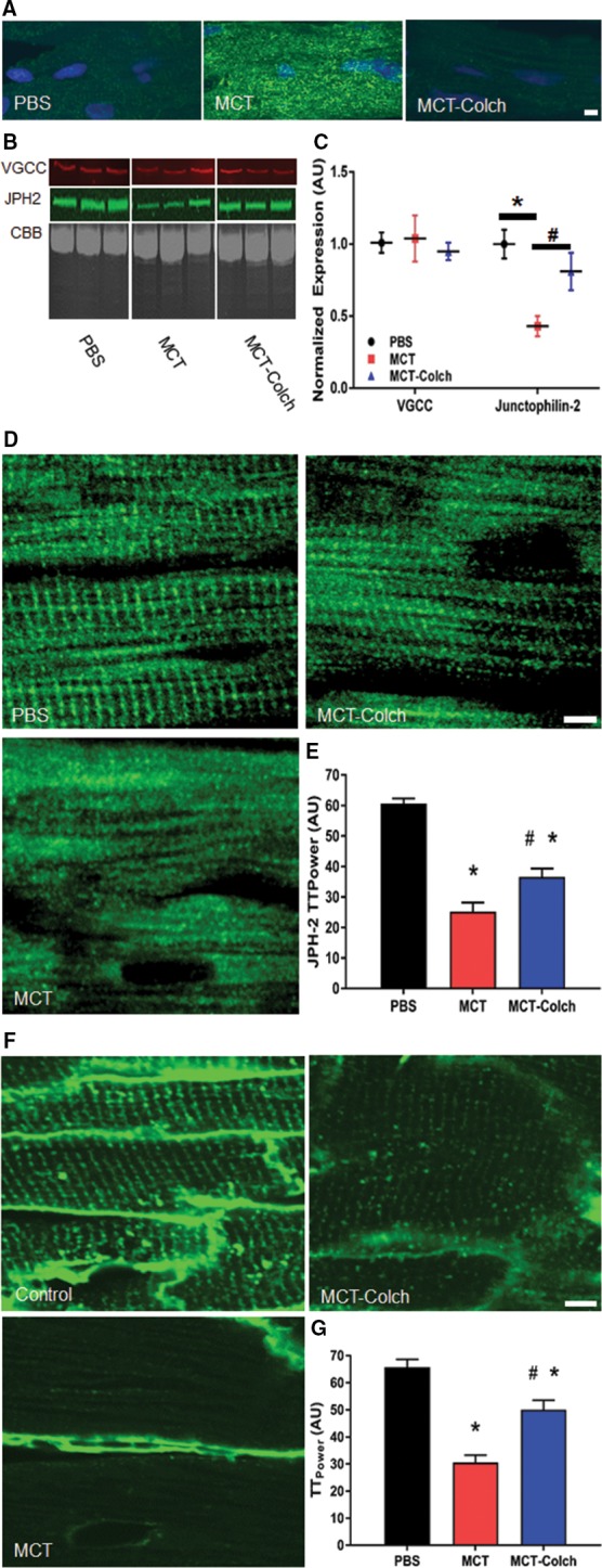
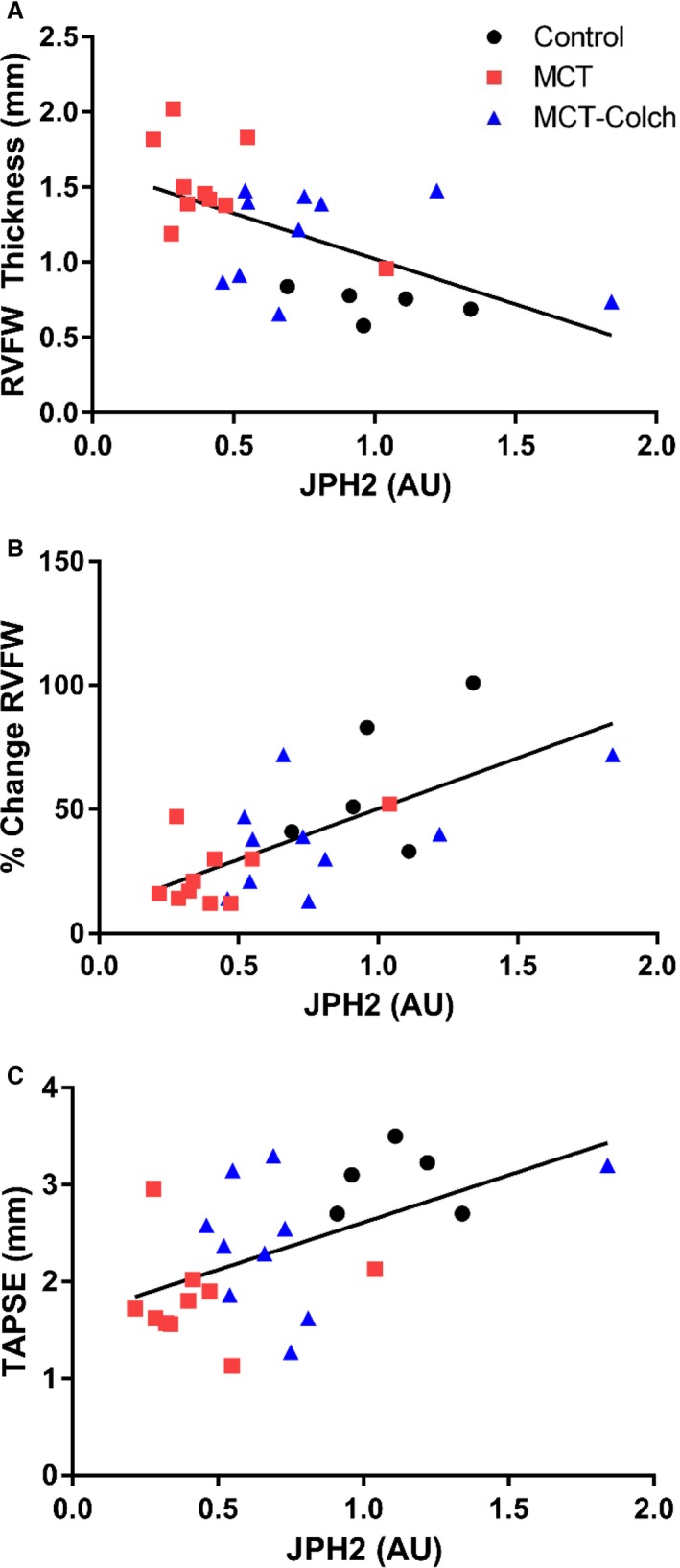
Background: Pulmonary arterial hypertension (PAH) is a lethal disease characterized by obstructive pulmonary vascular remodeling and right ventricular (RV) dysfunction. Although RV function predicts outcomes in PAH, mechanisms of RV dysfunction are poorly understood, and RV-targeted therapies are lacking. We hypothesized that in PAH, abnormal microtubular structure in RV cardiomyocytes impairs RV function by reducing junctophilin-2 (JPH2) expression, resulting in t-tubule derangements. Conversely, we assessed whether colchicine, a microtubule-depolymerizing agent, could increase JPH2 expression and enhance RV function in monocrotaline-induced PAH.

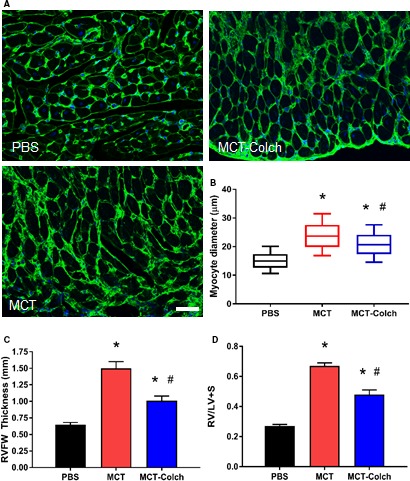
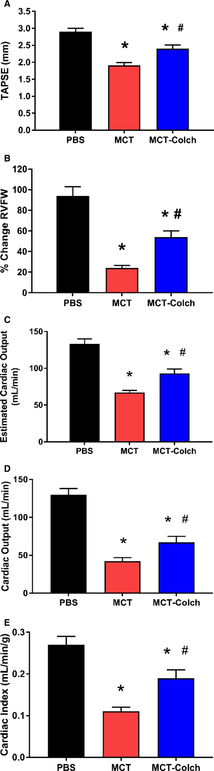
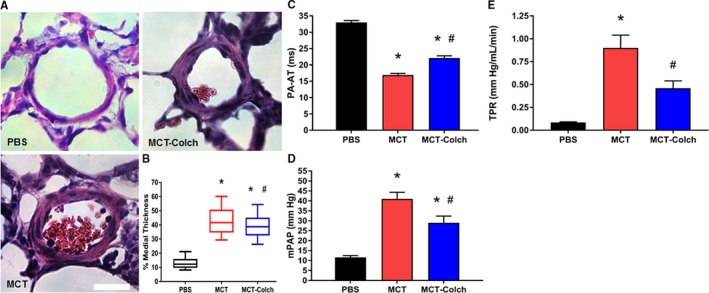
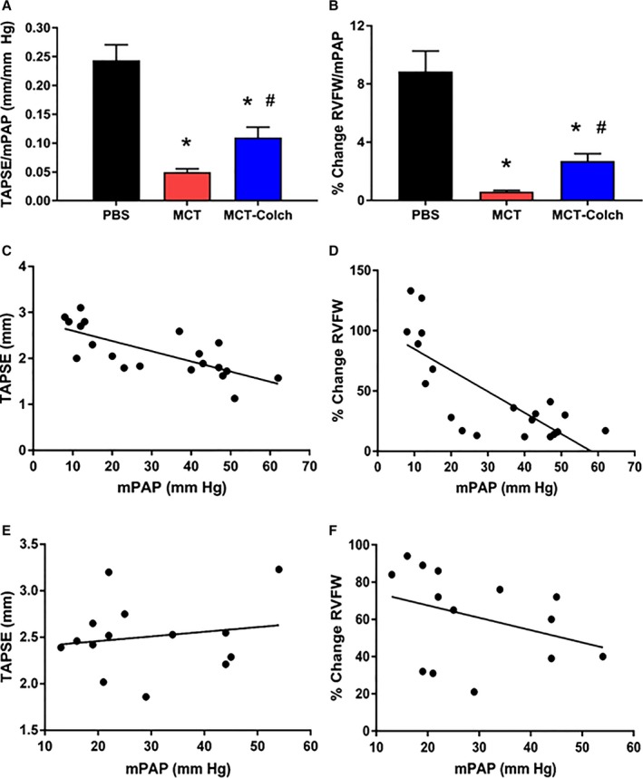
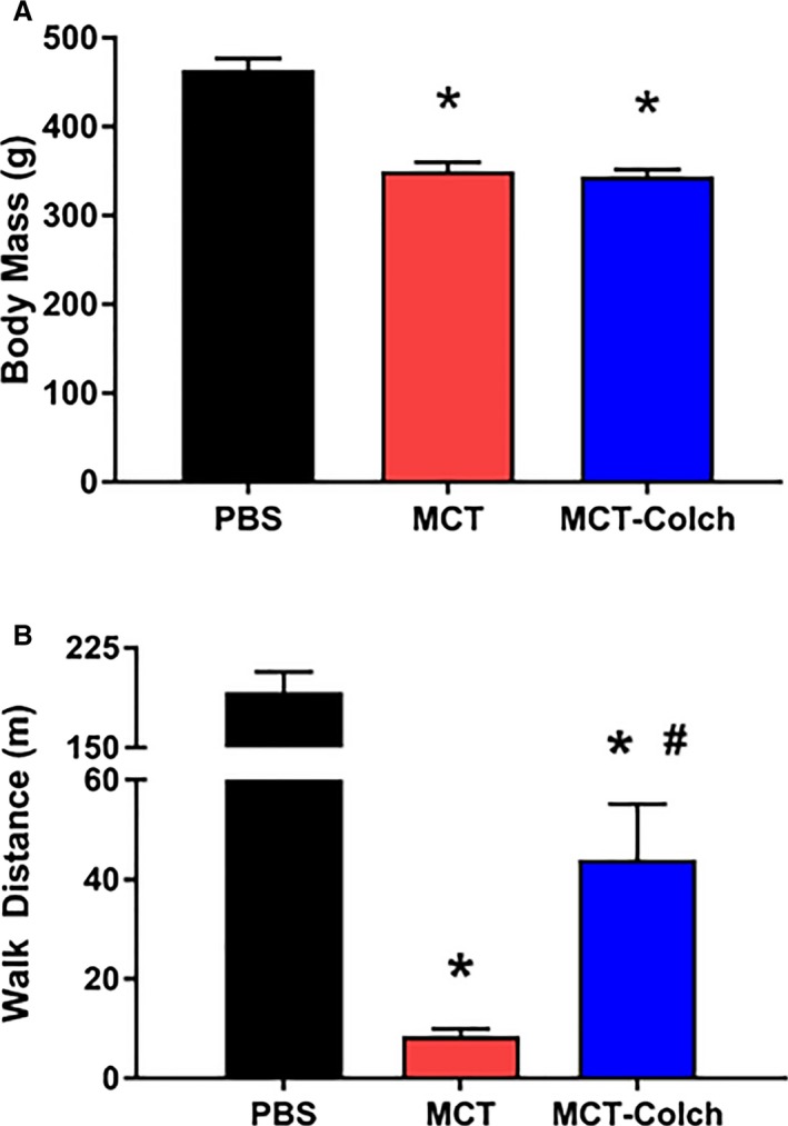
Methods and results: Immunoblots, confocal microscopy, echocardiography, cardiac catheterization, and treadmill testing were used to examine colchicine's (0.5 mg/kg 3 times/week) effects on pulmonary hemodynamics, RV function, and functional capacity. Rats were treated with saline (n=28) or colchicine (n=24) for 3 weeks, beginning 1 week after monocrotaline (60 mg/kg, subcutaneous). In the monocrotaline RV, but not the left ventricle, microtubule density is increased, and JPH2 expression is reduced, with loss of t-tubule localization and t-tubule disarray. Colchicine reduces microtubule density, increases JPH2 expression, and improves t-tubule morphology in RV cardiomyocytes. Colchicine therapy diminishes RV hypertrophy, improves RV function, and enhances RV-pulmonary artery coupling. Colchicine reduces small pulmonary arteriolar thickness and improves pulmonary hemodynamics. Finally, colchicine increases exercise capacity.

Conclusions: Monocrotaline-induced PAH causes RV-specific derangement of microtubules marked by reduction in JPH2 and t-tubule disarray. Colchicine reduces microtubule density, increases JPH2 expression, and improves both t-tubule architecture and RV function. Colchicine also reduces adverse pulmonary vascular remodeling. These results provide biological plausibility for a clinical trial to repurpose colchicine as a RV-directed therapy for PAH.

Keywords: T‐tubules; pulmonary hypertension; right ventricular failure; right ventricular pressure overload.


Figures

Figure 1
Figure 1
Colchicine increases JPH2 protein levels, restores JPH2 to t‐tubules, and mitigates pathological t‐tubule remodeling in the RV in MCT rats. A, Confocal micrographs of RV sections stained with a β‐tubulin antibody (green) to delineate microtubules. Microtubules are depolymerized in the MCT‐colchicine rats, and MCT rats have increased microtubule density as compared with PBS controls. B, Representative Western blots from 3 PBS, MCT, and MCT‐colchicine RV extracts and (C) quantification of VGCC (PBS 101±7 AU, n=5; MCT 104±16 AU, n=5; and MCT‐colchicine 95±6 AU, n=5) and JPH2 (PBS 100±11 AU, n=5, MCT 44±8 AU, n=10; and MCT‐colchicine 81±9 AU, n=10). Colchicine treatment significantly increases JPH2 expression. D, Confocal micrographs of RV sections stained for JPH2 (green). The striated pattern of JPH2 is partially restored with colchicine treatment. (PBS 70.6±1.6 AU, n=54 cells from 3 animals, MCT 25.2±3.0 AU, n=50 cells from 3 animals, MCT‐colchicine 36.6±2.7 AU, n=46 cells from 3 animals) (E). Confocal micrographs of RV sections stained with WGA (green) to label t‐tubules (F). Colchicine treatment improves t‐tubule architecture (PBS 65.7±2.9 AU, n=49 cells from 3 animals, MCT 30.6±2.7 AU, n=50 cells from 3 animals, MCT‐colchicine 50.1±3.5 AU, n=45 cells from 3 animals) (G). *Significantly different from PBS; #significantly different from MCT rats as determined by 1‐way ANOVA with Tukey post hoc analysis. Scale bar: 5 μm in all images. AU indicates arbitrary units; CBB, Coomassie brilliant blue; Colch, colchicine; JPH2, junctophilin‐2; MCT, monocrotaline; PBS, phosphate‐buffered saline; RV, right ventricle; VGCC, voltage‐gated calcium channel; WGA, wheat germ agglutinin.
Figure 2
Figure 2
Paclitaxel induces JPH2 mislocalization and t‐tubule disruptions in cultured control rat RV cardiomyocytes. A, Representative confocal micrographs of isolated RV cardiomyocytes stained with JPH2 antibody (green). B, Quantification of JPH2 localization patterns (DMSO 75.4±1.9, paclitaxel 68.1±1.9, P=0.01). Paclitaxel treatment partially disrupts the striated JPH2 staining pattern. C, Confocal micrographs of isolated cardiomyocytes stained with WGA (green) to delineate t‐tubules. D, Quantification of t‐tubule architecture (DMSO 55.2±1.6, paclitaxel 44.9±3.2, P=0.006). Paclitaxel treatment induces t‐tubule remodeling. Scale bar: 5 μm. *P<0.05 as determined by t test. DMSO indicates dimethyl sulfoxide; JPH2, junctophilin‐2; RV, right ventricle; WGA, wheat germ agglutinin.
Figure 3
Figure 3
Colchicine reduces right ventricular hypertrophy in MCT rats. A, WGA stained RVFW sections. B, Quantification of cardiomyocyte diameter using a box‐and‐whisker plot. Whiskers extend from 10th to 90th percentile. (PBS 15.1±0.2 μm, n=617 cells from 3 animals, MCT 23.9±0.3 μm, n=474 cells from 3 animals, MCT‐colchicine 21.0±0.2 μm, n=584 cells from 3 animals.) Colchicine significantly reduces RV cardiomyocyte diameter. C, Quantification of diastolic RVFW thickness on echocardiography. (PBS 0.6±0.03 mm, n=16, MCTPBS 1.5±0.10 mm, n=21, MCT‐colchicine 1.0±0.07 mm, n=22.) D, Fulton index in isolated hearts. (PBS 0.21±0.01, n=12, MCT 0.67±0.02, n=15, MCT‐colchicine 0.48±0.03, n=16. Colchicine blunts RV hypertrophy. *Significantly different from PBS; #significantly different from MCT rats as determined by 1‐way ANOVA with Tukey post hoc analysis. Scale bar: 50 μm. Colch indicates colchicine; MCT, monocrotaline; PBS, phosphate‐buffered saline; RVFW, right ventricle free wall; RV/LV+S, RV free wall mass/LV and septal mass; WGA, wheat germ agglutinin.
Figure 4
Figure 4
Colchicine therapy improves in vivo RV function in MCT rats. On echocardiography, TAPSE (PBS 2.9±0.01 mm, n=16, MCT 1.9±0.08 mm, n=21, MCT‐colchicine 2.4±0.10 mm, n=22) (A), percentage change in RVFW thickness (PBS 94±9%, n=16, MCT 24±3%, n=21, MCT‐colchicine 54±6%, n=22) (B), and estimated cardiac output (CO) (PBS 133±7 mL/min, n=16, MCT 67±3 mL/min, n=21, MCT‐colchicine 93±6 mL/min, n=22) (C) are all improved with colchicine treatment in MCT rats. Right heart catheterization‐derived thermodilution CO (PBS 130±8 mL/min, n=5, MCT 42±5 mL/min, n=10, MCT‐colchicine 67±8 mL/min, n=13) (D) and cardiac index (PBS 0.27±0.02 mL/[min·g], n=5, MCT 0.11±0.10 mL/[min·g], n=10, MCT‐colchicine 0.19±0.02 mL/[min·g], n=13) (E) are increased with colchicine treatment in MCT rats. *Significantly different from PBS, #significantly different from MCT rats as determined by 1‐way ANOVA with Tukey post hoc analysis. Colch indicates colchicine; MCT, monocrotaline; PBS, phosphate‐buffered saline; RVFW, right ventricle free wall; TAPSE, tricuspid annular plane systolic excursion.
Figure 5
Figure 5
Reduced JPH2 expression is associated with right ventricular hypertrophy and RV dysfunction in vivo. JPH2 levels are inversely associated with diastolic RVFW thickness (r=−0.58, P=0.002) (A) and are positively correlated with RV function as determined by percentage change in RVFW thickness (r=0.67, P=0.0003) (B) and TAPSE (r=0.54, P=0.005) (C). AU indicates arbitrary units; JPH2, junctophilin‐2; RVFW, right ventricle free wall; TAPSE, tricuspid annular plane systolic excursion.
Figure 6
Figure 6
Colchicine reduces severity of pulmonary vascular disease in MCT rats. A, Representative hematoxylin and eosin–stained lung sections showing pulmonary arterioles. B, Box‐and‐whisker plots of percentage medial thickness (PBS 13.5±0.6% n=85 arterioles from 3 different animals, MCT 43.4±1.5% n=78 arterioles from 3 animals, MCT‐colchicine 39.5±1.1% n=81 arterioles from 3 different animals). Whiskers extend from 10th to 90th percentiles. Colchicine reduces pulmonary arteriolar medial thickness. C, Colchicine increases PA acceleration time (PBS 33±1 milliseconds, n=16, MCT 17±1 milliseconds, n=21, MCT‐colchicine 22±1 milliseconds n=22). Quantification of (D) mPAP (PBS 12±1 mm Hg, n=5, MCT 41±3 mm Hg, n=13, MCT‐colchicine 29±3 mm Hg n=14) and (E) TPR (PBS 0.1±0.01 mm Hg/[mL·min], n=5, MCT 0.9±0.1 mm Hg/[mL·min], n=9, MCT‐colchicine 0.5±0.1 mm Hg/[mL·min], n=11) from right heart catheterization. Colchicine improves pulmonary hemodynamics. *Significantly different from PBS, #significantly different from MCT rats as determined by 1‐way ANOVA with Tukey post hoc analysis. Scale bar 25 μm. Colch indicates colchicine; MCT, monocrotaline; mPAP, mean pulmonary arterial pressure; PBS, phosphate‐buffered saline; TPR, total pulmonary resistance.
Figure 7
Figure 7
RVPA coupling is enhanced with colchicine treatment. Colchicine improves RVPA coupling as quantified by TAPSE/mPAP (PBS 0.24±0.03 mm/mm Hg, n=7, MCT 0.05±0.01 mm/mm Hg, n=13, MCT‐colchicine 0.11±0.02 mm/mm Hg, n=14) (A) or percentage change in RVFW thickness/mPAP (PBS 8.9±1.4%/mm Hg, n=7, MCT 0.6±0.1%/mm Hg, n=13, MCT‐colchicine 2.7±0.5%/mm Hg, n=14) (B). There are significant inverse relationships between (C) TAPSE (r=−0.73, P=0.0003) and mPAP and (D) percentage change in RVFW thickness and mPAP in control and MCT rats (r=−0.77, P<0.0001) but not in MCT‐colchicine rats: TAPSE and mPAP (r=0.17 P=0.56) (E) and percentage change in RVFW thickness and mPAP (r=−0.35 P=0.22) (F). *Significantly different from PBS, #significantly different from MCT rats as determined by 1‐way ANOVA with Tukey post hoc analysis. Colch indicates colchicine; MCT, monocrotaline; mPAP, mean pulmonary arterial pressure; PA, pulmonary artery; PBS, phosphate‐buffered saline; RVFW, right ventricle free wall; TAPSE, tricuspid annular plane systolic excursion.
Figure 8
Figure 8
Colchicine is well tolerated and increases exercise capacity in MCT rats. A, No significant difference in mass at end of study with colchicine treatment (PBS 464±13 g, MCT 350±10 g, and MCT‐colchicine 344±8 g); (B) treadmill walk distance is improved in colchicine‐treated MCT rats (PBS 192±15 m, n=7, MCT 9±2 m, n=20, MCT‐colchicine 44±11 m, n=14). Colch indicates colchicine; MCT, monocrotaline; PBS, phosphate‐buffered saline.

References

    1. Thenappan T, Shah SJ, Rich S, Tian L, Archer SL, Gomberg‐Maitland M. Survival in pulmonary arterial hypertension: a reappraisal of the NIH risk stratification equation. Eur Respir J. 2010;35:1079–1087. - PMC - PubMed
    1. Benza RL, Miller DP, Gomberg‐Maitland M, Frantz RP, Foreman AJ, Coffey CS, Frost A, Barst RJ, Badesch DB, Elliott CG, Liou TG, McGoon MD. Predicting survival in pulmonary arterial hypertension: insights from the Registry to Evaluate Early and Long‐Term Pulmonary Arterial Hypertension Disease Management (REVEAL). Circulation. 2010;122:164–172. - PubMed
    1. Humbert M, Sitbon O, Chaouat A, Bertocchi M, Habib G, Gressin V, Yaici A, Weitzenblum E, Cordier JF, Chabot F, Dromer C, Pison C, Reynaud‐Gaubert M, Haloun A, Laurent M, Hachulla E, Simonneau G. Pulmonary arterial hypertension in France: results from a national registry. Am J Respir Crit Care Med. 2006;173:1023–1030. - PubMed
    1. Farber HW, Loscalzo J. Pulmonary arterial hypertension. N Engl J Med. 2004;351:1655–1665. - PubMed
    1. Thenappan T, Prins KW, Pritzker MR, Scandurra J, Volmers K, Weir EK. The critical role of pulmonary arterial compliance in pulmonary hypertension. Ann Am Thorac Soc. 2016;13:276–284. - PMC - PubMed

MeSH terms

LinkOut - more resources