Review
. 2017 May;7(5):170069.
doi: 10.1098/rsob.170069.

Sphingolipids: membrane microdomains in brain development, function and neurological diseases

Affiliations
Review

Sphingolipids: membrane microdomains in brain development, function and neurological diseases

Anne S B Olsen et al. Open Biol. 2017 May.

Abstract

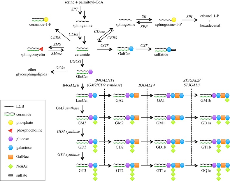
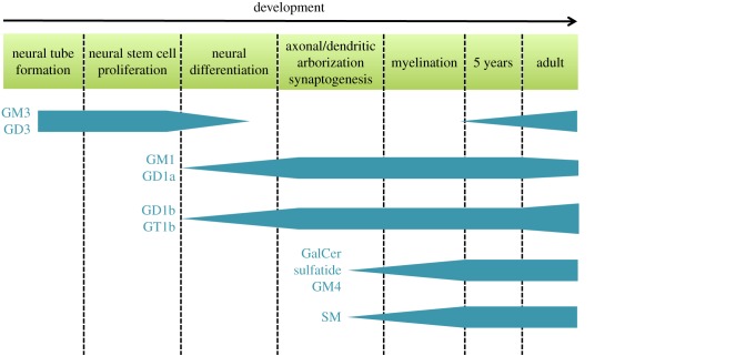
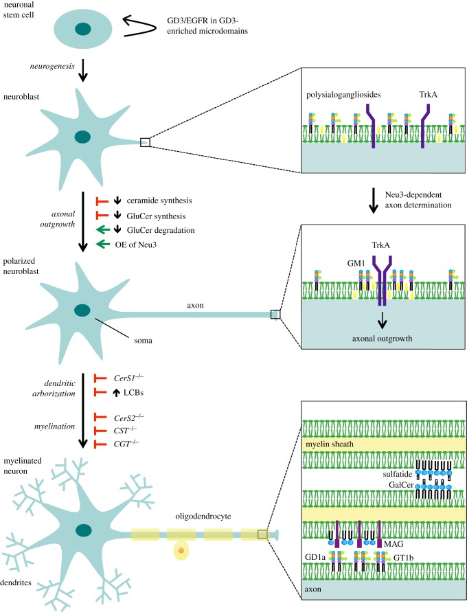
Sphingolipids are highly enriched in the nervous system where they are pivotal constituents of the plasma membranes and are important for proper brain development and functions. Sphingolipids are not merely structural elements, but are also recognized as regulators of cellular events by their ability to form microdomains in the plasma membrane. The significance of such compartmentalization spans broadly from being involved in differentiation of neurons and synaptic transmission to neuronal-glial interactions and myelin stability. Thus, perturbations of the sphingolipid metabolism can lead to rearrangements in the plasma membrane, which has been linked to the development of various neurological diseases. Studying microdomains and their functions has for a long time been synonymous with studying the role of cholesterol. However, it is becoming increasingly clear that microdomains are very heterogeneous, which among others can be ascribed to the vast number of sphingolipids. In this review, we discuss the importance of microdomains with emphasis on sphingolipids in brain development and function as well as how disruption of the sphingolipid metabolism (and hence microdomains) contributes to the pathogenesis of several neurological diseases.

Keywords: brain; ganglioside; membrane microdomain; neurological disease; raft; sphingolipid.


Conflict of interest statement

We declare we have no competing interests.

Figures

Figure 1.
Figure 1.
Overview of the sphingolipid metabolism. Sphingolipids encompass a broad spectrum of lipids. Ceramide is central in the sphingolipid metabolism as it serves as a precursor for the synthesis of more complex sphingolipids. Ceramide is synthesized de novo from serine and palmitoyl-CoA. Subsequently, complex sphingolipids are synthesized by attachment of different head groups to ceramide as indicated in the figure. In particular, ganglioside biosynthesis has been highlighted. Gangliosides are mono- or multi-sialosylated glycosphingolipids, which are highly abundant in the nervous system. Their synthesis is a multistep process of addition of sugars and sialic acids. Degradation of complex sphingolipids contributes to the pool of ceramide that can either be re-used for complex sphingolipid synthesis or alternatively be broken down. Degradation of glycosphingolipids by glycosidases and sialidases is not indicated in the figure. Abbreviations: beta-1,4-N-acetyl-galactosaminyl transferase 1 (B4GALNT1), beta-1,3-galactosyltransferase 4 (B3GALT4), beta-1,4-galactosyltransferase 6 (B4GALT6), ceramidase (CDase), ceramide galactosyltransferase (CGT), ceramide kinase (CERK), ceramide synthase (CERS), galactosylceramide sulfotransferase (CST), glycosphingolipid synthases (GCSs), serine palmitoyltransferase (SPT), sphingomyelin synthase (SMS), sphingomyelinase (SMase), sphingosine kinase (SK), sphingosine 1-phosphate phosphatase (SPP), sphingosine 1-phosphate lyase (SPL), ST3 beta-galactoside alpha-2,3-sialyltransferase 2 (ST3GAL2), ST3 beta-galactoside alpha-2,3-sialyltransferase 3 (ST3GAL3).
Figure 2.
Figure 2.
Outline of how key sphingolipids change during neurodevelopment and ageing. During development of the nervous system the ganglioside profile changes from the simple species (GM3 and GD3) early in embryogenesis to the more complex gangliosides (GM1, GD1a, GD1b and GT1b) later in embryogenesis. Concurrent with myelination, the levels of the myelin sphingolipids sphingomyelin (SM), galactosylceramide (GalCer), sulfatide and GM4 increase. In adulthood, the ganglioside profile changes again with increasing levels of GM3, GD3, GD1b and GT1b, while the levels of GM1 and GD1a decrease.
Figure 3.
Figure 3.
Roles of sphingolipids in neuronal and glial development and interaction. Sphingolipids are involved in multiple steps of the development of the nervous system. Ganglioside GD3 is important for neuronal stem cell proliferation during which it is found co-localizing with the epidermal growth factor receptor (EGFR) in microdomains. Inhibition of ceramide and glucosylceramide (GluCer) synthesis both inhibit axonal outgrowth, while inhibition of GluCer degradation and overexpression (OE) of the sialidase Neu3 stimulate axonal outgrowth. Neu3 stimulates breakdown of polysialogangliosides to GM1, which recruits the nerve growth factor receptor TrkA into microdomains thereby promoting axonal outgrowth. Dendritic arborization is reduced in CerS1−/ mice, which is likely to be caused by increase of long-chain bases (LCBs). Myelination defects have been found in mice deficient in ceramide synthase 2 (CERS2), GalCer sulfotransferase (CST) as well as UDP-galactose:ceramide galactosyltransferase (CGT). Sphingolipids are important for myelin stability. Galactosylceramide (GalCer) and sulfatide in microdomains in opposing membranes of the myelin sheath form glycosynapses important for long-term myelin stability. GD1a and GT1b in axonal membrane microdomains contribute to myelin stability by interacting with myelin-associated glycoprotein (MAG) residing in the myelin sheath.

Similar articles

Cited by

References

    1. van Echten-Deckert G, Herget T. 2006. Sphingolipid metabolism in neural cells. Biochim. Biophys. Acta 1758, 1978–1994. (doi:10.1016/j.bbamem.2006.06.009) - DOI - PubMed
    1. Piccinini M, et al. 2010. Deregulated sphingolipid metabolism and membrane organization in neurodegenerative disorders. Mol. Neurobiol. 41, 314–340. (doi:10.1007/s12035-009-8096-6) - DOI - PubMed
    1. Aureli M, Grassi S, Prioni S, Sonnino S, Prinetti A. 2015. Lipid membrane domains in the brain. Biochim. Biophys. Acta 1851, 1006–1016. (doi:10.1016/j.bbalip.2015.02.001) - DOI - PubMed
    1. O'Brien JS, Sampson EL. 1965. Lipid composition of the normal human brain: gray matter, white matter, and myelin. J. Lipid Res. 6, 537–544. - PubMed
    1. Svennerholm L, Vanier MT. 1973. The distribution of lipids in the human nervous system. IV. Fatty acid composition of major sphingolipids of human infant brain. Brain Res. 55, 413–423. (doi:10.1016/0006-8993(73)90306-5) - DOI - PubMed

Publication types